Understanding the Joint Centres of a Finger: Anatomy, Function, and Care
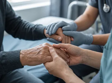
The human hand is a marvel of biological engineering, capable of executing movements ranging from the delicate plucking of a violin string to the robust grip required for lifting heavy objects. At the heart of this dexterity lies a complex network of bones, tendons, ligaments, and nerves. However, the true engines of this movement are the articulation points known as the joint centres of a finger. Understanding the anatomy and function of these specific structures is not merely an academic exercise; it is fundamental to maintaining hand health, preventing injury, and managing chronic conditions such as arthritis.
For many individuals, the significance of these joints goes unnoticed until pain or stiffness sets in. Whether due to age, occupational hazards, or systemic disease, the degradation of the joint centres of a finger can significantly impact quality of life. This comprehensive guide delves deep into the anatomical intricacies of these joints, exploring their biomechanics, common pathologies, and evidence-based strategies for preservation and care. By gaining a clearer understanding of how these joints function, you can take proactive steps to ensure your hands remain strong, flexible, and pain-free for years to come.
Anatomical Structure of the Finger Joints
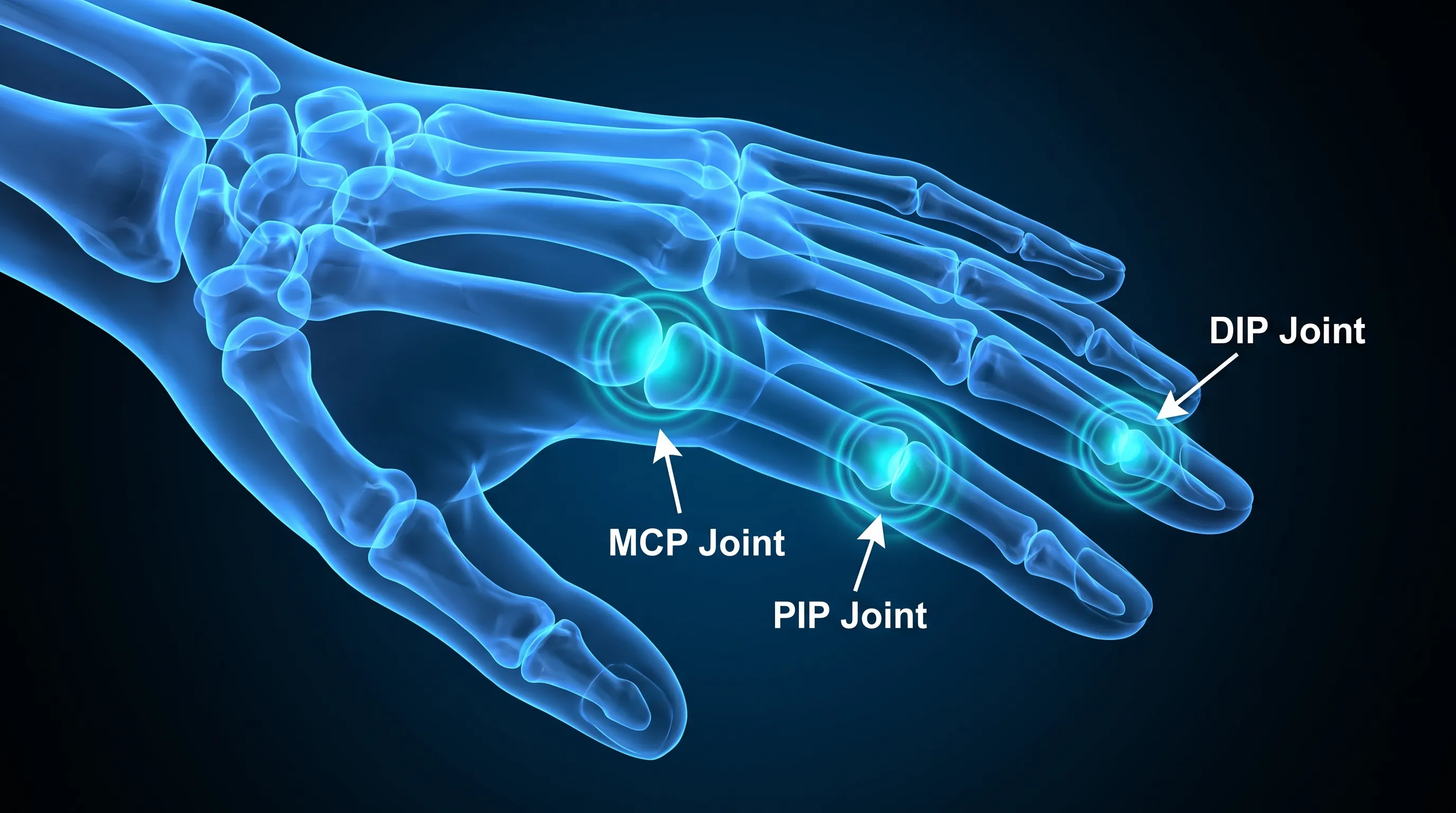
To fully appreciate the complexity of hand movement, one must first understand the skeletal framework that supports it. Each finger, excluding the thumb which has a unique structure, consists of three bones called phalanges: the proximal phalanx (closest to the hand), the middle phalanx, and the distal phalanx (at the fingertip). These bones are connected by three distinct types of joints, collectively referred to as the joint centres of a finger. Each joint type has a specific anatomical configuration designed to facilitate particular ranges of motion [1].
Free calculatorFoot Reflexology ChartFoot reflexology points and corresponding organsOpen the calculatorThe Metacarpophalangeal (MCP) Joints
The first of the joint centres of a finger is the Metacarpophalangeal (MCP) joint. This is the large knuckle visible when you make a fist. It forms the articulation between the head of the metacarpal bone in the palm and the base of the proximal phalanx of the finger. Anatomically, the MCP joint is classified as a condyloid joint, which allows for movement in two planes: flexion-extension (bending and straightening) and abduction-adduction (spreading the fingers apart and bringing them together).
The stability of the MCP joint is maintained by a complex system of ligaments, including the collateral ligaments on either side and the volar plate on the palm side. The volar plate is a crucial structure that prevents the joint from hyperextending backward. Because the MCP joints bear significant load during gripping activities, they are particularly susceptible to stress and inflammatory conditions. Understanding the mechanics of the MCP joint is essential for diagnosing issues related to grip strength and hand alignment [2].
The Proximal Interphalangeal (PIP) Joints
Moving distally along the finger, we encounter the second of the joint centres of a finger: the Proximal Interphalangeal (PIP) joint. This joint connects the proximal phalanx to the middle phalanx. Unlike the MCP joint, the PIP joint is a hinge joint (ginglymus), which primarily allows for flexion and extension only. It does not permit side-to-side movement. This restriction provides stability during powerful gripping actions, ensuring that the force applied by the fingers is directed efficiently.
The PIP joint is often considered the most stable of the finger joints due to its deep bony congruence and strong ligamentous support. However, this same stability makes it vulnerable to specific types of injuries, such as dislocations or fractures, if forced beyond its limited range of motion. The central slip of the extensor tendon attaches to the base of the middle phalanx at this joint, playing a vital role in straightening the finger. Damage to this tendon can lead to conditions like Boutonniere deformity, highlighting the critical importance of the PIP joint in overall hand function [3].
The Distal Interphalangeal (DIP) Joints
The final articulation point is the Distal Interphalangeal (DIP) joint, which connects the middle phalanx to the distal phalanx. Like the PIP joint, the DIP joint is a hinge joint, allowing for flexion and extension. This joint is responsible for the fine motor control needed for tasks such as typing, buttoning a shirt, or picking up small objects. The DIP joint is smaller and has less soft tissue coverage than the other joint centres of a finger, making it more exposed to external trauma.
Despite its small size, the DIP joint plays a disproportionate role in precision grip. The terminal tendon of the extensor mechanism inserts here, enabling the subtle adjustments necessary for delicate tasks. Osteoarthritis frequently affects the DIP joints, leading to the formation of Heberden’s nodes—bony enlargements that can cause pain and deformity. Understanding the specific vulnerabilities of the DIP joint helps in tailoring protective strategies for individuals engaged in fine manual work [4].
Biomechanics and Movement Dynamics
The functionality of the hand is not just about the individual joints but how they work in concert. The biomechanics of the joint centres of a finger involve a sophisticated interplay of bones, ligaments, tendons, and muscles. This synergy allows for a wide range of motion while maintaining the stability necessary for forceful activities.
The Role of Ligaments and the Volar Plate
Ligaments are dense bands of connective tissue that connect bones to other bones, providing stability to the joint centres of a finger. The collateral ligaments, located on the sides of each joint, tighten when the joint is extended and loosen slightly when flexed. This dynamic tension ensures that the joint remains stable throughout its range of motion.
The volar plate, mentioned earlier, is a specialized structure found in the MCP and PIP joints. It acts as a check-rein against hyperextension. Composed of fibrocartilage, the volar plate also contributes to the depth of the joint socket, enhancing stability. Injuries to the volar plate, such as tears or avulsions, can lead to chronic instability and pain, underscoring its critical role in the biomechanics of the finger [5].
Tendon Mechanics and Extensor Apparatus
Tendons connect muscles to bones, transmitting the force generated by muscle contraction to produce movement. The extensor apparatus of the finger is a complex network of tendons that covers the dorsal (back) aspect of the joint centres of a finger. This apparatus includes the extensor digitorum communis tendon, which splits into central and lateral slips.
The central slip inserts into the base of the middle phalanx, extending the PIP joint. The lateral slips converge to form the terminal tendon, which inserts into the distal phalanx, extending the DIP joint. This intricate arrangement allows for independent and coordinated movement of the different joint centres of a finger. Disruption of this balance, such as a rupture of the central slip, can lead to significant functional deficits, demonstrating the precision required in hand biomechanics [6].
Flexor Tendons and Pulley System
On the palmar side, the flexor tendons are responsible for bending the fingers. These tendons run through a series of fibrous tunnels called pulleys, which hold the tendons close to the bone. This proximity increases the mechanical advantage of the flexor muscles, allowing for powerful gripping. The A2 and A4 pulleys are particularly important for the function of the joint centres of a finger. Injury to these pulleys, common in rock climbing or heavy manual labor, can lead to "bowstringing," where the tendon pulls away from the bone, reducing efficiency and causing pain [7].
Common Conditions Affecting Finger Joints
Despite their robust design, the joint centres of a finger are susceptible to various pathological conditions. Recognizing the symptoms and underlying causes of these conditions is crucial for early intervention and effective management.
Osteoarthritis (OA)
Osteoarthritis is the most common form of arthritis affecting the hand. It is a degenerative joint disease characterized by the breakdown of cartilage, the smooth tissue that cushions the ends of bones. As cartilage wears away, the bones rub against each other, causing pain, stiffness, and swelling. OA frequently affects the DIP and PIP joints, leading to the formation of bony nodules known as Heberden’s nodes (at the DIP) and Bouchard’s nodes (at the PIP) [8].
The progression of OA in the joint centres of a finger can significantly impair hand function, making everyday tasks difficult. Risk factors include age, genetics, previous injury, and repetitive use. While there is no cure for OA, symptoms can be managed through a combination of medication, physical therapy, and lifestyle modifications.
Rheumatoid Arthritis (RA)
Rheumatoid arthritis is an autoimmune disorder in which the body’s immune system attacks the synovium, the lining of the membranes that surround the joints. This inflammation can eventually destroy cartilage and bone within the joint centres of a finger. RA typically affects the MCP and PIP joints symmetrically, meaning it occurs in the same joints on both hands [9].
Unlike OA, RA is a systemic condition that can affect other organs. Early diagnosis and treatment with disease-modifying antirheumatic drugs (DMARDs) are essential to prevent permanent joint damage and deformity. Symptoms include morning stiffness, swelling, warmth, and pain in the affected joints.
Trigger Finger (Stenosing Tenosynovitis)
Trigger finger is a condition that affects the tendons rather than the joint surfaces themselves, but it significantly impacts the movement of the joint centres of a finger. It occurs when the flexor tendon becomes inflamed or develops a nodule, making it difficult for the tendon to glide smoothly through the pulley system. This results in catching, locking, or popping sensations when bending or straightening the finger [10].
Trigger finger is more common in individuals with diabetes, rheumatoid arthritis, or those who perform repetitive gripping activities. Treatment options include rest, splinting, corticosteroid injections, and in severe cases, surgical release of the pulley.
Mallet Finger and Jersey Finger
These are acute injuries resulting from trauma to the extensor tendons. Mallet finger occurs when the terminal tendon is ruptured or avulsed from the distal phalanx, preventing the DIP joint from extending. Jersey finger involves the rupture of the flexor digitorum profundus tendon from the distal phalanx, preventing the DIP joint from flexing. Both conditions require prompt medical attention to restore function to the affected joint centres of a finger [11].
Diagnostic Approaches for Joint Issues
Accurate diagnosis is the cornerstone of effective treatment for conditions affecting the joint centres of a finger. Healthcare providers use a combination of clinical evaluation, imaging, and laboratory tests to determine the underlying cause of joint pain and dysfunction.
Clinical Examination
A thorough physical examination is the first step in diagnosing joint problems. The physician will assess the range of motion, stability, and strength of the joint centres of a finger. They will look for signs of swelling, redness, warmth, and deformity. Specific maneuvers, such as stressing the collateral ligaments, can help identify ligamentous injuries. The patient’s history, including the onset of symptoms, nature of pain, and any preceding trauma, provides valuable clues to the diagnosis [12].
Imaging Techniques
Imaging plays a crucial role in visualizing the internal structures of the finger. X-rays are the most common initial imaging modality, providing detailed images of the bones and joint spaces. They can reveal fractures, dislocations, joint space narrowing, and bony spurs associated with osteoarthritis.
For soft tissue injuries, such as ligament tears or tendon ruptures, magnetic resonance imaging (MRI) or ultrasound may be used. MRI provides high-resolution images of both bone and soft tissue, allowing for precise assessment of the extent of injury to the joint centres of a finger. Ultrasound is a dynamic imaging technique that can visualize tendon movement in real-time, making it useful for diagnosing trigger finger and tendonitis [13].
Laboratory Tests
In cases where inflammatory arthritis is suspected, blood tests may be ordered to check for markers of inflammation and autoimmunity. Rheumatoid factor (RF) and anti-cyclic citrullinated peptide (anti-CCP) antibodies are commonly tested for rheumatoid arthritis. Erythrocyte sedimentation rate (ESR) and C-reactive protein (CRP) levels can indicate the presence of systemic inflammation. These tests help differentiate between degenerative and inflammatory causes of joint pain [14].
Treatment and Management Strategies
Management of conditions affecting the joint centres of a finger is multifaceted, aiming to reduce pain, improve function, and prevent further damage. Treatment plans are tailored to the specific condition, severity of symptoms, and individual patient needs.
Conservative Management
Non-surgical treatments are usually the first line of defense. Rest and activity modification are essential to reduce stress on the affected joints. Splinting or bracing can provide support and immobilize the joint centres of a finger, allowing inflammation to subside. Over-the-counter nonsteroidal anti-inflammatory drugs (NSAIDs) can help manage pain and swelling [15].
Physical therapy is a cornerstone of conservative management. Therapists use various techniques, including heat and cold therapy, ultrasound, and manual therapy, to reduce pain and improve mobility. Customized exercise programs focus on strengthening the muscles around the joints, improving flexibility, and enhancing dexterity.
Pharmacological Interventions
For more severe pain or inflammatory conditions, prescription medications may be necessary. Corticosteroid injections can provide rapid relief from inflammation and pain in specific joints. Disease-modifying antirheumatic drugs (DMARDs) and biologic agents are used to treat rheumatoid arthritis, slowing disease progression and preventing joint destruction. Topical analgesics, such as capsaicin cream, can also be effective for localized pain relief [16].
Surgical Options
When conservative measures fail to provide adequate relief, surgery may be considered. Common procedures include arthrodesis (fusion) of the joint centres of a finger, which eliminates pain by fusing the bones together but sacrifices movement. Arthroplasty (joint replacement) involves replacing the damaged joint surfaces with artificial components, preserving some motion. Tendon repair or reconstruction may be necessary for traumatic injuries. The choice of surgical procedure depends on the specific joint involved, the patient’s age, and their functional requirements [17].
Preventive Care and Lifestyle Modifications
Prevention is always better than cure. Adopting healthy habits and making lifestyle changes can protect the joint centres of a finger from premature wear and tear.
Ergonomics and Workplace Adjustments
Many hand injuries and conditions are related to repetitive strain. Ensuring proper ergonomics at work is crucial. This includes using ergonomic keyboards and mice, adjusting chair height to maintain neutral wrist positions, and taking regular breaks to stretch and relax the hands. Using tools with larger handles can reduce the strain on the joint centres of a finger during gripping activities [18].
Exercise and Hand Hygiene
Regular hand exercises can maintain flexibility and strength. Simple stretches, such as making a fist and then spreading the fingers wide, can help keep the joints mobile. Strengthening exercises using putty or resistance bands can build muscle support around the joints. Keeping the hands warm can also improve circulation and reduce stiffness, especially in cold weather.

Nutrition and Joint Health
Diet plays a role in joint health. Consuming a balanced diet rich in anti-inflammatory foods, such as omega-3 fatty acids, fruits, and vegetables, can help reduce systemic inflammation. Maintaining a healthy weight reduces the overall load on the body’s joints, including those in the hands. Staying hydrated is also important, as water is a key component of synovial fluid, which lubricates the joint centres of a finger [19].
Comparison of Common Finger Joint Conditions
To better understand the differences between common ailments, the following table compares key features of Osteoarthritis, Rheumatoid Arthritis, and Trigger Finger.
| Feature | Osteoarthritis (OA) | Rheumatoid Arthritis (RA) | Trigger Finger |
|---|---|---|---|
| Primary Cause | Wear and tear of cartilage | Autoimmune inflammation of synovium | Inflammation/thickening of tendon sheath |
| Affected Joints | DIP, PIP, Base of Thumb | MCP, PIP (Symmetrical) | Flexor tendons at A1 pulley |
| Symptoms | Pain, stiffness, bony nodules | Swelling, warmth, morning stiffness >1 hr | Catching, locking, popping |
| Onset | Gradual, age-related | Can be sudden, any age | Gradual or sudden after overuse |
| Systemic Effects | None | Yes (fatigue, fever, organ involvement) | None |
| Treatment Focus | Pain management, joint protection | Immunosuppression, disease modification | Rest, splinting, steroid injection |
This comparison highlights the importance of accurate diagnosis, as the management strategies for these conditions differ significantly. Misdiagnosis can lead to inappropriate treatment and potential worsening of the condition affecting the joint centres of a finger.
Frequently Asked Questions
What are the three main joint centres of a finger?
The three main joint centres of a finger are the Metacarpophalangeal (MCP) joint, which connects the finger to the hand; the Proximal Interphalangeal (PIP) joint, located in the middle of the finger; and the Distal Interphalangeal (DIP) joint, located near the fingertip. Each plays a distinct role in hand movement and stability [1].
Why do the joint centres of a finger become stiff?
Stiffness in the joint centres of a finger is often caused by inflammation from conditions like osteoarthritis or rheumatoid arthritis, injury, overuse, or age-related wear and tear that reduces synovial fluid and cartilage integrity. Lack of movement can also contribute to stiffness [8].
Can damage to the joint centres of a finger be reversed?
While structural damage to cartilage is generally irreversible, symptoms can be managed effectively through physical therapy, medication, and lifestyle changes. Early intervention can prevent further deterioration and maintain function. In some cases, surgical interventions can restore alignment and reduce pain [15].
How can I protect the joint centres of my fingers during daily activities?
You can protect these joints by using ergonomic tools, avoiding repetitive gripping motions, taking frequent breaks during manual tasks, and performing regular strengthening and flexibility exercises for the hands. Keeping hands warm and maintaining a healthy diet also supports joint health [18].
What is the role of the volar plate in finger joint stability?
The volar plate is a thick fibrocartilaginous structure located on the palm side of the MCP and PIP joints. It prevents hyperextension of the joint centres of a finger and contributes significantly to joint stability and congruence, ensuring smooth movement during flexion [5].
Conclusion
The joint centres of a finger are intricate structures that enable the remarkable dexterity and strength of the human hand. From the broad movements of the MCP joints to the precise actions of the DIP joints, each component plays a vital role in our daily lives. Understanding the anatomy, biomechanics, and common pathologies of these joints empowers individuals to take proactive steps in maintaining hand health.
Whether through ergonomic adjustments, regular exercise, or timely medical intervention, caring for the joint centres of a finger is essential for preserving quality of life. By recognizing the early signs of trouble and adopting healthy habits, we can ensure that our hands remain capable, pain-free, and ready to engage with the world around us. Remember, your hands are your primary interface with the environment; treating their joint centres with respect and care is an investment in your long-term well-being.
References:
[1] American Academy of Orthopaedic Surgeons. (n.d.). Anatomy of the Hand. orthoinfo.aaos.org
[2] Mayo Clinic. (2023). Hand anatomy. mayoclinic.org
[3] Cleveland Clinic. (2022). Boutonniere Deformity. my.clevelandclinic.org
[4] National Institute of Arthritis and Musculoskeletal and Skin Diseases. (2021). Osteoarthritis of the Hand. niams.nih.gov
[5] MedlinePlus. (2023). Finger Injuries. medlineplus.gov
[6] Johns Hopkins Medicine. (n.d.). Extensor Tendon Injuries. hopkinsmedicine.org
[7] American Society for Surgery of the Hand. (n.d.). Flexor Tendon Injuries. assh.org
[8] Centers for Disease Control and Prevention. (2022). Arthritis Types. cdc.gov
[9] Arthritis Foundation. (n.d.). Rheumatoid Arthritis in the Hands. arthritis.org
[10] Mayo Clinic. (2023). Trigger finger. mayoclinic.org
[11] Cleveland Clinic. (2022). Mallet Finger. my.clevelandclinic.org
[12] National Institutes of Health. (2020). Diagnosing Arthritis. niams.nih.gov
[13] Radiological Society of North America. (2021). Ultrasound - Musculoskeletal. radiologyinfo.org
[14] American College of Rheumatology. (2021). Understanding Lab Tests. rheumatology.org
[15] Mayo Clinic. (2023). Arthritis treatment: A guide to easing pain and disability. mayoclinic.org
[16] Cleveland Clinic. (2023). Corticosteroid Injections. my.clevelandclinic.org
[17] American Academy of Orthopaedic Surgeons. (2022). Surgical Treatment of Arthritis of the Hand. orthoinfo.aaos.org
[18] Occupational Safety and Health Administration. (n.d.). Computer Workstations eTool. osha.gov
[19] World Health Organization. (2020). Healthy diet. who.int
How we work
Researched and drafted with AI assistance, then reviewed by a human editor against the cited sources.
This article is general health information, not medical advice. Consult a qualified healthcare professional about your own situation.