Loose Cartilage in Knee: Causes, Symptoms, and Evidence-Based Treatments

Imagine walking across a room, stepping off a curb, or simply turning to grab a coffee, when suddenly your knee catches, locks, or gives out with a sharp, unpredictable jolt. This unsettling sensation is one of the hallmark indicators that something is mechanically obstructing your joint. For millions of individuals worldwide, the underlying culprit is loose cartilage in knee joint spaces. Learn more about loose joint bodies Unlike soft tissue injuries that heal with time, a detached cartilage fragment behaves like a foreign object within a highly engineered hinge system, causing mechanical interference, recurrent inflammation, and progressive functional decline. Understanding the precise mechanics of how cartilage detaches, recognizing early clinical warning signs, and navigating evidence-based treatment pathways are essential for preserving long-term joint health. Whether you are an athlete pushing physical limits, a middle-aged adult managing early degenerative changes, or someone experiencing sudden joint instability, comprehending this condition empowers you to seek timely, targeted interventions that restore mobility and prevent irreversible structural damage.
Understanding Knee Cartilage and Joint Mechanics
To fully grasp why a loose cartilage fragment develops and causes symptoms, it is essential to first examine the intricate architecture and biomechanical role of knee cartilage. The knee joint is one of the most complex and heavily loaded synovial joints in the human body, bearing up to six times your body weight during activities like stair climbing or running. Cartilage serves as the primary biological shock absorber and low-friction articulating surface between the femur, tibia, and patella. Explore knee anatomy and cartilage function
Free calculatorBMI CalculatorCalculate Body Mass IndexOpen the calculatorTypes of Cartilage in the Knee
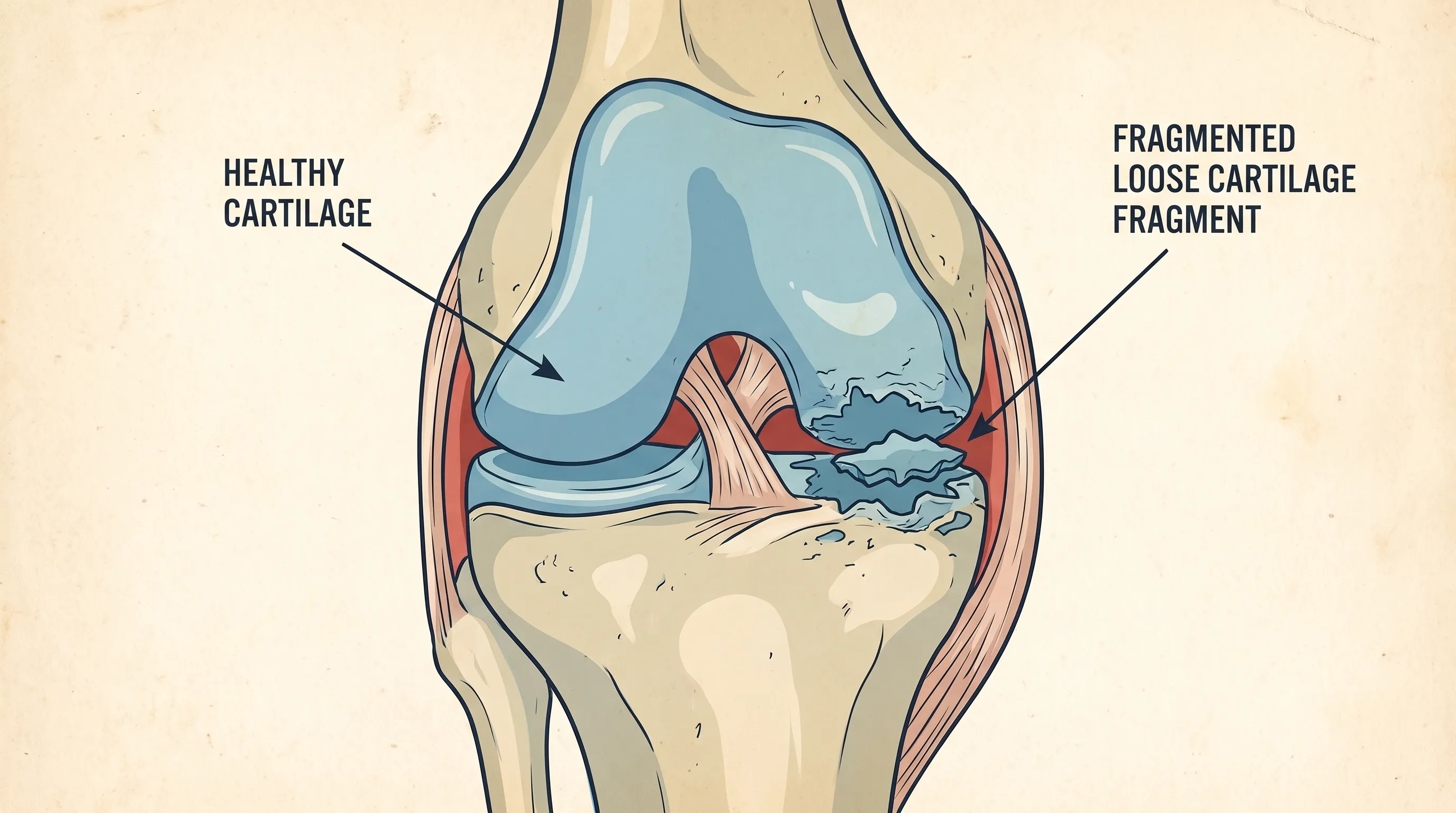
The knee relies on two distinct cartilage types: hyaline (articular) cartilage and fibrocartilage. Hyaline cartilage is a smooth, glassy tissue that covers the ends of the bones, enabling seamless gliding motion during flexion and extension. It contains a highly organized extracellular matrix rich in type II collagen and proteoglycans, which trap water molecules to provide compressive resistance. Fibrocartilage, primarily found in the menisci, consists of denser type I collagen fibers arranged in concentric rings. It stabilizes the joint, distributes load, and absorbs impact. When either of these structures sustains focal damage or experiences metabolic compromise, fragments can detach and migrate into the joint capsule.
How Cartilage Becomes Loose
Cartilage fragmentation rarely occurs spontaneously. It typically results from a cascade of biomechanical and biological stressors. High-velocity shear forces during sudden pivots can create focal defects. Repetitive microtrauma from occupational or athletic demands gradually degrades the extracellular matrix. Additionally, metabolic imbalances, synovial fluid abnormalities, and genetic predispositions can weaken collagen cross-linking. Once a critical defect forms, synovial fluid penetrates the subchondral bone, creating a cleavage plane that eventually allows a fragment to detach completely. This process explains why loose cartilage in knee presentations often emerge gradually before manifesting as acute mechanical symptoms.
Recognizing the Symptoms and Warning Signs
Identifying loose cartilage in knee conditions early is crucial because delayed intervention often leads to secondary complications like synovitis, chondromalacia, or accelerated osteoarthritic degeneration. Symptoms can range from subtle, intermittent discomfort to sudden, debilitating mechanical blocks.
Common Physical Symptoms
The clinical presentation typically revolves around three core phenomena: catching, locking, and swelling. Patients frequently report a distinct sensation that the knee has momentarily caught or that something is floating inside the joint space. True locking occurs when the fragment physically wedges between the femoral and tibial condyles, preventing full extension until the fragment shifts. Swelling often follows mechanical irritation, as the synovial membrane releases excess fluid in response to inflammatory cytokines. Grinding or crepitus may accompany weight-bearing, indicating uneven articular surfaces. Pain is usually localized to the joint line and exacerbates with activities requiring deep flexion or rotational torque.
Impact on Daily Mobility and Function
Beyond acute symptoms, loose cartilage fragments significantly alter gait patterns and functional capacity. The brain subconsciously restricts knee extension or flexion to avoid pain or locking, leading to muscle guarding and compensatory biomechanics. Over time, quadriceps atrophy, altered hip mechanics, and reduced proprioception can develop, increasing fall risk and decreasing overall activity levels. Many patients report abandoning previously enjoyed activities like hiking, cycling, or dancing due to unpredictable joint behavior. Early recognition of these functional limitations ensures timely referral to orthopedic specialists who can implement targeted rehabilitation or surgical management before irreversible compensatory changes take hold.
Primary Causes and Risk Factors
The etiology of cartilage detachment is multifactorial, encompassing traumatic, degenerative, developmental, and systemic variables. Understanding your individual risk profile helps in both diagnosis and preventive strategy formulation.
Traumatic Injuries and Sports-Related Damage
Acute trauma remains the leading precipitating factor, particularly in athletic populations. Sports requiring cutting, pivoting, or sudden deceleration place immense rotational shear stress on the knee. Ligamentous injuries, especially anterior cruciate ligament (ACL) tears, frequently coexist with cartilage damage due to the violent translational forces involved. Direct impacts from tackles, falls, or collisions can also cause focal osteochondral fractures where both bone and overlying cartilage fragment simultaneously. Even seemingly minor missteps can trigger detachment if pre-existing cartilage softening or matrix degradation is present.
Degenerative Changes and Osteoarthritis
In older adults, degenerative processes dominate. Years of cumulative load, combined with declining chondrocyte metabolic activity, reduce proteoglycan content and collagen integrity. The cartilage surface develops fibrillations, fissures, and eventually full-thickness defects. As subchondral bone sclerosis progresses, the weakened cartilage boundary becomes susceptible to fragment shedding. This is why loose cartilage in knee presentations frequently coexist with radiographic signs of osteoarthritis. The degenerative cascade is accelerated by obesity, sedentary lifestyles, and previous joint injuries, creating a compounding risk environment.
Congenital and Metabolic Factors
Certain individuals possess inherent structural or metabolic vulnerabilities. Osteochondritis dissecans (OCD) is a well-documented developmental condition where localized subchondral bone loses vascularization, causing the overlying cartilage to detach spontaneously during adolescence. Understand osteochondritis dissecans Additionally, metabolic disorders like gout, pseudogout, rheumatoid arthritis, or endocrine imbalances can alter synovial fluid composition, promoting enzymatic cartilage degradation. Genetic variations in collagen synthesis pathways may also predispose specific populations to earlier matrix breakdown.
Diagnostic Process and Medical Evaluation
Accurate diagnosis of loose cartilage in knee conditions requires a systematic clinical approach combining patient history, physical examination, and advanced imaging modalities. Self-diagnosis often proves unreliable because symptoms overlap with meniscal tears, patellofemoral syndrome, and synovial plica impingement.
Clinical Examination and Patient History
A comprehensive orthopedic evaluation begins with detailed symptom mapping. Clinicians assess onset, mechanism, frequency, and duration of locking, catching, and swelling episodes. Functional questionnaires evaluate pain scales, activity limitations, and quality-of-life impacts. The physical exam includes palpation of joint lines, assessment of effusion, measurement of range of motion, and specialized maneuvers like the McMurray test, Apley grind test, and patellar compression tests to isolate intra-articular pathology. Observing gait and single-leg balance provides additional biomechanical insights.
Imaging Techniques: MRI, X-Ray, and Ultrasound
Imaging confirms clinical suspicion and guides treatment planning. Weight-bearing X-rays remain the first-line modality for evaluating joint space narrowing, osteophyte formation, and calcified loose bodies. However, pure cartilage fragments are radiolucent and invisible on standard radiographs. View knee imaging and diagnostic guidelines Magnetic Resonance Imaging (MRI) is the diagnostic gold standard, utilizing proton density and T2-weighted sequences to visualize cartilage thickness, subchondral edema, and free-floating fragments with exceptional clarity. Ultrasound offers dynamic assessment of superficial cartilage defects and effusion but lacks deep joint visualization. Diagnostic arthroscopy remains the definitive standard when imaging is equivocal, allowing direct visualization and potential simultaneous intervention.
When to Seek Immediate Medical Attention
Certain symptoms warrant urgent evaluation. Complete mechanical locking that prevents knee extension, severe effusion developing within hours of injury, inability to bear weight, or acute instability following trauma should prompt immediate orthopedic consultation. These red flags indicate potential vascular compromise, large unstable fragments, or concomitant ligamentous disruption requiring expedited management to prevent secondary tissue damage.
Non-Surgical Treatment Options and Conservative Management
Not every cartilage fragment requires immediate surgical extraction. Many patients achieve meaningful symptom relief and functional restoration through structured conservative protocols, particularly when fragments are small, minimally symptomatic, or located in non-weight-bearing zones.
Physical Therapy and Targeted Strengthening
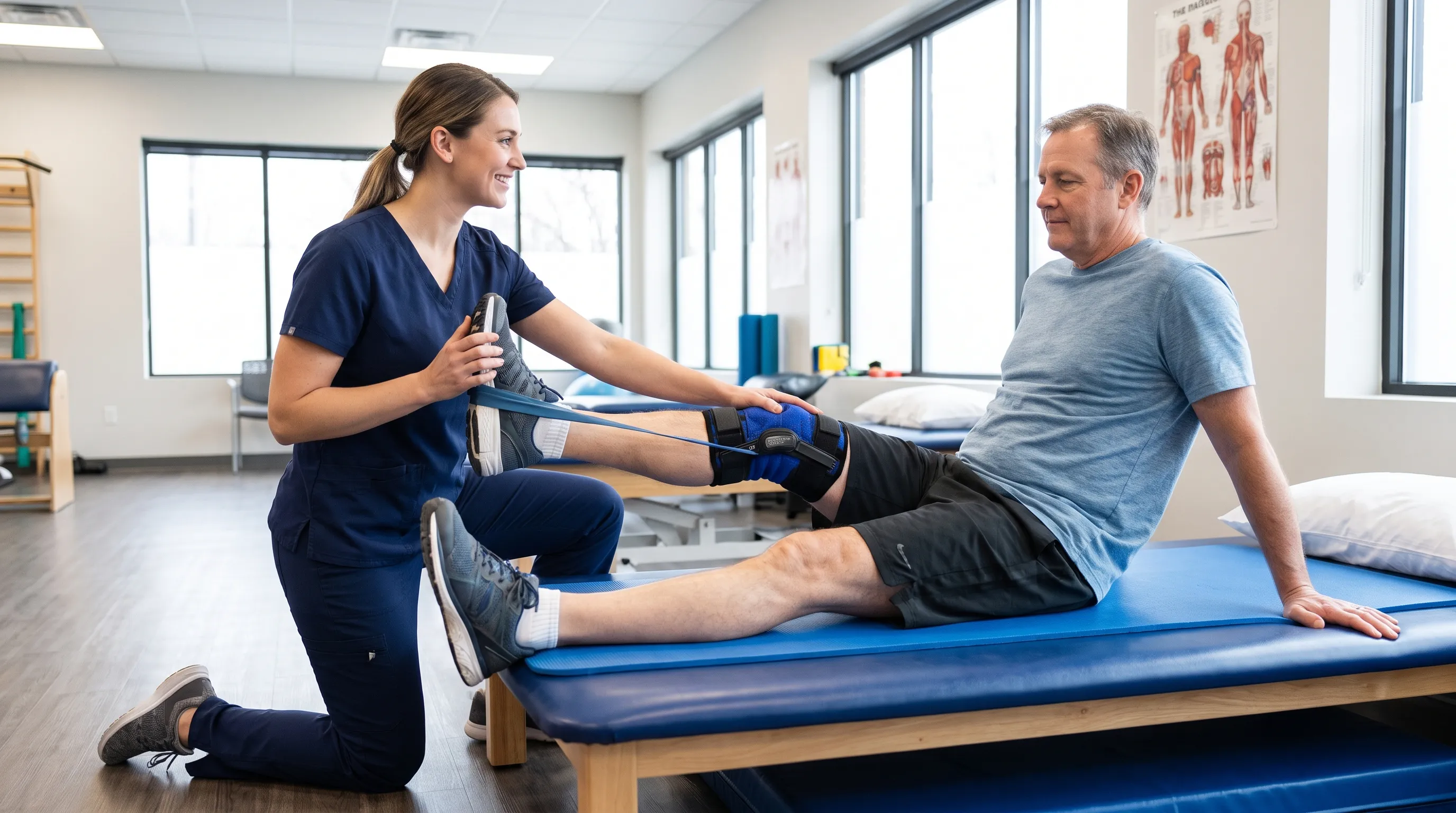
Physical therapy forms the cornerstone of non-operative management. View CDC guidelines on physical activity for arthritis The primary goal is to optimize neuromuscular control, restore balanced muscle activation, and improve joint kinematics to minimize abnormal loading. Programs emphasize quadriceps strengthening (vastus medialis oblique emphasis), hamstring flexibility, hip abductor activation, and core stability. Proprioceptive training on unstable surfaces, closed-chain kinetic exercises, and gait retraining help reestablish normal movement patterns. Evidence demonstrates that targeted strengthening reduces joint contact forces by up to thirty percent, significantly decreasing fragment irritation and synovial inflammation. Clinicians typically recommend three weekly supervised sessions combined with daily home exercises for a minimum of eight to twelve weeks.
Pain Management and Anti-Inflammatory Approaches
Symptomatic control is essential during rehabilitation. Nonsteroidal anti-inflammatory drugs (NSAIDs) like ibuprofen or naproxen reduce prostaglandin-mediated synovitis and pain. Read about arthritis pain management Topical diclofenac gel offers localized relief with minimal systemic absorption. Intra-articular corticosteroid injections provide rapid anti-inflammatory effects during acute flare-ups, though repeated use requires caution due to potential cartilage thinning. Hyaluronic acid (viscosupplementation) injections aim to restore synovial fluid viscosity, though clinical efficacy remains debated. Platelet-rich plasma (PRP) therapy is increasingly utilized for its growth factor-mediated healing potential, particularly in early degenerative stages.
Bracing, Taping, and Activity Modification
Mechanical offloading through bracing or taping can significantly reduce symptomatic fragment irritation. Unloader braces apply varus or valgus forces to shift load away from compromised compartments, while patellofemoral taping improves tracking and reduces anterior stress. Activity modification involves temporarily eliminating high-impact movements, deep squats, and rotational pivoting in favor of low-impact alternatives like swimming, stationary cycling, and elliptical training. This strategic load management prevents acute exacerbations while maintaining cardiovascular fitness and muscle tone.
| Treatment Modality | Primary Mechanism | Typical Duration | Efficacy Level | Best Suited For |
|---|---|---|---|---|
| Physical Therapy | Neuromuscular re-education, strength optimization | 8-12 weeks | High | Early-stage, stable fragments |
| NSAIDs & Topicals | Prostaglandin inhibition, localized pain relief | 1-3 months | Moderate | Acute inflammation phases |
| Hyaluronic Acid | Synovial viscosity restoration, lubrication | 3-6 months | Variable | Mild degeneration |
| PRP Therapy | Growth factor release, tissue modulation | 1-2 injections | Emerging | Early OA, active individuals |
| Unloader Bracing | Biomechanical load redistribution | Ongoing | High | Compartment-specific stress |
Surgical Interventions and Advanced Procedures
When conservative measures fail or mechanical symptoms significantly impair function, surgical intervention becomes necessary. Modern orthopedic surgery offers highly refined techniques tailored to fragment size, location, patient age, activity demands, and cartilage quality.
Arthroscopic Debridement and Loose Body Removal
Arthroscopic loose body extraction remains the most common procedure. Learn about arthroscopic knee procedures Using two to three small incisions, surgeons insert a high-definition camera and miniature instruments to visualize and retrieve the fragment. Chondroplasty, the process of smoothing roughened cartilage edges, often accompanies removal to prevent further fibrillation. The minimally invasive approach preserves healthy tissue, minimizes postoperative pain, and enables rapid rehabilitation. Hospital stays are typically outpatient, with most patients walking independently within twenty-four hours. This approach is ideal for isolated fragments without widespread degenerative changes.
Cartilage Restoration Techniques (Microfracture, OATS, ACI)
When fragments are large or involve critical weight-bearing surfaces, restoration becomes preferable to simple extraction. Microfracture involves drilling small holes into exposed subchondral bone, releasing marrow-derived mesenchymal stem cells that differentiate into fibrocartilage. While effective for small defects, fibrocartilage possesses inferior wear resistance compared to native hyaline tissue. Osteochondral Autograft Transfer System (OATS) plugs healthy cartilage-bone composite from non-weight-bearing zones into the defect, providing immediate structural integrity. Autologous Chondrocyte Implantation (ACI) involves a two-stage process: harvesting patient-specific chondrocytes, expanding them in vitro, and reimplanting them under a periosteal patch. MACI (Matrix-Induced ACI) utilizes a collagen scaffold, eliminating the need for periosteal grafts and accelerating integration. Selection depends on defect size, patient age, and rehabilitation compliance.
Post-Operative Rehabilitation Timeline
Successful surgical outcomes heavily depend on structured rehabilitation. Phase one (weeks 0-4) focuses on edema control, early range of motion, and protected weight-bearing using crutches or a brace. Phase two (weeks 4-8) introduces progressive strengthening, proprioceptive drills, and partial weight-bearing progression. Phase three (weeks 8-12) emphasizes full weight-bearing, gait normalization, and functional strengthening. Phase four (months 3-6) integrates sport-specific movements, agility training, and gradual return to impact. Adherence to weight-bearing restrictions and avoiding early rotational stress are critical to protecting newly integrated tissue.
Lifestyle Modifications and Long-Term Knee Health
Long-term management of loose cartilage in knee extends far beyond acute treatment or surgical recovery. Sustainable joint preservation requires proactive lifestyle optimization, biomechanical awareness, and consistent physiological support.
Weight Management and Biomechanical Load
Excess body weight dramatically accelerates cartilage degradation through both mechanical and metabolic pathways. Learn how weight affects knee osteoarthritis Every additional pound multiplies knee joint load by approximately fourfold during ambulation and significantly more during descent activities. Adipose tissue also secretes pro-inflammatory adipokines like leptin and resistin, which degrade cartilage matrix enzymes. Maintaining a body mass index within healthy ranges reduces compressive stress and systemic inflammation simultaneously. Even a five to ten percent reduction in body weight yields clinically meaningful improvements in pain scores and functional capacity.
Nutrition for Joint Support and Collagen Synthesis
Optimal nutrition provides the biochemical substrates necessary for tissue maintenance and repair. High-quality protein supplies essential amino acids for collagen synthesis. Omega-3 fatty acids from fatty fish, flaxseed, and walnuts downregulate inflammatory pathways. Vitamin D and calcium support subchondral bone mineralization, preventing secondary osteonecrosis. Antioxidant-rich foods like berries, leafy greens, and turmeric mitigate oxidative stress that damages chondrocytes. While supplements like glucosamine and chondroitin sulfate show mixed clinical trial results, some patients report modest symptomatic improvement. Collagen peptides and curcumin extracts demonstrate emerging evidence for modulating matrix metalloproteinase activity and supporting connective tissue integrity.
Preventive Exercise Routines and Low-Impact Activities
Prevention remains the most cost-effective intervention strategy. Regular low-impact cardiovascular conditioning maintains synovial fluid circulation, delivering nutrients to avascular cartilage through diffusion cycling. Swimming, water aerobics, and recumbent cycling strengthen periarticular musculature without generating destructive impact forces. Flexibility routines incorporating dynamic warm-ups and static stretching maintain optimal muscle-tendon compliance, preventing abnormal joint tracking. Incorporating balance and proprioceptive drills three times weekly enhances reactive stabilization, significantly reducing the risk of traumatic cartilage injury. Consistency, progressive overload, and proper biomechanical form form the foundation of lifelong knee resilience.
Frequently Asked Questions
What causes a piece of loose cartilage in knee to break off?
Loose cartilage in knee typically breaks off due to acute trauma like twisting injuries, degenerative wear from osteoarthritis, or osteochondritis dissecans, a condition where bone tissue beneath the cartilage loses blood supply. Repetitive microtrauma, sudden rotational forces, or metabolic degradation can all compromise the structural integrity of the extracellular matrix, allowing a fragment to detach and migrate into the joint space.
Can loose cartilage heal on its own without surgery?
Cartilage lacks blood vessels and a direct metabolic supply line, which means it cannot naturally regenerate or reattach once detached. While conservative treatments can effectively manage symptoms, reduce inflammation, and improve functional capacity, the loose fragment itself will not heal or reattach to its original bed. Surgical extraction or restoration is typically required if mechanical symptoms persist or if the fragment threatens adjacent healthy cartilage.
How do doctors diagnose a loose cartilage fragment in the knee?
Diagnosis begins with a comprehensive clinical examination focusing on joint stability, range of motion, and mechanical symptoms like catching or locking. Magnetic Resonance Imaging (MRI) serves as the diagnostic gold standard, providing high-resolution visualization of cartilage integrity and soft tissue structures. Plain X-rays may detect calcified loose bodies, while ultrasound can assess joint effusion and superficial abnormalities. When imaging remains inconclusive but clinical suspicion is high, diagnostic arthroscopy allows direct visualization and simultaneous therapeutic intervention.
What happens if you leave loose cartilage in knee untreated?
Leaving a loose cartilage fragment unaddressed can trigger a cascade of progressive joint complications. The fragment acts as an abrasive agent, repeatedly grinding against healthy articular surfaces during movement, which accelerates localized cartilage wear. Chronic synovial inflammation, recurrent effusions, and altered gait mechanics often follow, eventually leading to secondary osteoarthritis, quadriceps atrophy, and significant functional impairment. Early intervention significantly reduces the likelihood of irreversible degenerative changes.
What is the typical recovery time after arthroscopic removal of loose cartilage?
Arthroscopic loose body removal typically features a streamlined recovery protocol due to its minimally invasive nature. Most patients achieve independent ambulation within twenty-four to forty-eight hours. Basic daily activities resume within two to four weeks as swelling subsides and strength returns. Full return to high-impact sports or strenuous occupational tasks generally requires six to eight weeks, provided structured physical therapy protocols are consistently followed and weight-bearing restrictions are respected during the critical healing phase.
Conclusion
Managing loose cartilage in knee requires a balanced, evidence-based approach that prioritizes accurate diagnosis, targeted intervention, and proactive long-term joint stewardship. Whether the fragment originates from acute trauma, progressive degeneration, or developmental abnormalities, recognizing mechanical symptoms early prevents secondary structural compromise and preserves functional mobility. Conservative management through physical therapy, strategic load modification, and anti-inflammatory protocols remains highly effective for many individuals, while modern arthroscopic and restorative techniques offer reliable solutions when mechanical obstruction dictates. By integrating weight optimization, targeted nutrition, consistent low-impact conditioning, and proper biomechanical awareness, patients can significantly extend joint longevity and maintain an active, fulfilling lifestyle. If you experience persistent catching, recurrent swelling, or unpredictable knee instability, consult a board-certified orthopedic specialist to explore personalized treatment pathways and safeguard your long-term mobility.
How we work
Researched and drafted with AI assistance, then reviewed by a human editor against the cited sources.
This article is general health information, not medical advice. Consult a qualified healthcare professional about your own situation.