Pain in Between Shoulder Blades: Causes, Relief, and Expert Care Guide

Living with persistent discomfort in the upper back can significantly disrupt your daily routine, turning simple tasks like reaching for a coffee mug or sitting at your desk into sources of daily frustration. Many individuals experience aching, stiffness, or sharp sensations directly along the thoracic region, yet the root causes often remain misunderstood or hastily dismissed. When left unaddressed, pain in between shoulder blades can progress from a mild nuisance into a chronic condition that impacts sleep quality, posture, and overall functional mobility. Understanding the complex interplay of muscles, nerves, joints, and sometimes even internal organs is the first critical step toward finding lasting relief. This comprehensive guide explores the anatomical foundations, evidence-based treatments, and proactive lifestyle adjustments necessary to resolve upper back discomfort safely and effectively. By combining peer-reviewed clinical research with practical self-care strategies, you will gain a clear, actionable roadmap to recovery and long-term spinal health.
Understanding the Anatomy of Your Upper Back
The upper back, medically referred to as the thoracic region, comprises a complex network of osseous structures, musculature, ligaments, and neural pathways designed to provide both stability and dynamic movement. Spanning twelve vertebrae (T1-T12), the thoracic spine naturally exhibits a gentle outward curvature known as kyphosis, which works in tandem with the cervical and lumbar curves to distribute mechanical load evenly during upright posture and physical activity. Learn more about the thoracic spine structure. Unlike the highly mobile cervical or lumbar segments, the thoracic vertebrae articulate with the rib cage, which inherently restricts excessive flexion, extension, and lateral bending in favor of structural protection and respiratory mechanics.
Free calculatorBMI CalculatorCalculate Body Mass IndexOpen the calculator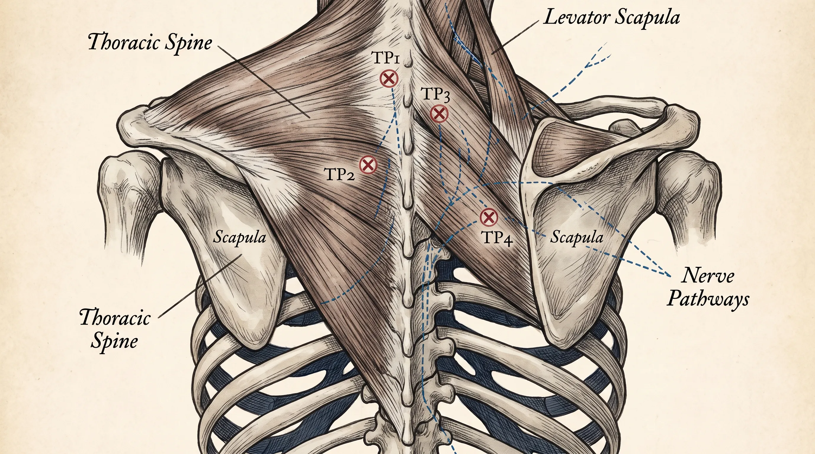
The scapulae, or shoulder blades, function as floating articulations anchored primarily by muscular and fascial connections rather than bony joints. The rhomboids, trapezius (particularly the middle and lower fibers), and levator scapulae serve as primary stabilizers, coordinating with the serratus anterior and pectoralis minor to facilitate smooth scapulothoracic rhythm. Disruptions in this intricate synergy often manifest as pain in between shoulder blades, particularly when prolonged static positioning or repetitive overhead movements create muscle imbalances. The thoracic paraspinal muscles, including the erector spinae and multifidus, further reinforce segmental stability, while the dorsal rami of thoracic spinal nerves provide sensory and motor innervation to the posterior thorax. Fascial continuity along the thoracolumbar region also means that dysfunction in the cervical spine, lumbar spine, or even the pelvic girdle can transmit compensatory tension upward or downward, making localized symptoms a potential reflection of broader kinetic chain imbalances. Clinicians emphasize that understanding this interconnected architecture is essential before initiating any targeted intervention, as isolated treatments frequently fail to address the underlying biomechanical drivers of chronic thoracic discomfort.
Common Causes of Pain in Between Shoulder Blades
Identifying the precise etiology of upper back discomfort requires a systematic evaluation of both mechanical and non-mechanical factors. The most prevalent cause is undoubtedly postural strain resulting from prolonged sitting, forward head posture, and rounded shoulders, often termed upper crossed syndrome. Read about posture-related back pain. In this pattern, tightness in the pectorals and upper trapezius coincides with weakness in the lower trapezius, rhomboids, and deep cervical flexors. The resulting imbalance forces the scapulae into protraction and downward rotation, continuously overloading the posterior thoracic musculature and triggering localized myofascial trigger points. Over time, these adaptive changes lead to chronic tissue remodeling, reduced blood flow, and heightened nociceptive signaling, which collectively sustain discomfort even in the absence of acute injury.
Degenerative disc disease and thoracic facet joint osteoarthritis represent another significant category, particularly among adults over fifty. While less common than lumbar or cervical spondylosis, thoracic disc herniations and annular fissures can still compress dorsal root ganglia, producing sharp, radiating sensations that wrap around the rib cage or concentrate directly between the scapulae. Explore degenerative disc conditions. Joint degeneration further compromises segmental mobility, leading to stiffness, crepitus, and activity-related discomfort that typically improves with gentle movement and worsens after prolonged rest.
Nerve-related etiologies include thoracic radiculopathy and dorsal scapular nerve entrapment. Compression or irritation of the C5-C8 nerve roots due to cervical pathology can refer discomfort distally, while entrapment of the dorsal scapular nerve as it passes through or near the middle scalene muscle can produce deep, aching discomfort localized precisely to the medial scapular border. Conditions such as scoliosis, Scheuermann’s disease, and postural kyphosis alter the natural load distribution across the thoracic spine, creating asymmetrical muscle recruitment patterns that inevitably lead to chronic fatigue and tissue breakdown in overworked regions.
Visceral referral patterns must also be carefully considered. Gallbladder pathology frequently manifests as right-sided interscapular discomfort, often accompanied by fatty food intolerance and episodic nausea. Peptic ulcer disease, pancreatitis, and esophageal spasms can similarly project discomfort to the mid-thoracic region through shared sympathetic innervation pathways. Pulmonary conditions such as pleuritis, pneumonia, and in rare instances, pulmonary embolism, may present with pleuritic upper back discomfort exacerbated by deep inspiration or coughing. Understanding referred pain from internal organs. Recognizing these systemic connections ensures that clinicians and patients alike do not prematurely assume a purely musculoskeletal origin when evaluating persistent pain in between shoulder blades. Early differentiation between mechanical and visceral sources dramatically influences treatment trajectories and clinical outcomes.
Identifying Warning Signs and Red Flags
While the majority of thoracic discomfort originates from benign, self-limiting musculoskeletal sources, certain clinical presentations demand immediate medical evaluation. Red flag symptoms serve as critical indicators that the discomfort may stem from life-threatening or rapidly progressive conditions. Chest pressure, shortness of breath, diaphoresis, nausea, or sudden onset of severe interscapular pain should never be dismissed as simple muscle strain, as these are hallmark presentations of acute coronary syndrome or aortic dissection. Recognize heart attack warning signs. Women and older adults frequently experience atypical cardiac symptoms, making upper back discomfort a crucial diagnostic clue in cardiovascular triage.
Neurological deficits represent another urgent category. Progressive weakness in the arms or legs, bowel or bladder dysfunction, gait disturbances, or saddle anesthesia suggest spinal cord compression, which requires immediate imaging and potential surgical decompression to prevent permanent neurological impairment. When to seek emergency care for back pain. Similarly, unexplained weight loss, persistent fever, night sweats, or a history of malignancy raise suspicion for metastatic spinal disease or systemic infection. These constitutional symptoms, when paired with unrelenting pain in between shoulder blades, warrant comprehensive laboratory workup and advanced imaging such as whole-body MRI or PET-CT.
Trauma-related presentations, including falls from height, motor vehicle collisions, or direct blows to the thoracic region, necessitate radiographic evaluation to rule out vertebral compression fractures, particularly in osteoporotic populations. Learn about osteoporosis and spinal fractures. Age-related bone density reduction significantly increases fracture risk, and minor axial loading can sometimes produce structural compromise that initially presents as localized posterior discomfort. Differentiating between benign myofascial strain and structural compromise relies heavily on clinical palpation, range-of-motion assessment, and targeted orthopedic maneuvers. A thorough history taking session, including occupation, activity patterns, previous injuries, and medication use, provides essential context for risk stratification. Patients should always err on the side of caution when symptoms deviate from typical mechanical patterns or fail to improve with conservative measures within two to three weeks.
Diagnostic Approaches for Persistent Discomfort
A structured diagnostic pathway begins with a comprehensive clinical evaluation, incorporating subjective history, physical examination, and targeted functional assessments. Clinicians utilize standardized screening tools such as the Oswestry Disability Index and Numeric Pain Rating Scale to quantify functional impairment and establish baseline metrics for tracking progress. Physical examination focuses on posture analysis, palpation for trigger points, assessment of active and passive range of motion, and neurological screening including reflex testing, dermatomal sensation mapping, and myotomal strength grading. Specialized orthopedic tests, such as the scapular assistance test and thoracic extension-rotation mobilization, help differentiate between joint hypomobility and muscular inhibition.
Imaging modalities are reserved for cases where red flags are present, conservative management has failed, or structural pathology is strongly suspected. Diagnostic imaging guidelines for back pain. Plain radiographs (X-rays) effectively identify vertebral alignment abnormalities, degenerative changes, fractures, and gross structural deformities. Magnetic resonance imaging (MRI) remains the gold standard for visualizing soft tissue structures, including intervertebral discs, spinal cord, nerve roots, and paraspinal musculature, providing unparalleled detail for diagnosing herniations, stenosis, or inflammatory conditions. Computed tomography (CT) scans offer superior bony resolution, making them valuable for assessing complex fractures or post-traumatic anatomy.
Electromyography (EMG) and nerve conduction studies (NCS) become essential when radiculopathy or peripheral neuropathy is suspected, allowing clinicians to map nerve irritation, conduction blockages, and axonal damage. Functional movement assessments, increasingly integrated into physical therapy protocols, evaluate kinetic chain efficiency, breathing mechanics, and core-stabilization patterns. Tools like the Functional Movement Screen (FMS) and Selective Functional Movement Assessment (SFMA) identify compensatory movement strategies that perpetuate tissue overload and delay recovery. Diagnostic ultrasound is also gaining traction for real-time visualization of myofascial tissue, guiding targeted interventions like dry needling or corticosteroid injections with precision. By combining structural imaging with dynamic functional analysis, clinicians develop a multidimensional understanding of each patient’s unique presentation, ensuring that interventions address both localized symptoms and systemic movement dysfunctions.
Evidence-Based Treatment Strategies
Addressing upper back discomfort requires a multimodal approach tailored to the underlying etiology, symptom chronicity, and individual functional goals. Physical therapy serves as the cornerstone of conservative management, emphasizing scapular stabilization, thoracic mobility restoration, and postural re-education. Physical therapy for musculoskeletal pain. Evidence consistently supports targeted strengthening of the middle and lower trapezius, serratus anterior, and deep cervical flexors to counteract anterior dominance and restore neutral scapular alignment. Manual therapy techniques, including joint mobilizations, soft tissue myofascial release, and instrument-assisted techniques, effectively reduce tissue adhesions, improve fascial glide, and restore segmental mobility in hypomobile thoracic segments.

Pharmacological management focuses on short-term symptom modulation to facilitate active rehabilitation. Nonsteroidal anti-inflammatory drugs (NSAIDs) reduce localized inflammation and modulate nociceptive signaling, while muscle relaxants provide temporary relief for acute muscle spasms that inhibit proper movement patterns. Topical analgesics containing menthol, capsaicin, or lidocaine offer localized relief without systemic side effects, making them suitable for prolonged use in chronic presentations. For refractory cases involving nerve compression or severe inflammation, epidural steroid injections or facet joint blocks administered under fluoroscopic guidance provide targeted anti-inflammatory effects, often enabling patients to engage more effectively in therapeutic exercise programs.
Integrative therapies continue to demonstrate strong clinical efficacy in multidisciplinary pain management. Dry needling, performed by trained physical therapists, deactivates hyperirritable myofascial trigger points, restoring local blood flow and reducing referred pain patterns. Therapeutic massage, acupuncture, and cupping therapy modulate autonomic nervous system activity, decreasing sympathetic tone and promoting parasympathetic-driven tissue repair. Cognitive behavioral therapy (CBT) addresses the psychological components of chronic pain, helping patients reframe pain catastrophizing, develop stress resilience, and maintain adherence to rehabilitation protocols despite temporary discomfort fluctuations. The integration of these modalities into a cohesive, patient-centered plan consistently outperforms isolated treatments, yielding superior long-term functional outcomes and reduced recurrence rates.
Ergonomics and Daily Lifestyle Modifications
Optimizing environmental factors and daily habits plays a decisive role in both preventing and resolving upper back strain. Workplace ergonomics directly impact thoracic loading, with poorly designed stations forcing prolonged forward flexion and sustained scapular protraction. CDC workplace ergonomics guidelines. Monitor height should be adjusted so the top third aligns with eye level, eliminating the need to crane the neck upward or downward. Chair selection requires adequate lumbar support, adjustable armrests, and a reclining backrest that maintains a 100-110 degree hip angle to reduce posterior spinal compression. Keyboard and mouse placement should allow elbows to rest at 90-110 degrees with wrists in neutral alignment, preventing compensatory shoulder elevation and scapular winging.
Implementing micro-breaks every thirty to forty-five minutes disrupts sustained muscle contraction and restores tissue perfusion. Simple seated thoracic extensions, shoulder blade retractions, and chin tucks take less than two minutes but significantly reduce cumulative strain over an eight-hour workday. Standing desks and active seating options introduce postural variability, which research demonstrates is superior to static positioning for long-term musculoskeletal health. When selecting sleep systems, mattress firmness should match individual body weight and preferred sleep position, while pillow loft must maintain cervical neutrality without forcing shoulder elevation. Optimal sleeping positions for back pain. Memory foam or contoured cervical pillows often outperform traditional flat pillows by distributing pressure evenly along the posterior neck and upper trapezius.
| Work Setup Factor | Poor Configuration | Optimized Configuration |
|---|---|---|
| Monitor Height | Screen at or below desk level | Top edge aligned with eye level, 20-30 inches away |
| Chair Support | Flat backrest, no lumbar contour | Adjustable lumbar support, recline to 105 degrees |
| Keyboard/Mouse Position | Elbows extended, wrists flexed | Elbows at 90-100 degrees, forearms parallel to floor |
| Break Frequency | 2+ hours continuous sitting | Micro-movements every 30-45 minutes |
| Lighting | Overhead glare, screen reflection | Ambient indirect lighting, anti-glare screen filter |
Hydration and nutrition further influence tissue resilience and recovery capacity. Adequate water intake maintains intervertebral disc hydration and fascial pliability, while anti-inflammatory dietary patterns rich in omega-3 fatty acids, turmeric, ginger, and leafy greens modulate systemic inflammatory markers. Reducing refined carbohydrate and trans-fat intake minimizes chronic low-grade inflammation that exacerbates tissue sensitivity and delays muscular repair. Consistency in these daily modifications creates compounding protective effects, transforming passive recovery into active spinal preservation.
Proactive Prevention and Long-Term Wellness
Transitioning from reactive symptom management to proactive spinal wellness requires consistent, structured movement programming and sustained stress regulation. Building resilient upper back strength focuses on progressive overload of scapular stabilizers, thoracic extensors, and posterior shoulder girdle musculature. Foundational movements such as scapular push-ups, prone Y-T-W-L raises, banded face pulls, and cable rows effectively target the posterior kinetic chain while promoting neuromuscular control and joint centration. Incorporating dynamic mobility drills like thoracic foam rolling extensions, quadruped thread-the-needle rotations, and seated cat-cow progressions maintains segmental flexibility and prevents fascial restrictions from accumulating over time.
Stress management remains equally critical, as chronic psychological tension directly manifests as elevated muscle tone in the posterior thoracic region. Evidence-based stress management techniques. Diaphragmatic breathing techniques stimulate the vagus nerve, shifting autonomic balance toward parasympathetic dominance and reducing cortisol-mediated muscle guarding. Mindfulness meditation, progressive muscle relaxation, and guided imagery protocols have demonstrated measurable reductions in perceived pain intensity and improved functional capacity in clinical trials tracking chronic musculoskeletal discomfort. Tracking movement consistency through wearable devices or structured training logs enhances accountability, allowing individuals to identify patterns between stress spikes, sleep disruptions, and symptom flare-ups.
Long-term wellness also demands periodic reassessment of training programs, footwear selection, and occupational demands, as aging and lifestyle transitions continually alter biomechanical requirements. Engaging in cross-training activities like swimming, Pilates, and yoga provides multidirectional spinal loading, promoting balanced muscular development and proprioceptive refinement. Avoiding rapid training progression, prioritizing movement quality over maximal loading, and integrating deload weeks prevent cumulative tissue fatigue that frequently precedes chronic upper back dysfunction. By viewing pain in between shoulder blades as a modifiable outcome of lifestyle, movement quality, and physiological resilience rather than an inevitable consequence of aging or desk work, individuals maintain agency over their long-term spinal health and functional longevity.
Frequently Asked Questions
How long does pain in between shoulder blades typically last?
Acute muscular discomfort usually resolves within 7 to 14 days with proper rest, gentle movement, and targeted stretching. If the discomfort persists beyond three to four weeks, worsens despite conservative care, or is accompanied by neurological symptoms like numbness, tingling, or radiating arm pain, clinical evaluation is strongly recommended to rule out disc involvement or systemic conditions.
Can heart issues cause discomfort between the shoulder blades?
Yes. Cardiac events such as myocardial ischemia or aortic dissection can manifest as referred discomfort along the interscapular region. This is particularly common in women and older adults. CDC guidance on cardiovascular symptoms. If upper back discomfort is accompanied by chest pressure, shortness of breath, nausea, diaphoresis, or dizziness, seek emergency medical attention immediately.
What sleeping position is best for upper back discomfort?
Side sleeping with a supportive pillow that maintains cervical alignment and a thin pillow or rolled towel between the shoulder blades is generally optimal. Stomach sleeping should be avoided as it forces excessive cervical rotation and thoracic extension. Back sleepers can place a small cushion under the knees to reduce lumbar and thoracic strain.
Are there specific exercises that worsen upper back pain?
Heavy overhead pressing, improper deadlift form, excessive bench pressing without adequate scapular control, and repetitive forward-flexion movements can aggravate the rhomboids and trapezius. Always prioritize neutral spine positioning, progressive loading, and proper breathing mechanics during resistance training.
How does stress contribute to upper back tension?
Chronic psychological stress elevates circulating cortisol and adrenaline, triggering sustained low-grade muscle contraction, particularly in the trapezius and levator scapulae. This creates a feedback loop where physical tension amplifies mental fatigue, and vice versa. Mindfulness practices, diaphragmatic breathing, and progressive muscle relaxation effectively interrupt this neurophysiological cycle.
Key Takeaways
Addressing pain in between shoulder blades requires a comprehensive approach that bridges anatomical understanding, clinical diagnostics, targeted rehabilitation, and sustainable lifestyle modifications. By recognizing the intricate relationship between posture, stress, tissue health, and systemic function, individuals can move beyond temporary symptom suppression toward genuine structural resilience. Implementing ergonomic adjustments, committing to consistent mobility and strengthening protocols, and prioritizing stress regulation create compounding protective effects that significantly reduce recurrence rates. While self-care strategies effectively resolve the majority of benign cases, prompt medical evaluation remains essential when red flag symptoms emerge or conservative management fails to yield improvement. With evidence-based guidance, professional oversight when necessary, and proactive daily habits, you can restore upper back comfort, enhance functional mobility, and maintain optimal spinal wellness for years to come.
How we work
Researched and drafted with AI assistance, then reviewed by a human editor against the cited sources.
This article is general health information, not medical advice. Consult a qualified healthcare professional about your own situation.