Red Color on Bottom of Feet: Causes, Symptoms, and Medical Management

Key points
- Onset and Duration: Sudden onset often suggests infection, acute trauma, or allergic reaction. Gradual, progressive redness typically correlates with chronic vascular, metabolic, or neurological conditions.
- Distribution: Diffuse redness spanning the entire sole frequently indicates systemic or autonomic causes. Localized redness near joints or pressure points points toward mechanical stress, gout, or early ulceration.
- Temperature and Sensation: Warm, hot skin combined with burning pain is characteristic of neurovascular dysregulation. Cold, pale skin that turns red upon dependency suggests arterial insufficiency. Numbness or tingling alongside erythema strongly points to peripheral neuropathy.
- Skin Integrity: The presence of scaling, blistering, cracking, or maceration indicates dermatological or infectious processes. Smooth, shiny, taut skin may reflect edema or deep tissue involvement.
Noticing a sudden or persistent change in the appearance of your feet can be alarming, particularly when you observe a red color on bottom of feet. The plantar surface is highly vascularized and densely innervated, making it exceptionally responsive to systemic health shifts, localized trauma, inflammatory processes, and neurological changes. While occasional mild redness may simply result from friction, heat exposure, or prolonged standing, persistent or symptomatic erythema often signals an underlying medical condition that requires professional evaluation. Understanding the distinction between benign physiological responses and clinically significant pathology is essential for maintaining long-term mobility, preventing complications, and preserving overall vascular and neurological health.
The feet serve as the foundational pillars of the human musculoskeletal system, bearing immense mechanical stress while simultaneously functioning as highly sensitive sensory organs. When the skin on the soles changes color, it often reflects alterations in microcirculation, inflammatory mediator release, autonomic nerve dysfunction, or infectious processes. A red color on bottom of feet can manifest uniformly across the plantar surface, appear in patchy distributions, or localize around specific anatomical landmarks like the metatarsal heads or heel pads. The accompanying symptoms—ranging from burning pain and warmth to numbness, swelling, or skin breakdown—provide crucial diagnostic clues. This comprehensive guide explores the multifactorial etiology of plantar erythema, outlines evidence-based management strategies, and clarifies when immediate medical intervention is necessary to prevent severe outcomes.
The Anatomy of Foot Redness: Why the Soles React
The skin on the bottom of the feet is structurally unique compared to other body regions. It features a thick, specialized stratum corneum designed to withstand constant weight-bearing pressure, a dense network of sweat glands for thermoregulation, and a rich capillary bed that dynamically responds to temperature and positional changes. When blood vessels in the plantar dermis dilate, increased blood flow becomes visibly apparent as erythema. This vasodilation is primarily mediated by the autonomic nervous system and local inflammatory pathways.
Free calculatorA1C CalculatorA1C to average blood glucoseOpen the calculatorSeveral physiological mechanisms can trigger this vascular response. Heat exposure prompts the hypothalamus to direct increased peripheral blood flow for cooling. Mechanical friction stimulates local prostaglandin and histamine release, causing capillary engorgement. Autonomic neuropathy disrupts the normal vasoconstriction/vasodilation balance, leading to unregulated blood pooling. Inflammatory cytokines from infection or autoimmune activity further increase vascular permeability and redness. Recognizing these underlying pathways helps contextualize why a red color on bottom of feet is rarely an isolated phenomenon and usually points to a broader physiological imbalance requiring targeted intervention.
Decoding the Symptoms: What a Red Color on the Bottom of Feet Indicates
Clinical evaluation of plantar erythema always begins with a thorough symptom analysis. The presentation, onset, duration, and associated complaints guide differential diagnosis and treatment planning. Key characteristics to monitor include:
- Onset and Duration: Sudden onset often suggests infection, acute trauma, or allergic reaction. Gradual, progressive redness typically correlates with chronic vascular, metabolic, or neurological conditions.
- Distribution: Diffuse redness spanning the entire sole frequently indicates systemic or autonomic causes. Localized redness near joints or pressure points points toward mechanical stress, gout, or early ulceration.
- Temperature and Sensation: Warm, hot skin combined with burning pain is characteristic of neurovascular dysregulation. Cold, pale skin that turns red upon dependency suggests arterial insufficiency. Numbness or tingling alongside erythema strongly points to peripheral neuropathy.
- Skin Integrity: The presence of scaling, blistering, cracking, or maceration indicates dermatological or infectious processes. Smooth, shiny, taut skin may reflect edema or deep tissue involvement.
When assessing a red color on bottom of feet, clinicians correlate these physical findings with medical history, medication profiles, glycemic control, and occupational or lifestyle factors. A systematic approach ensures that transient, benign causes are distinguished from conditions requiring urgent pharmacological or surgical intervention.
Primary Medical Conditions Associated with Plantar Erythema
Understanding the specific pathologies that manifest with foot redness is critical for accurate diagnosis and effective management. The following sections detail the most prevalent and clinically significant conditions.
Erythromelalgia: A Neurovascular Paradox
Erythromelalgia is a rare, poorly understood neurovascular disorder characterized by intense burning pain, visible redness, and markedly increased skin temperature, predominantly affecting the feet and occasionally the hands. The Mayo Clinic outlines clinical criteria and symptom patterns for erythromelalgia to aid in recognition. The condition is classified as primary (idiopathic or genetic) or secondary to underlying systemic diseases, hematological disorders, or medication side effects.
Recent genetic research has identified mutations in the SCN9A gene, which encodes the Nav1.7 voltage-gated sodium channel in peripheral neurons, as a key driver in primary erythromelalgia. This mutation leads to hyperexcitability of small-diameter C-fibers and A-delta fibers, resulting in exaggerated pain signaling and abnormal autonomic control of peripheral blood vessels. Patients typically describe flare-ups triggered by ambient heat, physical exertion, stress, alcohol consumption, spicy foods, or tight footwear. During episodes, the affected skin becomes vividly red, warm, and swollen. Post-flare, the skin may paradoxically feel cold and exhibit a cyanotic or grayish hue due to reactive vasoconstriction.
Management strategies prioritize trigger avoidance and gentle symptom modulation. Patients are advised to cool affected areas using fans or room-temperature gel packs, avoiding direct ice or extreme cold water exposure which can cause microvascular injury or paradoxical pain exacerbation. Elevation during flare-ups reduces hydrostatic pressure and venous pooling. Pharmacological interventions, prescribed by neurologists or pain specialists, may include topical lidocaine, gabapentinoids, sodium channel blockers, or in refractory cases, intravenous prostacyclin analogs. Lifestyle modifications, including stress reduction techniques and careful hydration, play a supportive role in minimizing episode frequency and severity.
Cellulitis: When Bacteria Breach the Skin Barrier
Cellulitis represents a bacterial infection of the dermis and subcutaneous tissues, most commonly caused by Streptococcus pyogenes (Group A Streptococcus) or Staphylococcus aureus. The CDC provides detailed guidance on recognizing and treating cellulitis to prevent severe complications. The condition occurs when microorganisms penetrate compromised skin through microtears, fissures, surgical wounds, insect bites, or chronic fungal infections like tinea pedis.
A red color on bottom of feet caused by cellulitis typically presents as an expanding, ill-defined erythematous plaque that is warm, tender, and often swollen. Systemic symptoms such as fever, chills, malaise, and regional lymphadenopathy frequently accompany localized signs. Without prompt antibiotic therapy, cellulitis can progress to abscess formation, necrotizing fasciitis, osteomyelitis, or sepsis. Individuals with diabetes, lymphedema, venous insufficiency, or immunosuppressive conditions face significantly higher complication rates.
Standard treatment involves targeted oral antibiotics (e.g., cephalexin, amoxicillin-clavulanate) for mild to moderate cases, with intravenous therapy reserved for severe, rapidly progressing, or systemic presentations. Source identification is crucial; treating concurrent fungal infections, repairing skin breakdown, and managing edema reduce recurrence risk. Prevention emphasizes meticulous foot hygiene, prompt wound care, moisturizing to prevent fissures, and avoiding walking barefoot in high-risk environments.
Diabetic Peripheral Neuropathy: Metabolic Nerve Damage
Diabetic peripheral neuropathy is a progressive microvascular and metabolic complication affecting approximately 33-50% of individuals with diabetes mellitus. The NIH/NIDDK explains how chronic hyperglycemia damages peripheral nerves and microvasculature leading to distal axonal degeneration. Advanced glycation end-products (AGEs), and dyslipidemia (particularly elevated triglycerides) induce oxidative stress and microvascular ischemia within the vasa nervorum, leading to distal axonal degeneration.
While classically associated with distal symmetrical sensory loss, tingling, and neuropathic pain, autonomic neuropathy profoundly impacts foot physiology. Sympathetic denervation disrupts normal vasomotor control, causing unregulated arteriovenous shunting, increased cutaneous blood flow, and temperature dysregulation. This autonomic dysfunction frequently manifests as a persistent red color on bottom of feet, often accompanied by dry, atrophic skin, reduced sweating, and abnormal thermal perception.
The clinical significance of neuropathic erythema cannot be overstated. The combination of sensory loss and altered perfusion creates a perfect storm for unrecognized trauma, delayed wound healing, and Charcot neuroarthropathy—a devastating condition characterized by joint destruction and foot deformity. Rigorous glycemic management (target HbA1c <7% for most adults), daily structured foot inspections, professional podiatric care, and custom orthotic support are foundational to preventing catastrophic outcomes. Early recognition of color changes and temperature asymmetry allows for intervention before ulceration occurs.
Peripheral Artery Disease (PAD) and Dependent Rubor
Peripheral artery disease results from atherosclerotic plaque accumulation in the lower extremity arteries, progressively restricting oxygenated blood flow to distal tissues. While early PAD often presents with claudication (exercise-induced calf pain), advanced disease manifests in critical ischemia. The Cleveland Clinic details the vascular mechanisms behind dependent rubor and critical limb ischemia for clinical reference.
A classic sign of severe arterial insufficiency is dependent rubor: the foot appears pale when elevated but turns a deep, violaceous red within minutes of dependency (lowering the legs). This phenomenon occurs because dilated collateral vessels fail to regulate perfusion pressure adequately, leading to stagnant, deoxygenated blood pooling in the capillary beds. The skin may appear thin, shiny, and hairless, with diminished or absent pedal pulses.
Diagnosis relies on the ankle-brachial index (ABI), duplex ultrasonography, and sometimes CT or MR angiography. Management encompasses cardiovascular risk factor modification (smoking cessation, statin therapy, antiplatelet agents), supervised exercise programs, and, in critical cases, endovascular revascularization or surgical bypass. Recognizing dependent rubor early prevents progression to tissue necrosis and limb-threatening ischemia.
Inflammatory, Allergic, and Mechanical Triggers
Beyond systemic and vascular pathologies, localized environmental and immunological factors frequently cause plantar erythema. Contact dermatitis, either irritant or allergic, arises from prolonged exposure to footwear dyes, synthetic sock materials, topical analgesics, or cleaning agents. The Mayo Clinic describes diagnostic approaches for allergic and irritant contact dermatitis affecting the lower extremities. Presentation includes sharply demarcated redness, intense pruritus, scaling, and sometimes vesiculation.
Gout and other inflammatory arthritides can cause acute, localized plantar redness when inflammatory crystals or autoimmune activity target the metatarsophalangeal joints or midfoot structures. The affected area becomes exquisitely tender, hot, and swollen, often disrupting normal gait patterns.
Mechanical stress represents the most common non-pathological cause. Prolonged standing on hard surfaces, repetitive high-impact activities, or improperly fitted footwear generates repetitive microtrauma, increasing local metabolic demand and triggering compensatory vasodilation. Friction blisters, callus formation, and plantar fasciitis often accompany this redness. Addressing ergonomic factors, utilizing cushioned insoles, and implementing rest periods typically resolve mechanically induced erythema without medical intervention.
Clinical Evaluation and Diagnostic Pathways
Accurate diagnosis of a red color on bottom of feet requires a systematic clinical approach integrating patient history, physical examination, and targeted diagnostic testing. Healthcare providers follow established protocols to differentiate benign causes from progressive pathology.
The initial assessment includes a comprehensive medical history focusing on symptom onset, trigger patterns, pain characteristics, systemic illnesses, medication use, and occupational habits. A detailed physical examination evaluates skin integrity, capillary refill time, peripheral pulses, temperature gradients, sensory thresholds (using monofilament testing), and joint mobility. Clinicians often compare bilateral symmetry, as unilateral redness strongly suggests localized infection, trauma, or deep vein thrombosis, while bilateral involvement points toward systemic, metabolic, or neurological etiologies.
Diagnostic testing is guided by clinical suspicion. Laboratory studies may include complete blood count (CBC) with differential to identify leukocytosis in infection, C-reactive protein (CRP) and erythrocyte sedimentation rate (ESR) for systemic inflammation, fasting blood glucose and HbA1c for metabolic screening, and lipid panels for cardiovascular risk assessment. Imaging modalities such as plain radiographs evaluate bony alignment and rule out stress fractures or Charcot changes. Doppler ultrasound assesses arterial and venous flow dynamics. In suspected erythromelalgia or complex neuropathies, nerve conduction studies, skin biopsies for intraepidermal nerve fiber density, or genetic panels may be indicated.
Collaboration among primary care physicians, podiatrists, dermatologists, vascular surgeons, and neurologists ensures comprehensive care. Multidisciplinary evaluation is particularly crucial for patients with overlapping conditions like diabetes and PAD, where therapeutic priorities must be carefully balanced to optimize tissue perfusion and nerve preservation.
Red Flags Requiring Immediate Medical Intervention
While some plantar erythema resolves with conservative measures, certain presentations demand urgent clinical evaluation to prevent irreversible damage or systemic complications. Healthcare guidelines emphasize that delaying treatment for high-risk symptoms significantly increases morbidity and amputation risk.
Seek immediate medical care if you experience:
- Rapidly spreading erythema, especially with streaking patterns or lymphatic involvement
- Fever, chills, rigors, or systemic malaise accompanying foot redness
- Severe, unrelenting pain disproportionate to visible injury
- Open wounds, ulcerations, purulent drainage, or foul odor
- Skin discoloration progressing to blue, purple, gray, or black (indicating ischemia or necrosis)
- Loss of protective sensation, sudden motor weakness, or inability to bear weight
- History of diabetes, peripheral vascular disease, immunosuppression, or chronic kidney disease
These red flags signal potential deep tissue infection, acute arterial occlusion, severe neuropathic deterioration, or systemic inflammatory response. Prompt intervention, including advanced imaging, intravenous antimicrobials, surgical debridement, or vascular bypass, can be limb-saving. Never attempt home remedies for rapidly worsening or complicated foot symptoms.
Comprehensive Management and Evidence-Based Self-Care
Effective management of a red color on bottom of feet hinges on accurate diagnosis, targeted therapy, and consistent self-care practices. The CDC emphasizes evidence-based diabetic foot care protocols to prevent ulceration and infection. Guidelines from the American Diabetes Association (ADA), American Academy of Dermatology (AAD), and podiatric medical societies emphasize a multimodal approach.
Footwear and Daily Foot Hygiene Protocols
Proper footwear selection is foundational to plantar health. Shoes must provide adequate toe box width, arch support, and shock absorption to minimize mechanical stress and friction. Breathable materials like leather or advanced mesh promote air circulation, reducing moisture accumulation and bacterial proliferation. Moisture-wicking socks, preferably seamless blends of merino wool or synthetic fibers, prevent maceration and maintain optimal skin barrier function.
Daily foot hygiene should be systematic but gentle. Wash feet with lukewarm water and mild, fragrance-free soap. Avoid prolonged soaking, which compromises the lipid barrier and increases fissure risk. Pat dry thoroughly, especially between toes. Apply fragrance-free emollients containing ceramides, urea, or glycerin to maintain hydration and prevent hyperkeratosis. Inspect feet daily using a mirror or smartphone camera to visualize the plantar surfaces, noting any new redness, swelling, or skin breakdown.
Temperature Regulation and Trigger Avoidance
For neurovascular and inflammatory conditions, temperature management directly impacts symptom frequency and severity. Maintain a consistent, moderate ambient temperature in living and working spaces. During flare-ups, elevate the feet above heart level for 20-30 minutes to facilitate venous and lymphatic return, reducing hydrostatic pressure and erythema intensity.
Utilize passive cooling methods such as ceiling fans, floor fans, or commercially available cooling mats. Avoid direct ice application, cold water immersion below 50°F (10°C), or extreme temperature shifts, which can trigger rebound vasodilation or cause thermal injury in insensate feet. Track environmental and dietary triggers using a symptom journal. Limit alcohol consumption, spicy foods, and excessive caffeine if they correlate with flare patterns. Practice structured stress management, including mindfulness meditation, diaphragmatic breathing, and progressive muscle relaxation, to modulate autonomic hyperactivity.
Systemic Health and Chronic Disease Management
Addressing underlying systemic conditions is paramount for long-term resolution of plantar erythema. For diabetic patients, maintaining glycemic targets through balanced nutrition, regular aerobic and resistance exercise, and adherence to prescribed pharmacotherapy reduces neuropathic progression. Routine A1C monitoring, lipid management, and blood pressure control mitigate microvascular and macrovascular damage.
Individuals with confirmed PAD should participate in supervised walking programs to stimulate collateral vessel development and improve claudication distance. Smoking cessation is non-negotiable; nicotine induces potent vasoconstriction and accelerates endothelial dysfunction. Patients with autoimmune or hematological secondary erythromelalgia require specialized immunomodulatory or cytoreductive therapies under guidance. Regular follow-up appointments ensure treatment efficacy, allow for medication adjustments, and facilitate early detection of complications.
Lifestyle Adjustments for Long-Term Foot Health
Sustainable foot health extends beyond clinical interventions into daily habits and environmental modifications. Ergonomic adjustments at workstations, including anti-fatigue mats for prolonged standing, reduce mechanical strain. Incorporating low-impact cross-training activities like swimming, cycling, or yoga maintains cardiovascular fitness without excessive plantar loading.
Nutritional support plays a complementary role. The WHO highlights the importance of cardiovascular and metabolic health in preventing peripheral complications through balanced diets rich in antioxidants, omega-3 fatty acids, B-complex vitamins, and magnesium to support endothelial health and nerve myelination. Adequate hydration maintains tissue elasticity and optimal blood viscosity. Conversely, excessive sodium intake exacerbates dependent edema, while chronic alcohol consumption worsens neuropathy and impairs microcirculation.
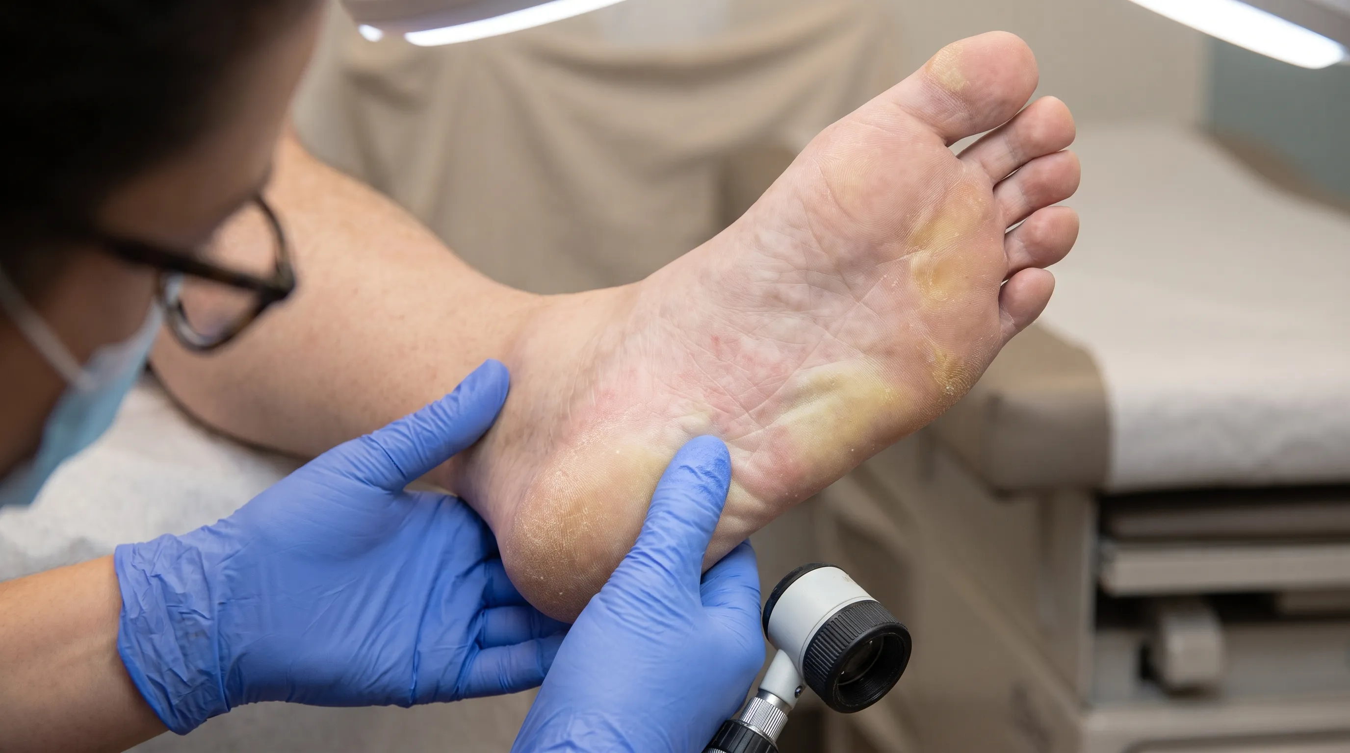
Prioritizing preventive care includes annual comprehensive foot examinations by qualified professionals, even in the absence of symptoms. Early intervention for minor abnormalities prevents cascade complications. Educating family members on proper foot inspection techniques fosters accountability and early detection, particularly for elderly or cognitively impaired individuals. Building a proactive relationship with healthcare providers ensures personalized, continuous care tailored to evolving physiological needs.

Frequently Asked Questions
Why do the bottoms of my feet turn red after standing for long periods?
Prolonged standing increases hydrostatic pressure in the lower extremity venous system, causing capillary engorgement and dependent redness. This physiological response is compounded by gravity, reduced calf muscle pump activity, and sometimes underlying venous insufficiency or early peripheral nerve dysfunction. Elevating the feet, wearing compression socks (if arterial flow is adequate), and taking periodic movement breaks typically restore normal coloration.
Can diabetes cause a red color on the bottom of feet?
Yes, diabetic peripheral neuropathy and autonomic dysfunction frequently manifest as plantar erythema. Chronic hyperglycemia damages microvessels and peripheral nerves, disrupting normal vasomotor regulation and thermal perception. This leads to abnormal blood pooling, dry skin, and increased infection susceptibility. Strict glycemic control, daily inspections, and professional podiatric care are essential to prevent ulceration and amputation.
How is erythromelalgia diagnosed and managed?
Diagnosis relies on clinical criteria: characteristic burning pain, visible redness, heat sensitivity, and symptom exacerbation with warmth or exercise. Genetic testing for SCN9A mutations may confirm primary forms. Management focuses on trigger avoidance, gentle cooling, elevation, and medications like gabapentin or topical agents prescribed by neurologists. Lifestyle modifications, including stress reduction and hydration, significantly reduce flare frequency.
When should I see a doctor for red soles?
Seek immediate medical attention if redness spreads rapidly, is accompanied by fever, severe pain, warmth, swelling, open wounds, or discoloration turning blue/grey. Patients with diabetes, peripheral vascular disease, or compromised immune systems should never ignore sudden or persistent plantar erythema, as it may indicate severe infection or ischemia requiring urgent intervention.
Is it safe to use ice packs for red, burning feet?
Direct ice application is generally discouraged, particularly for individuals with neuropathy or vascular disease, as it can cause tissue damage, frostbite, or paradoxical pain without the patient feeling it. Instead, use cool (not icy) gel packs, room-temperature fans, or mild cool water exposure. Always monitor skin response and avoid extreme temperature shifts that trigger vascular instability.
Conclusion
Observing a red color on bottom of feet serves as an important clinical signal that warrants careful attention and systematic evaluation. Whether stemming from benign mechanical stress, neurovascular dysregulation, infectious processes, or metabolic complications like diabetic neuropathy, plantar erythema reflects the intricate interplay between circulation, nerve function, and tissue integrity. Understanding the underlying etiology enables targeted interventions that alleviate symptoms, prevent progression, and preserve functional mobility. By adhering to evidence-based foot care protocols, maintaining rigorous chronic disease management, and recognizing critical warning signs, individuals can effectively safeguard their lower extremity health. Always consult qualified healthcare professionals for personalized diagnosis and treatment plans, ensuring that every step toward recovery is guided by medical expertise and scientific accuracy.
How we work
Researched and drafted with AI assistance, then reviewed by a human editor against the cited sources.
This article is general health information, not medical advice. Consult a qualified healthcare professional about your own situation.