Understanding Feet with Black Spots: Causes, Warning Signs & Care Guide

Noticing unusual pigmentation or dark discoloration on your lower extremities can be deeply unsettling, yet understanding feet with black spots is a crucial step toward proactive health management. The human foot is subjected to constant pressure, friction, and environmental exposure, making it a frequent site for various dermatological changes. While many dark spots originate from harmless, everyday occurrences like minor trauma or benign skin growths, others may signal underlying medical conditions that require immediate clinical attention. The complexity of foot dermatology lies in the fact that visual similarities often mask vastly different etiologies. A seemingly innocent dark patch could be a healed bruise, a viral wart, or an early-stage malignant melanoma. Because the soles and nail beds are areas people rarely inspect in detail, dangerous lesions frequently progress unnoticed until they reach advanced stages. This comprehensive guide explores the medical causes, diagnostic pathways, and evidence-based management strategies for foot pigmentation. By integrating clinical research, dermatological best practices, and actionable self-care routines, readers will gain the knowledge needed to monitor their skin effectively, recognize early warning signs, and seek timely medical intervention when necessary. Protecting your foot health requires vigilance, education, and a partnership with qualified healthcare professionals who can differentiate between routine cosmetic variations and serious pathological developments.
Understanding the Causes of Black Spots on Feet
The development of pigmented lesions on the lower extremities stems from a diverse array of biological mechanisms, ranging from localized trauma and viral infections to complex genetic mutations driving cellular proliferation. When evaluating feet with black spots, clinicians consider both intrinsic dermatological conditions and extrinsic environmental factors. Understanding these underlying causes is the foundation for accurate self-assessment and appropriate medical consultation. The differential diagnosis spans benign dermatoses that require minimal intervention to aggressive malignancies demanding multidisciplinary treatment protocols.
Free calculatorSun Protection GuideSPF guide and sun protection recommendationsOpen the calculatorAcral Lentiginous Melanoma: The Serious Culprit
Acral lentiginous melanoma (ALM) represents a rare but highly consequential subtype of melanoma that originates specifically on the palms, soles, and subungual regions (under the nails). Unlike the more common superficial spreading melanoma, which correlates strongly with ultraviolet radiation damage, ALM exhibits a distinct pathogenesis that remains independent of cumulative sun exposure. According to research published by DermNet NZ, the etiology of ALM is strongly associated with mechanical stress, repetitive shearing forces, and somatic genetic mutations affecting pathways such as KIT, BRAF, NRAS, NF1, cyclin D1, CDKN2A, and MYC. Clinically, ALM often presents initially as a flat, dark brown or black macule with irregular borders and heterogeneous pigmentation. Over time, without intervention, the lesion may thicken, develop nodular components, ulcerate, or exhibit central hypopigmentation indicative of spontaneous tumor regression.
The epidemiological profile of ALM underscores its disproportionate impact on populations with darker skin tones. While it accounts for only two to three percent of melanoma cases globally, it represents approximately forty to sixty percent of melanoma diagnoses among Asian and African-American patients. The average age at diagnosis hovers around sixty-three years, with equal distribution across biological sexes. Because melanoma in these concealed locations often evades routine visual screening, it is frequently diagnosed at more advanced Breslow depths compared to sun-exposed melanomas. The Mayo Clinic emphasizes that melanomas occurring in hidden areas are particularly dangerous precisely because they escape everyday detection during casual observation. Recognizing the subtle early manifestations of ALM remains paramount for improving long-term survival outcomes.
Benign and Trauma-Related Causes
A significant proportion of discolorations on the feet originate from mechanical injury rather than neoplastic transformation. Subungual hemorrhage, commonly referred to as a subungual hematoma, occurs when minor blunt trauma or repetitive microtrauma from poorly fitted shoes causes blood vessels beneath the toenail plate to rupture. The pooled blood appears as a dark purple or black spot that gradually migrates distally as the nail grows. While visually alarming, subungual hematomas are typically asymptomatic and resolve spontaneously. Differentiating them from subungual melanoma involves assessing uniformity, growth pattern, and history of trauma.
Talon noir, or black heel, is another benign trauma-induced condition frequently observed in athletes and active individuals. Rapid acceleration, deceleration, or sudden directional changes generate intense shear stress within the stratum corneum, resulting in capillary rupture and intradermal hemorrhage. The resulting pinpoint black or reddish-brown macules cluster along the lateral heel and often resolve without treatment. Plantar warts (verruca plantaris) represent a third common benign etiology. Caused by human papillomavirus (HPV) infection, these hyperkeratotic lesions frequently harbor thrombosed capillaries that manifest as tiny black dots scattered throughout the wart's surface. Unlike malignant lesions, plantar warts typically exhibit a rough, cauliflower-like texture, are tender upon lateral compression, and display interrupted dermatoglyphics (skin lines).
Medical Conditions and Infections
Systemic health disorders and localized infections can also manifest as dark foot pigmentation. Fungal nail infections (onychomycosis) occasionally cause severe discoloration, transforming nails from yellowish-brown to near-black as keratin degradation and pigment deposition progress. Diabetic patients may experience acanthosis nigricans, a condition characterized by velvety, hyperpigmented patches typically located in flexural areas but occasionally extending to the feet. Additionally, diabetic dermopathy presents as round or oval, reddish-brown macules that eventually darken and fade, often going unnoticed due to their asymptomatic nature. Benign melanocytic nevi, while uncommon on the soles, can occur and generally maintain symmetrical borders and uniform coloration over decades. However, any nevus displaying rapid morphological changes warrants immediate clinical reassessment.
| Condition | Primary Cause | Typical Appearance | Pain/Discomfort | Progression Pattern | Key Diagnostic Feature |
|---|---|---|---|---|---|
| Acral Lentiginous Melanoma | Genetic mutations, mechanical stress | Irregular dark brown/black macule, variegated colors | Often painless initially; may ulcerate later | Slow but persistent growth, potential nodular transformation | Asymmetrical borders, Hutchinson's sign, dermoscopic atypia |
| Subungual Hemorrhage | Trauma, repetitive friction | Solid dark purple/black patch under nail plate | Tender acutely, otherwise painless | Migrates outward with nail growth | Uniform color, clear trauma history, moves with nail |
| Plantar Warts | HPV infection | Hyperkeratotic plaque with pinpoint black dots | Painful upon direct or lateral pressure | Gradual enlargement, may cluster (mosaic warts) | Disrupted skin lines, thrombosed capillaries, rough surface |
| Talon Noir | Shear stress from activity | Pinpoint black/reddish macules on lateral heel | Usually asymptomatic | Fades over weeks to months | Punctate hemorrhages, common in athletes, benign dermoscopy |
How to Identify Warning Signs and Clinical Red Flags
Early detection fundamentally alters the prognosis of pigmented foot lesions. Because many individuals lack formal dermatological training, relying on standardized clinical frameworks ensures consistent, objective evaluation. The medical community universally advocates the ABCDE rule for initial self-assessment, supplemented by location-specific indicators that account for the unique anatomical and physiological characteristics of acral skin.
The ABCDE Rule Explained
Dermatologists worldwide utilize the ABCDE mnemonic as a foundational screening tool for pigmented lesions. Each criterion addresses a distinct morphological characteristic that correlates with malignant transformation potential:
A - Asymmetry: Benign moles and trauma-induced marks typically mirror themselves when bisected visually. Malignant lesions frequently exhibit structural imbalance, where one quadrant differs markedly in contour or pigment distribution from the opposite half.
B - Border Irregularity: Non-threatening spots generally possess smooth, well-defined perimeters. Suspicious lesions demonstrate scalloped, notched, or blurred margins where pigmentation fades unevenly into surrounding healthy tissue.
C - Color Variation: Uniformity indicates benignity, whereas melanoma frequently displays chaotic pigment architecture. A single lesion containing multiple hues—deep black, tan, white, blue, or red—suggests active melanocyte dysregulation and possible tumor heterogeneity.
D - Diameter: While traditional guidelines emphasize a six-millimeter threshold (approximately the size of a pencil eraser), contemporary dermatology recognizes that malignant melanomas can present at smaller dimensions. Nevertheless, larger lesions warrant heightened scrutiny.
E - Evolution: Temporal changes represent the most critical warning sign. Any lesion that alters in size, elevation, texture, or symptomatology over weeks to months demands professional evaluation. Spontaneous bleeding, crusting, or persistent pruritus further elevate clinical suspicion.
ALM-Specific Red Flags to Watch For
The acral skin environment necessitates additional observational parameters. Subungual melanoma frequently manifests as a longitudinal melanonychia—a vertical dark band extending from the proximal nail fold to the distal free edge. Benign bands typically remain narrow, uniform in width, and parallel to the nail axis. Malignant variants progressively widen, darken centrally, and may demonstrate pigment extension onto the adjacent periungual skin, clinically recognized as Hutchinson's sign. This phenomenon occurs when melanoma cells migrate beyond the nail matrix, providing a highly specific clinical indicator of malignancy.
Plantar melanoma often begins as a subtle, lightly pigmented patch that gradually darkens and thickens. Unlike common calluses, malignant lesions do not soften with regular exfoliation or emollient therapy. Instead, they may develop raised, firm nodules or central ulceration resistant to standard wound care. Numbness, tingling, or localized pain beneath a pigmented patch can indicate deeper tissue invasion or perineural involvement, signaling the need for urgent histopathological investigation.
When to Seek Immediate Medical Attention
Timing dramatically influences treatment success and survival rates. You should schedule a dermatological consultation promptly if you notice feet with black spots that emerge without preceding injury, fail to fade within four to six weeks, or demonstrate any single ABCDE criterion. Rapid expansion, recurrent bleeding without provocation, satellite pigmented macules surrounding the primary lesion, or unexplained systemic symptoms (such as unexplained weight loss or profound fatigue) require expedited evaluation. Delaying professional assessment for presumed benign trauma, especially when the injury mechanism is unclear or absent, remains a common diagnostic pitfall. The National Cancer Institute consistently stresses that early-stage melanoma confined to the epidermis boasts significantly higher five-year survival rates compared to invasive or metastatic disease.
Diagnosis, Evaluation, and the Pathway to Care
Accurate diagnosis bridges the gap between clinical suspicion and definitive management. When patients present with concerning foot pigmentation, healthcare providers follow a structured diagnostic algorithm designed to minimize uncertainty and optimize patient safety. This process integrates visual assessment, advanced imaging techniques, laboratory testing, and collaborative multidisciplinary planning.
The Role of Dermatology and Dermoscopy
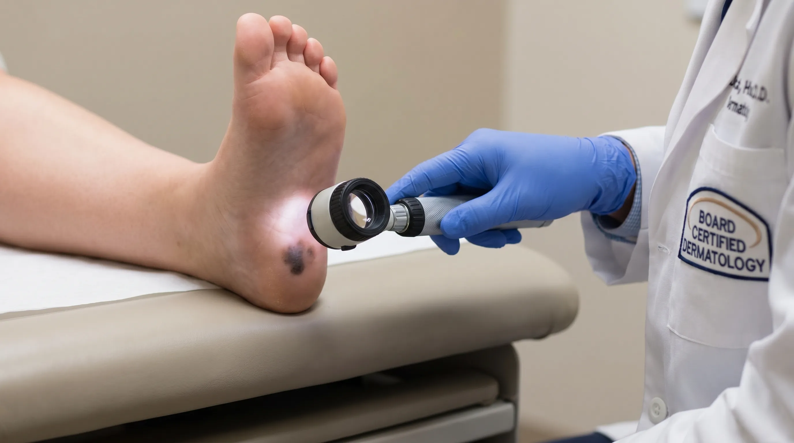
Board-certified dermatologists serve as the primary diagnostic authorities for complex cutaneous presentations. During a comprehensive examination, the clinician evaluates the entire integumentary system, recognizing that individuals with suspicious acral lesions often harbor concurrent atypical nevi or actinic damage elsewhere. The cornerstone of modern dermatological diagnosis is dermoscopy, a non-invasive technique utilizing a handheld magnifying device equipped with polarized light. Dermoscopy eliminates surface reflection and penetrates the stratum corneum, revealing subsurface pigment networks, vascular architectures, and structural anomalies invisible to the naked eye. In acral sites, dermoscopic patterns such as parallel ridge (highly suspicious for ALM) versus parallel furrow (benign) provide critical diagnostic clarity. Studies consistently demonstrate that dermoscopy increases melanoma detection sensitivity by up to thirty percent while reducing unnecessary biopsies of benign lesions.
Biopsy Procedures and What to Expect
When clinical and dermoscopic findings raise suspicion for malignancy, a skin biopsy becomes indispensable. The gold standard for acral lesions involves a complete excisional biopsy whenever anatomically feasible. This approach removes the entire lesion along with a narrow margin of surrounding tissue, allowing pathologists to assess the complete architecture, measure invasive depth (Breslow thickness), and identify ulceration or mitotic activity—all critical prognostic factors. If complete excision is impractical due to lesion size or location constraints, an incisional or punch biopsy targets the most atypical region. The tissue specimen undergoes histopathological processing, where specialized stains highlight melanocytic proliferation and cellular atypia. Immunohistochemical markers such as S100, Melan-A, and HMB-45 further confirm melanocytic lineage. Patients should anticipate minimal discomfort during the procedure, localized anesthesia, and a recovery period involving wound care instructions and suture removal within one to two weeks.
Genetic and Risk Factor Assessment
Following a confirmed diagnosis, clinicians conduct comprehensive risk stratification. Family history significantly influences surveillance protocols, as germline mutations in CDKN2A, BAP1, and MITF predispose individuals to familial atypical mole-melanoma syndromes. Additionally, personal history of immunosuppression, extensive prior UV exposure (for non-ALM melanomas), or numerous dysplastic nevi elevates baseline risk. Genetic counseling may be recommended for patients meeting high-risk criteria. Contemporary research also explores the molecular landscape of acral melanoma, identifying actionable mutations in NRAS and KIT that may qualify patients for targeted clinical trials. Understanding these genetic profiles enables precision medicine approaches tailored to individual tumor biology.

Practical Prevention, Self-Care, and Daily Habits
While genetic predisposition and spontaneous mutations fall outside voluntary control, numerous lifestyle modifications and routine practices significantly reduce the risk of trauma-induced pigmentation and enhance early detection of pathological changes. Implementing systematic self-care protocols empowers individuals to maintain optimal foot health and recognize abnormalities before they progress.
Monthly Self-Examination Routine
Consistent self-monitoring remains the most accessible and effective screening method. Establish a fixed schedule, ideally aligning with the first weekend of each month, to conduct thorough foot inspections. Position yourself in a well-lit room and utilize a handheld mirror or a full-length mirror to visualize the plantar surfaces, interdigital spaces, and lateral aspects of the feet. Carefully examine each toenail for longitudinal pigmentation, noting any new bands or darkening of existing ones. Document your findings by taking high-resolution photographs under consistent lighting conditions, annotating the date, and storing them securely in a dedicated health journal or digital folder. Serial photography provides objective evidence of morphological changes, enabling you and your dermatologist to evaluate evolution accurately. The American Academy of Dermatology strongly recommends incorporating acral areas into routine full-body skin checks, particularly for individuals with elevated melanoma risk.
Footwear and Lifestyle Modifications
Mechanical trauma represents a preventable trigger for numerous benign black spots. Select footwear that accommodates your natural foot dimensions, ensuring adequate toe box space to prevent repetitive compression against the nail plate during ambulation or athletic activities. Replace worn-out shoes before midsole breakdown compromises shock absorption, as inadequate cushioning amplifies impact forces transmitted to the plantar skin. Moisture-wicking socks reduce friction and minimize the risk of maceration, which can alter skin integrity and exacerbate pigmentation. For runners, dancers, and military personnel, alternating between multiple pairs of shoes distributes shear stress across different contact points, reducing localized tissue damage. If you frequently participate in high-impact sports, consider custom orthotics to optimize gait mechanics and minimize abnormal pressure distribution on the lateral heel or forefoot.
Sun Protection for Exposed Areas
Although acral lentiginous melanoma demonstrates minimal correlation with ultraviolet radiation, other melanoma subtypes can develop on the dorsal (top) surfaces of the feet, which receive direct sunlight during warm weather or outdoor activities. Applying broad-spectrum sunscreen with SPF 30 or higher to exposed feet, ankles, and the dorsal aspects of toes provides essential photoprotection. Reapply every two hours or immediately after swimming, sweating, or towel drying. Wearing breathable, UV-protective footwear during extended outdoor exposure further minimizes cumulative radiation damage. Integrating sun safety into daily routines complements trauma prevention and establishes a comprehensive skin defense strategy.
Treatment Pathways and Long-Term Management
Effective management of pigmented foot lesions hinges upon accurate diagnosis, stage-appropriate intervention, and diligent post-treatment surveillance. Treatment paradigms vary dramatically based on lesion histology, depth of invasion, and patient comorbidities. Modern oncology emphasizes personalized care, balancing curative intent with quality-of-life preservation.
Early Intervention and Surgical Excision
For confirmed melanoma in situ and early invasive stages, wide local excision remains the primary therapeutic modality. Surgical margins are determined by Breslow thickness: one-centimeter margins for thin tumors (≤1 mm) and two-centimeter margins for thicker lesions, per established clinical guidelines. Mohs micrographic surgery, utilizing staged excision with complete margin assessment, may be employed in anatomically constrained areas where tissue preservation is critical. Sentinel lymph node biopsy (SLNB) evaluates regional metastatic spread for intermediate or high-risk tumors, guiding subsequent adjuvant therapy decisions. Precise histopathological staging following excision dictates the necessity for additional interventions.
Advanced Therapies for Malignant Cases
When melanoma progresses beyond localized disease, systemic therapies revolutionize survival outcomes. Immunotherapy utilizing checkpoint inhibitors (anti-PD-1, anti-CTLA-4 antibodies) reactivates the host immune system to recognize and destroy metastatic melanoma cells. Targeted therapies address specific genetic alterations; BRAF and MEK inhibitors demonstrate remarkable efficacy in tumors harboring V600E BRAF mutations, rapidly shrinking advanced disease. The American Cancer Society reports that melanoma mortality rates declined substantially between 2013 and 2022, largely attributable to these pharmacological breakthroughs. For localized but unresectable disease or satellite lesions, localized therapies such as intralesional injections, radiation therapy, or isolated limb perfusion may be integrated into the treatment algorithm.
Long-Term Monitoring and Follow-Up
Survivors of melanoma face elevated lifetime risks for recurrence, new primary melanomas, and secondary malignancies. Structured follow-up protocols mandate regular dermatological examinations, typically every three to six months for the first two years, then annually thereafter. Patients should continue monthly self-exams and maintain meticulous documentation. Psychological support plays an integral role in survivorship care, as anxiety regarding recurrence frequently impacts mental well-being. Integrating survivorship clinics, mental health resources, and peer support networks fosters holistic recovery. Education regarding warning signs, lifestyle optimization, and sun safety remains continuous, ensuring patients remain active participants in their long-term health maintenance.
Frequently Asked Questions
Can a black spot on the foot go away on its own?
Yes, certain benign conditions resolve spontaneously. Subungual hemorrhages and talon noir typically fade as the skin or nail grows outward, replacing damaged tissue with new, healthy cells. However, lesions that persist beyond four to six weeks, exhibit progressive enlargement, or display irregular morphological features require professional dermatological evaluation to exclude malignancy.
How does acral lentiginous melanoma differ from common skin cancer?
Acral lentiginous melanoma develops in sun-protected regions such as the soles, palms, and nail beds, whereas most melanomas correlate strongly with cumulative UV radiation exposure. Research indicates ALM arises from genetic driver mutations and mechanical stress rather than photodamage. It disproportionately affects darker-skinned populations and frequently presents at later stages due to its concealed location, necessitating specialized diagnostic attention.
What does a dermatologist look for during a foot examination?
Dermatologists evaluate structural symmetry, border definition, color distribution, size, and temporal changes. They utilize dermoscopy to visualize subsurface pigment networks and vascular patterns, distinguishing benign parallel furrow arrangements from suspicious parallel ridge patterns characteristic of ALM. The presence of Hutchinson's sign, ulceration, or atypical vascular architecture further guides clinical decision-making and biopsy recommendations.
Should I wear shoes to prevent black spots on my feet?
Proper footwear significantly mitigates mechanical trauma responsible for benign pigmentation. Shoes with adequate toe clearance, supportive cushioning, and breathable materials reduce repetitive friction, pressure points, and shear forces that cause subungual hematomas and talon noir. Investing in well-fitted athletic and casual shoes protects foot integrity and minimizes preventable skin discoloration.
Is a skin biopsy necessary for every black spot on the foot?
No, biopsies are reserved for clinically suspicious or unexplained lesions. Stable, clearly trauma-related, or uniformly pigmented spots with documented benign history may be monitored via serial photography and clinical observation. However, any new lesion lacking a clear traumatic origin, or one demonstrating asymmetry, color variation, border irregularity, or evolution, warrants prompt dermatological assessment where biopsy serves as the definitive diagnostic standard.
Conclusion
Recognizing and addressing feet with black spots demands a balanced approach combining patient vigilance, clinical expertise, and evidence-based intervention. The spectrum of possible etiologies ranges from self-limiting mechanical trauma to aggressive malignancies requiring specialized oncological care. By internalizing the ABCDE criteria, understanding location-specific warning signs like Hutchinson's sign, and establishing consistent self-examination habits, individuals empower themselves to detect abnormalities at the most treatable stages. Dermatology and dermoscopy provide the technological and clinical foundation necessary to differentiate benign dermatoses from early melanoma, while surgical and systemic therapies offer robust management pathways for confirmed diagnoses. Ultimately, proactive foot health transcends cosmetic concerns; it represents a fundamental component of comprehensive cancer prevention and early intervention strategies. Prioritizing regular skin checks, selecting appropriate footwear, protecting exposed areas from unnecessary environmental stress, and maintaining open communication with qualified healthcare providers significantly enhances long-term outcomes. When in doubt, seeking professional evaluation remains the safest and most prudent course of action, ensuring that every pigmented lesion receives the accurate diagnosis and timely care it deserves.
How we work
Researched and drafted with AI assistance, then reviewed by a human editor against the cited sources.
This article is general health information, not medical advice. Consult a qualified healthcare professional about your own situation.