Black Spots on Bottom of Feet: Causes, Diagnosis & Treatment Guide

Key points
- Wear protective footwear in communal environments including swimming pools, gym showers, and hotel rooms
- Maintain thorough daily foot washing followed by complete drying between digital spaces
- Rotate moisture-wicking socks and avoid sharing personal textiles or pedicure instruments
- Inspect feet systematically each evening, utilizing mirrors or caregiver assistance for posterior visualization
- Address callus formation and minor trauma promptly to maintain epidermal integrity
- Optimize footwear fit to minimize focal pressure points and shear forces
Noticing an unexpected dark mark on the sole of your foot can immediately trigger concern. The plantar surface bears constant pressure, friction, and environmental exposure, making it vulnerable to a wide range of dermatological changes. Whether you recently started wearing new shoes, walked barefoot in a public locker room, or simply discovered the mark during a routine self-examination, understanding what you are looking at is the critical first step toward peace of mind and appropriate care. Black spots on bottom of feet are remarkably common, yet they encompass a broad clinical spectrum ranging from entirely benign viral growths to conditions that require urgent oncological intervention. This comprehensive guide examines the underlying pathophysiology, diagnostic pathways, evidence-based treatments, and preventive strategies you need to make informed decisions about your foot health.
Understanding the Primary Causes of Dark Plantar Lesions
The appearance of dark pigmentation or distinct lesions on the soles rarely occurs randomly. Each presentation reflects a specific biological mechanism, whether driven by viral invasion, melanocytic proliferation, localized hemorrhage, or environmental pathogens. Recognizing the distinct visual and symptomatic patterns of these conditions helps patients navigate next steps safely and effectively.
Free calculatorA1C CalculatorA1C to average blood glucoseOpen the calculatorPlantar Warts: Viral Origins and Misconceptions
Plantar warts, medically termed verruca plantaris, represent the most frequent diagnosis for individuals presenting with dark plantar lesions. These growths develop when specific strains of the human papillomavirus penetrate the epidermis through microscopic abrasions. HPV types 1, 2, 4, 60, and 63 demonstrate a strong predilection for the thick keratinized tissue of the soles. Unlike common cutaneous warts that protrude outward, plantar warts typically grow inward due to continuous body weight and pressure.
The hallmark visual feature involves small, pinpoint black dots scattered across the lesion. Despite widespread folklore suggesting these are "wart seeds," dermatological examination reveals they are actually thrombosed capillaries. As the viral infection stimulates rapid epidermal proliferation, it disrupts local microvasculature, causing tiny vessels to clot and appear dark. You can learn more about the viral mechanism at the American Academy of Dermatology. These lesions frequently localize to weight-bearing zones such as the metatarsal heads and calcaneal regions. Patients typically report a sharp, punctate pain during ambulation, often describing the sensation as stepping on a small pebble. Approximately 7 to 12 percent of the general population experiences plantar warts, with peak incidence occurring during adolescence and young adulthood when immune surveillance and environmental exposure intersect. Spontaneous resolution occurs in roughly 65 percent of pediatric cases within two years, but adult persistence is significantly higher, often requiring clinical intervention. Mayo Clinic provides detailed guidance on symptom progression and conservative management.
Acral Lentiginous Melanoma: A Serious Concern
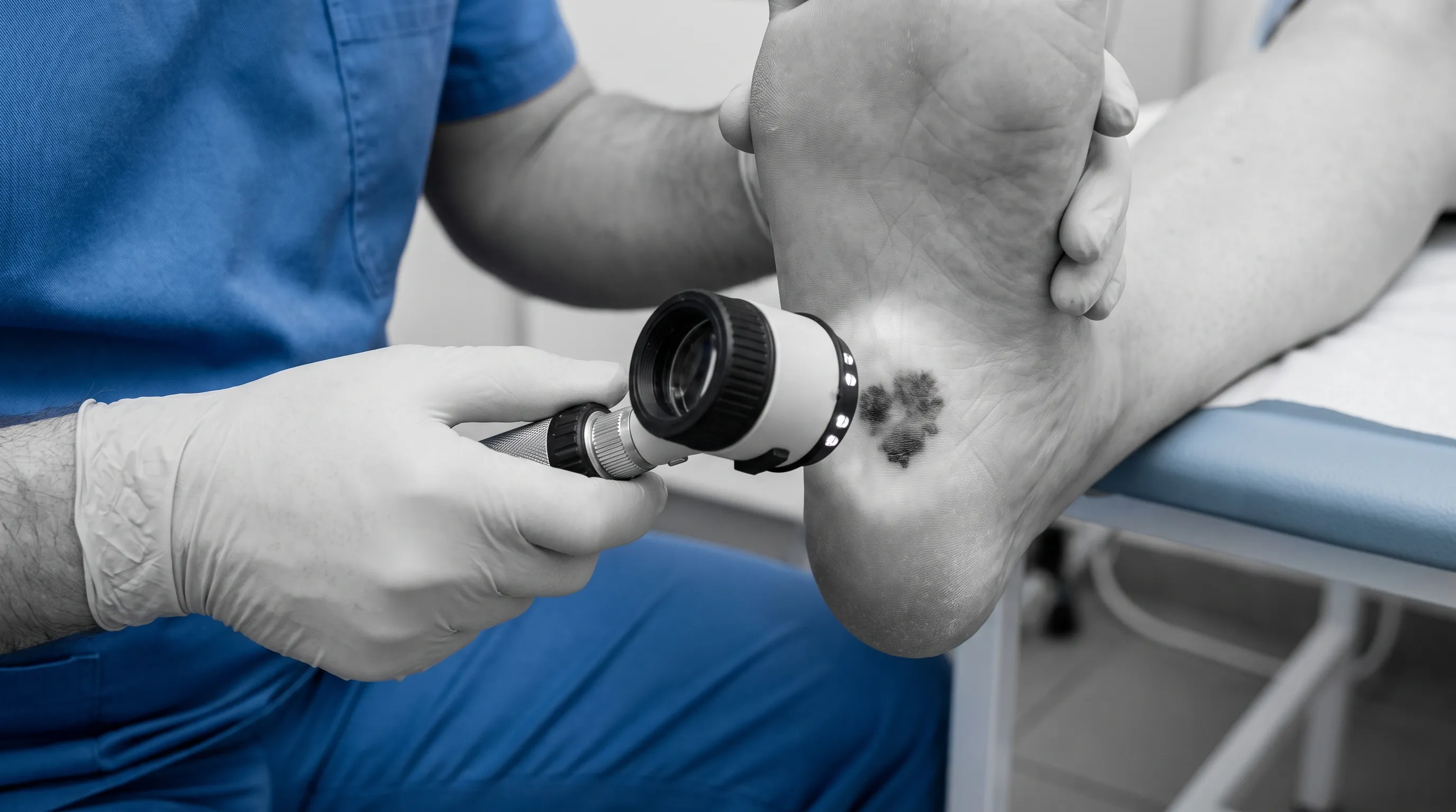
While far less common, acral lentiginous melanoma (ALM) demands immediate recognition due to its aggressive biological behavior and potential for metastatic spread. Unlike cutaneous melanomas strongly linked to ultraviolet radiation, ALM develops on glabrous skin surfaces—palms, soles, and subungual regions—regardless of cumulative sun exposure. Epidemiological data reveals a striking demographic pattern: although ALM accounts for only 2 to 3 percent of all melanomas in Caucasian populations, it represents 60 to 75 percent of melanoma diagnoses among African American and Asian individuals. The American Cancer Society emphasizes that ALM frequently presents in later stages because patients and clinicians alike may overlook lesions on less-examined surfaces.
Clinically, ALM manifests as a progressively enlarging, irregularly pigmented patch. The classic ABCDE framework serves as a critical screening tool: asymmetry where one half diverges significantly from the other, border irregularity characterized by scalloped or blurred edges, color variegation displaying multiple shades of brown, black, tan, or blue, diameter exceeding six millimeters, and evolutionary changes in morphology over weeks or months. Early detection dramatically improves five-year survival rates, which is why any rapidly evolving pigmentation warrants professional evaluation without delay.
Secondary and Environmental Triggers to Consider
Beyond viral and neoplastic processes, several external and mechanical factors contribute to localized plantar discoloration. These conditions typically follow a predictable timeline and respond well to targeted conservative measures when identified correctly.
Subcorneal Hemorrhages and Friction-Induced Blisters
Repetitive mechanical stress or acute trauma frequently causes localized bleeding beneath the stratum corneum, creating what patients commonly refer to as blood blisters. Ill-fitting footwear, sudden increases in athletic training volume, or accidental impact can rupture superficial capillaries without compromising the overlying epidermal barrier. The trapped deoxygenated blood initially appears dark red, gradually transitioning to purple, brown, and ultimately black as hemoglobin degrades. Unlike infectious or neoplastic lesions, traumatic hemorrhages remain confined to the site of injury and gradually migrate upward as the epidermis renews itself. Most resolve spontaneously within two to four weeks, provided the mechanical insult is removed and secondary infection is prevented. Cleveland Clinic offers practical guidance on differentiating trauma-related lesions from infectious growths.
Tinea Nigra: A Rare Fungal Presentation
Tinea nigra represents a superficial mycotic infection caused by Hortaea werneckii, a halophilic dematiaceous fungus commonly found in soil and decaying organic matter in tropical and subtropical climates. The organism adheres to the stratum corneum, secreting melanin-like pigments that stain the superficial keratin. Clinically, it presents as a solitary, painless, well-demarcated macule with brown to black coloration. Unlike deeper fungal infections, tinea nigra causes no tissue invasion, inflammation, or systemic symptoms. Geographic distribution heavily influences prevalence, with higher incidence reported in South America, Africa, and coastal regions. Topical antifungal agents such as clotrimazole or terbinafine applied twice daily typically clear the infection within fourteen to twenty-eight days. The NIH MedlinePlus portal provides additional context on distinguishing fungal macules from other pigmented lesions.
Foreign Bodies and Penetrating Injuries
Penetrating trauma introduces organic or inorganic material into the plantar tissue, triggering localized inflammation, minor hemorrhage, and subsequent hyperpigmentation. Common culprits include glass fragments, wooden splinters, thorns, or metal shards. The body responds by isolating the foreign material with granulation tissue, which often darkens over time due to hemosiderin deposition and localized oxidative processes. Patients typically report focal tenderness that intensifies with direct pressure. Prompt removal using sterile technique, combined with appropriate wound cleansing and tetanus prophylaxis when indicated, prevents chronic inflammation, secondary bacterial infection, and granuloma formation.
The Critical Role of Underlying Health Conditions
Systemic health profoundly influences skin integrity, microcirculation, and wound healing capacity. Certain chronic conditions transform minor plantar changes into potentially limb-threatening complications.
Diabetes, Neuropathy, and Vascular Complications
Individuals living with diabetes mellitus face significantly elevated risks for foot pathology. Peripheral neuropathy diminishes protective sensation, allowing minor trauma or ill-fitting footwear to progress unrecognized into ulceration. Concurrent microvascular disease impairs tissue perfusion, delaying healing and increasing susceptibility to infection. Dark discoloration on diabetic feet may signify ischemic changes, hemorrhagic ulcer precursors, or early necrosis. WebMD highlights that any new cutaneous alteration in diabetic patients requires immediate podiatric evaluation to prevent amputation. Glycemic control, daily foot inspection, and appropriate footwear modifications form the cornerstone of preventive management.
Immune System Status and Lesion Progression
Immunosuppression—whether medication-induced, HIV-related, or genetically determined—alters the natural history of plantar lesions. Viral clearance slows dramatically, leading to persistent, recalcitrant plantar warts that frequently cluster and coalesce into mosaic patterns. Similarly, immune surveillance against atypical melanocytes weakens, potentially accelerating malignant transformation. Patients undergoing chemotherapy, organ transplant recipients, and those with autoimmune conditions treated with biologics should maintain heightened vigilance and establish routine dermatological screening intervals.
Clinical Diagnostic Pathways
Accurate diagnosis relies on systematic evaluation rather than visual assumption alone. Dermatologists and podiatrists employ a tiered diagnostic approach to differentiate benign from malignant processes safely and efficiently.
Dermatoscopic Examination and Visual Assessment
Dermatoscopy utilizes a handheld polarized light device to magnify skin structures ten to thirty times, revealing pigment architecture and microvascular patterns invisible to the naked eye. In plantar warts, dermoscopy displays characteristic dotted vessels arranged in a papillary distribution, often surrounded by white scaling halos. Conversely, melanoma exhibits irregular pigment networks, asymmetric globules, radial streaks, and blue-white veils. Skin Cancer Foundation provides visual references for understanding dermatoscopic criteria.
Skin Scraping, Biopsy, and Laboratory Testing
When dermatoscopy remains inconclusive or malignancy suspicion rises, clinicians proceed to histopathological confirmation. A superficial scraping examined under potassium hydroxide preparation identifies fungal hyphae in tinea nigra cases. If melanoma cannot be excluded, a punch or excisional biopsy provides definitive tissue architecture analysis, measuring Breslow thickness, ulceration status, and mitotic rate. This gold-standard diagnostic procedure guides subsequent oncological staging and therapeutic planning.
Evidence-Based Treatment and Management Protocols
Therapeutic strategies must align precisely with diagnostic findings. Inappropriate treatment delays care, exacerbates pathology, or causes unnecessary tissue damage. The following protocols reflect current clinical guidelines.
Stepwise Therapies for Plantar Warts
First-line management typically begins with over-the-counter salicylic acid formulations containing seventeen to forty percent concentration. Daily application softens hyperkeratotic tissue and stimulates local immune response through mild chemical irritation. If conservative measures fail after twelve weeks, clinical interventions include cryotherapy with liquid nitrogen, cantharidin application, immunotherapy using contact sensitizers like squaric acid or diphenylcyclopropenone, pulsed dye laser ablation targeting neovascularization, or surgical curettage combined with electrodesiccation. NHS UK outlines stepwise treatment algorithms for refractory cases.
Oncological and Surgical Interventions
Confirmed acral lentiginous melanoma requires multidisciplinary oncological management. Wide local excision with appropriate surgical margins remains the cornerstone of treatment. Sentinel lymph node biopsy determines regional metastatic involvement and guides adjuvant therapy selection. Advanced or metastatic cases increasingly utilize immune checkpoint inhibitors and targeted BRAF/MEK inhibitors, dramatically improving survival outcomes compared to historical chemotherapy protocols.
Conservative and Topical Approaches for Infections
Tinea nigra responds reliably to twice-daily topical antifungals for two to four weeks. Blood blisters require protective dressings, pressure redistribution, and time for natural epithelial migration. Foreign bodies necessitate sterile extraction followed by wound monitoring. All conservative approaches emphasize preventing secondary bacterial superinfection through meticulous hygiene and appropriate barrier protection.
[Clinical Dermatology Video: Plantar Lesion Differentiation]
Proactive Prevention and Daily Foot Hygiene
Implementing consistent preventive measures dramatically reduces lesion recurrence and complications. The following strategies integrate seamlessly into daily routines:
- Wear protective footwear in communal environments including swimming pools, gym showers, and hotel rooms
- Maintain thorough daily foot washing followed by complete drying between digital spaces
- Rotate moisture-wicking socks and avoid sharing personal textiles or pedicure instruments
- Inspect feet systematically each evening, utilizing mirrors or caregiver assistance for posterior visualization
- Address callus formation and minor trauma promptly to maintain epidermal integrity
- Optimize footwear fit to minimize focal pressure points and shear forces
| Condition | Primary Visual Signs | Pain Level | Contagious | Typical Resolution Timeline |
|---|---|---|---|---|
| Plantar Warts | Rough surface, thrombosed capillaries, inward growth | Moderate to severe with pressure | Highly contagious | 6-24 months (variable) |
| Acral Melanoma | Irregular borders, color variegation, asymmetry | Usually painless early, painful if ulcerated | No | Requires surgical/oncological treatment |
| Blood Blister | Uniform dark red/black, well-defined, flat | Mild to moderate initially | No | 2-4 weeks with epithelial turnover |
| Tinea Nigra | Smooth brown/black macule, superficial stain | Painless | No | 2-4 weeks with antifungals |
| Foreign Body | Focal tenderness, possible visible fragment | Sharp on pressure | No | Immediate with removal |

Frequently Asked Questions
Are black dots in plantar warts actually seeds?
No. This is a persistent myth that has circulated for generations without scientific basis. The tiny black dots visible within plantar warts are thrombosed capillaries, meaning small blood vessels have clotted due to viral disruption of the local microvasculature. They play no role in reproduction or spread and simply indicate active tissue involvement.
When should I worry about a black spot on my foot?
Immediate medical consultation is necessary when lesions display asymmetry, irregular or blurred borders, multiple color variations, diameter exceeding six millimeters, or rapid morphological evolution. Additional red flags include spontaneous bleeding, crusting, ulceration, persistent pain unresponsive to pressure offloading, or new onset in individuals with diabetes or immunosuppression.
Can black spots on bottom of feet disappear on their own?
Certain etiologies follow natural resolution pathways without intervention. Blood blisters resolve as epidermal cells regenerate and migrate outward. Approximately sixty-five percent of pediatric plantar warts undergo spontaneous immune clearance within twenty-four months. Tinea nigra may clear gradually but typically requires topical antifungals for reliable eradication. Conversely, malignant melanoma never resolves spontaneously and demands urgent oncological management.
How do doctors tell the difference between a wart and melanoma?
Board-certified dermatologists rely on dermatoscopic visualization to evaluate pigment distribution and vascular architecture. Warts display characteristic dotted vessels arranged in clustered patterns with overlying hyperkeratosis. Melanoma reveals atypical pigment networks, asymmetric globules, radial streaming, and blue-white structural alterations. When visual differentiation proves uncertain, histopathological biopsy provides definitive cellular diagnosis.
Is it safe to use over-the-counter acid treatments on dark foot spots?
Only after professional confirmation that the lesion is a benign plantar wart. Applying salicylic acid to undiagnosed melanoma can cause significant tissue damage, obscure visual markers, delay definitive diagnosis, and potentially accelerate disease progression. Self-treatment of dark plantar lesions without clinical evaluation carries substantial risk.
Conclusion
Discovering dark lesions on the plantar surface requires careful observation and, when appropriate, professional evaluation. Black spots on bottom of feet span a wide clinical spectrum, from common viral growths that respond well to conservative therapy to serious malignancies demanding immediate intervention. Understanding the distinct visual characteristics, symptom patterns, and underlying pathophysiology empowers patients to make timely, informed decisions. Prioritize regular foot inspection, maintain protective hygiene practices, and never dismiss evolving pigmentation. Early consultation with a dermatologist or podiatrist ensures accurate diagnosis, prevents unnecessary tissue damage from inappropriate self-treatment, and ultimately safeguards long-term foot health and mobility.
How we work
Researched and drafted with AI assistance, then reviewed by a human editor against the cited sources.
This article is general health information, not medical advice. Consult a qualified healthcare professional about your own situation.