Subcutaneous Nodules: Causes, Diagnosis, and Evidence-Based Treatment

Discovering a small, firm lump beneath your skin can immediately trigger concern, yet understanding subcutaneous nodules reveals that the vast majority of these growths are entirely benign and manageable. These discrete, solid masses reside in the subcutaneous tissue—the fatty and connective tissue layer situated just beneath the dermis. Ranging from a few millimeters to several centimeters, they can feel soft or firm, mobile or fixed, and may or may not cause discomfort. While most develop gradually without serious health implications, a subset can signal underlying systemic conditions, localized infections, or, rarely, malignancy. Proper clinical evaluation, informed by modern dermatological and oncological guidelines, ensures accurate identification and appropriate intervention. This comprehensive guide explores the pathophysiology, epidemiological trends, diagnostic pathways, and evidence-based management strategies for these growths. By combining authoritative medical research with practical patient guidance, you will gain a clear understanding of when monitoring is sufficient and when professional intervention is necessary. Whether you are a patient navigating a new discovery or a caregiver seeking reliable information, this resource provides a structured, medically sound overview of everything you need to know.
What Are Subcutaneous Nodules? Understanding the Basics
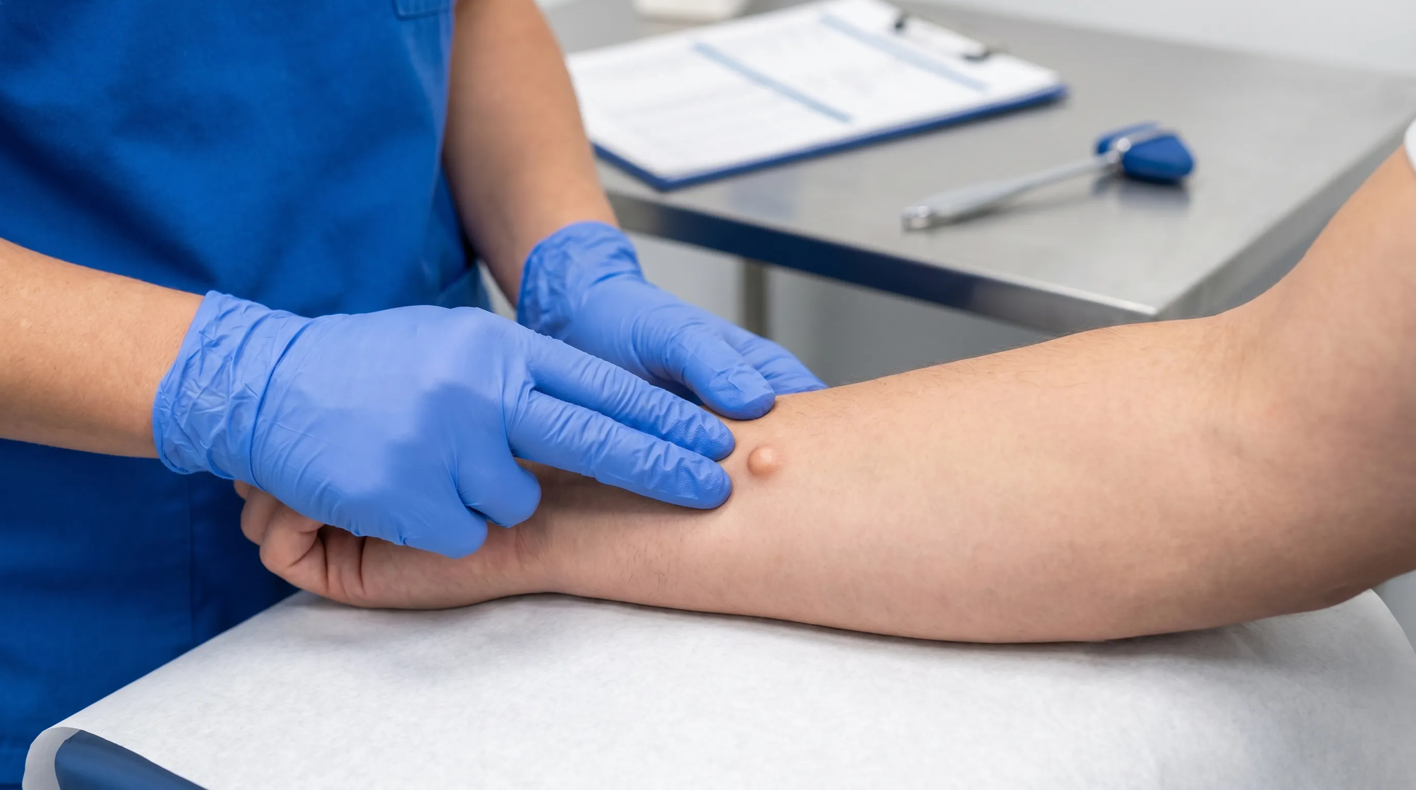
Subcutaneous nodules are localized, palpable masses that develop within the hypodermis, commonly referred to as the subcutaneous layer. This anatomical compartment consists primarily of adipose tissue, collagenous fibers, blood vessels, nerves, and lymphatics, serving as both an insulator and a shock absorber for the underlying musculoskeletal structures. When cellular proliferation, fluid accumulation, inflammatory processes, or foreign material deposition occurs in this region, the body may encapsulate the material, forming a discrete lump that can be easily felt during palpation.
Free calculatorSun Protection GuideSPF guide and sun protection recommendationsOpen the calculatorAnatomy of the Subcutaneous Layer
The hypodermis is distinct from the overlying epidermis and dermis, which are primarily epithelial and connective tissues responsible for barrier function and sensation. Unlike superficial skin lesions such as papules, pustules, or plaques, subcutaneous nodules sit deeper, meaning they often do not alter the surface texture or color of the skin in their early stages. As they enlarge, however, they may push upward, creating visible contours or stretching the overlying epidermis. The vascular and lymphatic networks within this layer play a critical role in how these masses grow, resolve, or spread. For instance, highly vascularized inflammatory nodules may appear reddened or warm to the touch, while avascular fatty tumors typically maintain the normal skin tone.
How They Form and Develop
The pathogenesis of these growths varies widely depending on the underlying etiology. Benign adipose tumors arise from the clonal expansion of mature fat cells, often influenced by genetic predispositions and localized trauma. Cystic formations develop when hair follicles or sebaceous glands become obstructed, trapping keratin and desquamated epithelial cells within a fibrous capsule. Immune-mediated processes, such as rheumatoid arthritis or delayed hypersensitivity reactions, trigger macrophage and fibroblast infiltration, leading to organized inflammatory masses. Infectious agents, metabolic crystal deposits, and nerve sheath proliferations follow distinct biological pathways but share the common endpoint of forming a discrete, subcutaneous mass. Understanding these mechanisms is essential for clinicians when determining the appropriate diagnostic algorithm and treatment trajectory.
Key Facts and Epidemiology
Subcutaneous nodules represent one of the most frequent reasons patients seek evaluation from primary care physicians, dermatologists, and general surgeons. The epidemiological data surrounding these growths provides valuable context for risk stratification and reassurance.
Prevalence Across Demographics
Benign soft tissue tumors are exceedingly common in the general population. Lipomas alone affect approximately 1 to 2 percent of individuals, with incidence peaking between the ages of 40 and 60, according to the Cleveland Clinic. Genetic syndromes such as familial multiple lipomatosis or Gardner syndrome can increase susceptibility, while metabolic factors like obesity may influence their presentation and growth rate. Inflammatory variants exhibit distinct demographic patterns; erythema nodosum, for example, disproportionately affects women at a ratio of 3 to 1 compared to men, typically presenting between ages 20 and 40, as detailed by NIH MedlinePlus. Autoimmune-associated growths, such as rheumatoid nodules, develop in 20 to 30 percent of patients with established rheumatoid arthritis, frequently localizing to extensor surfaces and pressure-bearing areas like the elbows and forearms, per World Health Organization (WHO) data.
When to Worry: Benign vs. Malignant Statistics
Despite the alarming nature of discovering an unexplained lump, malignancy remains rare. Comprehensive clinical studies indicate that less than 1 percent of all palpable subcutaneous masses are cancerous, as reported by the National Cancer Institute (NIH). The most concerning malignant entities include soft tissue sarcomas, metastatic carcinomas, and certain lymphomas. However, specific clinical features significantly raise the index of suspicion. Rapid enlargement, dimensions exceeding 5 centimeters, deep fixation to fascia or muscle, and persistent pain are recognized red flag indicators that warrant advanced imaging and histopathological evaluation. Spontaneous resolution is common in reactive and inflammatory variants, particularly those triggered by transient infections, medication reactions, or minor trauma, which often regress over weeks to months without intervention.
Common Types and Etiological Classifications
Categorizing subcutaneous nodules by their underlying pathology streamlines diagnosis and guides targeted therapy. Each variant presents with characteristic clinical features that aid clinicians in forming an accurate differential diagnosis before definitive testing.
Lipomas and Fatty Tumors
Lipomas are the most prevalent benign mesenchymal neoplasms encountered in clinical practice, as outlined by Mayo Clinic. They typically present as soft, doughy, highly mobile, and painless masses with smooth borders. Histologically, they consist of mature adipocytes arranged in lobules, separated by delicate fibrous septa. While usually solitary, multiple lipomas can occur in familial patterns or alongside metabolic disorders. Surgical literature notes that they rarely undergo malignant transformation, though careful clinical monitoring is advised if atypical features emerge.
Cysts and Keratin-Filled Masses
Epidermoid and sebaceous cysts arise from follicular infundibulum obstruction. They feel firm or rubbery and frequently exhibit a central punctum—a small, dark opening where trapped keratin and cellular debris may occasionally drain. These growths are prone to secondary infection, inflammation, and rupture, which can trigger a significant localized immune response. Complete surgical excision of the cyst wall is necessary to prevent recurrence, as leaving behind capsule fragments significantly increases the likelihood of regrowth. Management guidelines from the Cleveland Clinic emphasize professional removal over home attempts.
Inflammatory and Autoimmune Nodules
Immune-mediated growths represent a complex category requiring systemic evaluation. Rheumatoid nodules are firm, non-tender, and histologically characterized by central fibrinoid necrosis surrounded by palisading histiocytes and lymphocytes. Their presence often correlates with severe or long-standing disease activity, necessitating optimization of disease-modifying antirheumatic drug (DMARD) therapy. Erythema nodosum, a delayed hypersensitivity vasculitis, presents as tender, erythematous to violaceous nodules predominantly on the anterior shins. It is frequently triggered by streptococcal pharyngitis, sarcoidosis, tuberculosis, or oral contraceptives.
Metabolic and Infectious Variants
Gouty tophi form due to the deposition of monosodium urate crystals in the subcutaneous tissue, particularly around joints, bursae, and the helix of the ear. They appear chalky or yellowish and can erode through the skin, discharging a crumbly white material if left untreated, according to CDC resources. Dermatofibromas, often triggered by minor insect bites or trauma, are firm dermal-to-subcutaneous nodules that characteristically dimple when laterally pinched. Neurofibromas, associated with neurofibromatosis type 1, are soft, compressible tumors arising from peripheral nerve sheaths that may be accompanied by café-au-lait macules and Lisch nodules.
| Type | Clinical Features | Typical Etiology | Primary Management |
|---|---|---|---|
| Lipoma | Soft, doughy, highly mobile, painless | Benign adipocyte proliferation; genetic predisposition | Observation or elective excision if symptomatic |
| Epidermoid Cyst | Firm, central punctum, may drain keratinous material | Follicular occlusion and infundibulum blockage | Complete surgical excision of cyst capsule |
| Rheumatoid Nodule | Firm, non-tender, over pressure points/extensor surfaces | Immune complex deposition in established RA | Optimize DMARD or biologic therapy |
| Erythema Nodosum | Tender, red/purple, anterior shins | Delayed hypersensitivity (infections, drugs, sarcoidosis) | NSAIDs, potassium iodide, corticosteroids, treat trigger |
| Dermatofibroma | Firm, hyperpigmented, positive dimple sign | Benign fibrohistiocytic proliferation (post-trauma) | Observation; excision if cosmetically concerning |
| Gouty Tophus | Chalky, firm, periarticular or auricular | Monosodium urate crystal deposition | Urate-lowering therapy (allopurinol, febuxostat) |
Clinical Evaluation and Diagnostic Pathways
Accurate diagnosis of subcutaneous nodules relies on a systematic approach that integrates detailed patient history, meticulous physical examination, targeted imaging, and selective tissue sampling.
The Physical Examination and Patient History
Clinicians begin by characterizing the lesion using precise descriptors. Key historical points include onset, rate of growth, associated symptoms (pain, tenderness, systemic fever, unexplained weight loss), and relevant medical history such as autoimmune disease, prior trauma, recent travel, or medication changes. During palpation, providers assess dimensions in three planes, mobility relative to overlying skin and underlying fascia, consistency, and temperature. Skin changes like erythema, ulceration, or tethering provide crucial diagnostic clues. As noted in the American Family Physician guidelines, most cases encountered in primary care are benign, but recognizing red flag features dictates whether advanced workup is necessary.
Imaging Modalities: Ultrasound and MRI
High-frequency ultrasonography serves as the first-line imaging modality due to its accessibility, cost-effectiveness, and ability to rapidly differentiate cystic from solid lesions. Ultrasound can assess internal echogenicity, posterior acoustic enhancement, vascularity on Doppler interrogation, and relationship to surrounding structures. When lesions are deep, atypical, or suspicious for malignancy, magnetic resonance imaging (MRI) becomes the gold standard. MRI provides exceptional soft tissue contrast, accurately delineating fascial planes, muscular involvement, neurovascular displacement, and internal architecture. It is particularly valuable in preoperative planning and differentiating benign fatty tumors from atypical lipomatous neoplasms or sarcomas, per NIH Radiology guidelines.
Laboratory Testing and Biopsy Procedures
Laboratory evaluation is indicated when systemic or inflammatory etiologies are suspected. Complete blood count, erythrocyte sedimentation rate (ESR), and C-reactive protein (CRP) assess systemic inflammation. Rheumatoid factor (RF), anti-cyclic citrullinated peptide (anti-CCP), antinuclear antibodies (ANA), and serum uric acid help confirm autoimmune or metabolic diagnoses. When clinical and radiological findings raise suspicion for malignancy, tissue diagnosis is mandatory. Fine-needle aspiration (FNA) may provide cytological data, but core needle biopsy or excisional biopsy is preferred for soft tissue masses to preserve architectural context for histopathological grading and immunohistochemical staining.
Evidence-Based Management and Treatment Strategies
Management of subcutaneous nodules is highly individualized, balancing lesion characteristics, symptom burden, patient preferences, and underlying systemic conditions. Treatment ranges from conservative monitoring to targeted pharmacotherapy and surgical intervention.
Observation and Watchful Waiting
For asymptomatic, clinically classic benign growths such as small lipomas or dormant cysts, active intervention is rarely warranted. Evidence supports a watchful waiting approach, with clinicians advising patients to monitor for changes in size, shape, consistency, or symptomatology. Regular self-examination and periodic clinical follow-up every 6 to 12 months are sufficient for stable lesions. This strategy minimizes unnecessary procedures, reduces healthcare costs, and avoids surgical complications such as infection, scarring, or nerve injury.
Medical Interventions and Pharmacotherapy
Inflammatory and metabolic nodules often respond to systemic or targeted medical therapy. Erythema nodosum typically resolves with nonsteroidal anti-inflammatory drugs (NSAIDs) or, in refractory cases, short courses of oral corticosteroids or potassium iodide. Treating the underlying trigger, such as streptococcal infection or sarcoidosis, accelerates resolution. Rheumatoid nodules require optimization of systemic RA management, including methotrexate, biologic response modifiers, or targeted synthetic DMARDs. Gouty tophi necessitate strict urate-lowering therapy with medications like allopurinol or febuxostat, maintaining serum uric acid below 6 mg/dL to promote gradual crystal dissolution. Expert dermatological literature emphasizes that addressing the root pathophysiology often eliminates the need for surgical removal, as supported by Mayo Clinic treatment guidelines.
Surgical Excision and Minimally Invasive Techniques
Surgical intervention is indicated for lesions causing pain, functional impairment, rapid enlargement, cosmetic distress, or diagnostic uncertainty. Procedures are routinely performed in-office or outpatient surgical centers under local anesthesia with excellent safety profiles. Lipoma excision involves sharp dissection to remove the encapsulated fatty tissue intact. Cyst removal requires meticulous extraction of the entire sac wall to prevent recurrence, which studies show occurs in 15 to 30 percent of cases when rupture or incomplete removal happens. Minimally invasive options such as steroid injections, laser-assisted drainage, or endoscopic removal may be considered for select superficial cysts, though complete excision remains the standard of care for definitive management.
Patient Self-Care, Lifestyle Modifications, and Monitoring
Empowering patients with evidence-based self-care strategies and clear monitoring protocols enhances outcomes and reduces anxiety.
Daily Skin and Lifestyle Habits
Maintaining optimal skin hygiene, using gentle cleansers, and avoiding excessive friction or trauma to prone areas can reduce the risk of follicular irritation and secondary cyst formation. A balanced diet rich in anti-inflammatory foods supports immune regulation and may benefit individuals prone to inflammatory or autoimmune-related nodules. Maintaining a healthy weight reduces mechanical stress on adipose-rich areas and can lower the overall incidence of fatty tumors. Avoiding tight clothing or repetitive mechanical pressure on bony prominences minimizes trauma-induced inflammatory responses.
Tracking Changes and Knowing When to Seek Care
Patients should document baseline dimensions using a flexible measuring tape or compare against common household objects for scale. Periodic photographs under consistent lighting and distance provide objective records for clinical review. If a nodule remains stable, painless, and unchanged over several months, continued observation is appropriate. However, any deviation from baseline warrants timely re-evaluation. Keeping a symptom journal that notes pain levels, mobility changes, or associated systemic signs facilitates accurate communication with healthcare providers during follow-up visits.
Red Flag Symptoms Requiring Immediate Attention
Certain clinical features should never be ignored. Seek prompt medical evaluation if a mass exceeds 5 centimeters, demonstrates rapid or exponential growth, becomes fixed to deeper tissues or bone, causes unrelenting night pain, or is accompanied by constitutional symptoms such as unexplained fever, drenching night sweats, fatigue, or unintentional weight loss, according to NIH cancer warning guidelines. These indicators may suggest malignancy, deep-seated infection, or aggressive inflammatory disease. Additionally, signs of secondary infection—including spreading erythema, purulent drainage, warmth, or systemic fever—require urgent clinical intervention to prevent cellulitis or abscess formation.
Frequently Asked Questions
What exactly causes a subcutaneous nodule to form?
The underlying cause depends entirely on the specific type. Most commonly, they develop from localized overgrowth of fat cells (lipomas), blocked hair follicles trapping keratin (cysts), immune complex deposition in autoimmune conditions, or delayed hypersensitivity reactions to infections or medications. Genetic predispositions and minor localized trauma frequently act as initiating factors.
Are subcutaneous nodules dangerous or cancerous?
The overwhelming majority are completely benign and pose no threat to overall health. Malignant transformation is exceedingly rare, occurring in less than 1 percent of cases. However, certain clinical features such as rapid growth, size over 5 centimeters, deep fixation, or severe pain warrant prompt imaging and biopsy to rule out soft tissue sarcomas or metastatic disease, as outlined by the National Cancer Institute.
Can I safely drain or pop a cyst-like nodule at home?
Absolutely not. Attempting to squeeze, lance, or puncture a subcutaneous mass at home significantly increases the risk of severe bacterial infection, permanent scarring, and tissue damage. Incomplete removal of the cyst capsule almost guarantees recurrence, often with worse inflammation. Always seek professional medical evaluation for safe and effective treatment.
How long does it typically take for these nodules to resolve?
Resolution depends entirely on the etiology. Inflammatory nodules triggered by transient infections or medications often regress spontaneously within 2 to 8 weeks. Metabolic deposits like gouty tophi may gradually dissolve over months to years with strict urate-lowering therapy. Benign tumors such as lipomas do not shrink on their own and require elective excision if they become bothersome.
What diagnostic tests will my doctor likely recommend?
Initial evaluation typically involves a thorough physical examination followed by a high-resolution ultrasound to determine whether the mass is cystic or solid. If atypical features are present, your provider may order blood work to check inflammatory markers or autoimmune antibodies. For suspicious lesions, an MRI and core needle biopsy provide definitive characterization and guide treatment planning.
Conclusion
Discovering a lump beneath the skin can understandably cause concern, but understanding subcutaneous nodules reveals a highly predictable clinical landscape where benign growths vastly outnumber serious pathology. By recognizing characteristic features, understanding diagnostic pathways, and adhering to evidence-based management strategies, patients and clinicians can effectively differentiate harmless masses from those requiring intervention. Whether managed through watchful waiting, targeted pharmacotherapy for underlying systemic conditions, or precise surgical excision, modern medicine offers highly effective solutions tailored to each patient’s unique presentation. Prioritizing regular monitoring, avoiding unproven home interventions, and promptly reporting red flag symptoms ensures optimal skin health and overall well-being. For personalized evaluation and guidance, always consult a qualified healthcare professional or visit authoritative resources like the Mayo Clinic or CDC.
How we work
Researched and drafted with AI assistance, then reviewed by a human editor against the cited sources.
This article is general health information, not medical advice. Consult a qualified healthcare professional about your own situation.