Understanding Cysts on Testicles Pictures: A Complete Medical Guide

Noticing an unfamiliar lump or swelling in the scrotal area can be an unsettling experience for any individual. Whether you are reviewing medical resources, comparing clinical images, or preparing for a urologist appointment, understanding what to expect when analyzing cysts on testicles pictures can significantly reduce anxiety and promote informed decision-making. Scrotal masses are surprisingly common, and the majority are entirely benign. Epididymal cysts, spermatoceles, and simple fluid-filled sacs account for a large percentage of clinical presentations. By exploring the underlying anatomy, diagnostic imaging standards, clinical evaluation processes, and evidence-based management strategies, you can navigate this health concern with confidence. The following comprehensive guide breaks down everything you need to know about scrotal cysts, how medical professionals interpret imaging, what differentiates harmless growths from serious conditions, and actionable steps you can take to protect your reproductive health. When searching for cysts on testicles pictures, it is essential to contextualize what you are viewing, as medical photographs often depict ultrasound results, anatomical diagrams, or surgical illustrations rather than surface-level photographs. This guide will walk you through the clinical reality, ensuring you separate myth from medical fact while maintaining a proactive approach to your wellness journey.
Understanding Testicular and Epididymal Cysts
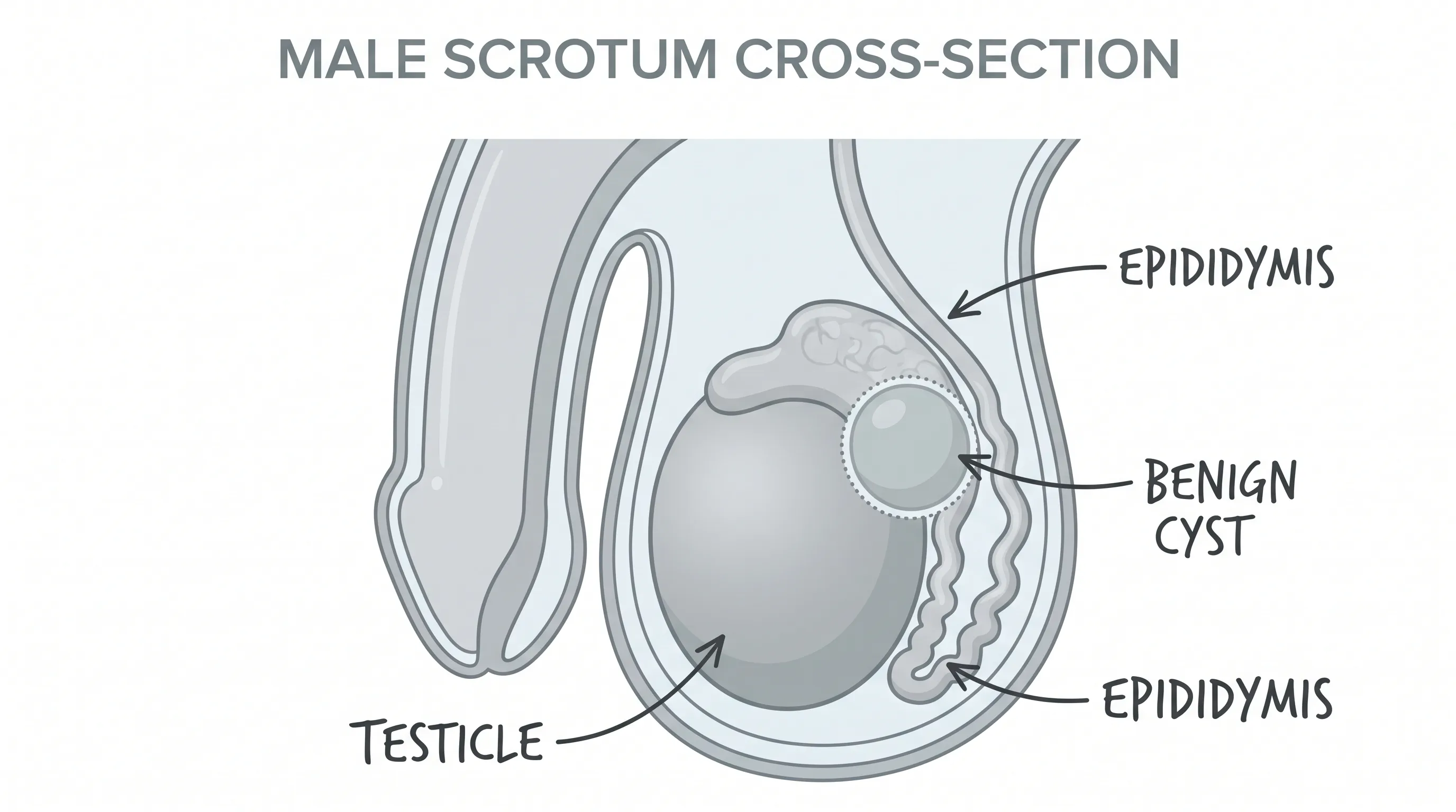
To fully comprehend what medical imagery reveals, you must first understand the anatomy of the male reproductive tract and the specific locations where cysts typically develop. The scrotum is a dual-layered sac of skin and muscle that houses the testicles, epididymides, and lower portions of the spermatic cords. Each testicle is encased in a tough, fibrous capsule known as the tunica albuginea, which protects delicate seminiferous tubules responsible for sperm production. Adjacent to the upper and posterior aspect of each testicle lies the epididymis, a coiled tube network that matures and stores sperm before transport. Cysts most frequently originate in the epididymis, which is why they are clinically referred to as epididymal cysts or spermatoceles.
Anatomy of the Scrotum
The scrotal environment is highly vascularized and temperature-regulated, maintaining an optimal thermal range slightly below core body temperature to support spermatogenesis. Blood flow is managed by the pampiniform plexus, while lymphatic drainage occurs through the inguinal nodes. The tunica vaginalis, a serous membrane derived from the abdominal peritoneum, covers the anterior and lateral surfaces of the testicle. Fluid accumulation in this space typically results in a hydrocele, which is distinct from true parenchymal cysts. Recognizing these structural layers is crucial when interpreting clinical photographs or reviewing cysts on testicles pictures, as the precise anatomical location directly influences diagnostic classification and treatment pathways. The epididymis contains three primary segments: the head (caput), body (corpus), and tail (cauda). Most benign cysts emerge from the head, where the efferent ductules converge.
Common Types of Cysts Found on or Near the Testicles
Several cystic formations can present in the scrotal region, each with unique pathophysiology and clinical characteristics. Epididymal cysts are clear fluid-filled sacs that develop when the efferent ductules become obstructed. They typically contain lymphatic fluid and are usually painless unless they enlarge significantly. Spermatoceles, while often grouped with epididymal cysts, contain milky fluid rich in sperm cells and form from blocked spermatic tubules. Both conditions share similar imaging features and management protocols. Intratesticular cysts, though less common, occur within the testicular parenchyma and often result from prior trauma, infection, or tunica albuginea cysts. Unlike extratesticular formations, intratesticular cysts require closer monitoring due to the higher statistical association with underlying pathology. Simple cysts remain benign in the vast majority of cases, but any complex features such as septations, solid components, or irregular borders warrant immediate specialist evaluation. Understanding these distinctions helps contextualize why clinical guidelines strongly advise against self-diagnosis based solely on surface observations or online cysts on testicles pictures.
What to Look for in Cysts on Testicles Pictures
Visual documentation of scrotal abnormalities serves multiple purposes in clinical practice, from patient education to diagnostic confirmation. When reviewing cysts on testicles pictures, you are typically viewing one of three formats: high-resolution clinical photographs, ultrasound imaging stills, or anatomical cross-sectional diagrams. Each modality highlights different structural characteristics and provides distinct diagnostic clues.
How Medical Imaging Represents Testicular Cysts
Medical professionals rarely rely on external photography for definitive diagnosis. Instead, scrotal ultrasonography remains the gold standard for evaluating cystic lesions. On a standard grayscale ultrasound, a benign cyst appears as a well-defined, anechoic (completely dark) round or oval structure with smooth posterior acoustic enhancement, indicating fluid transmission. The walls are thin and regular, measuring less than two millimeters. Doppler ultrasound further confirms the absence of internal blood flow, distinguishing cystic formations from vascular or solid tumors. When patients examine cysts on testicles pictures sourced from medical journals, they are usually viewing ultrasound stills that highlight these characteristic acoustic patterns. External photographs may only reveal visible scrotal swelling, asymmetry, or skin changes, which provide limited diagnostic value without imaging correlation.
Interpreting Ultrasound and MRI Visuals
Magnetic resonance imaging (MRI) is occasionally utilized when ultrasound findings are equivocal or when surgical planning requires precise anatomical mapping. T2-weighted MRI sequences vividly display cysts as hyperintense (bright white) lesions against the darker background of testicular tissue. This contrast allows urologists to measure exact dimensions, assess proximity to critical structures like the epididymal head or vas deferens, and rule out complex masses. Radiologists document size measurements, wall thickness, internal debris, and surrounding tissue reaction. If you are reviewing cysts on testicles pictures from clinical case studies, you will often see side-by-side comparisons of ultrasound and MRI modalities, illustrating how each imaging technique contributes to a comprehensive diagnostic picture. Always remember that imaging interpretation requires specialized training, and laypersons should not attempt to self-diagnose based on radiographic visuals alone.
Clinical Features vs. Visual Findings
While medical imagery provides objective structural data, clinical evaluation integrates patient history, physical examination findings, and symptom progression. Understanding the relationship between what appears on imaging and what patients actually experience is essential for accurate diagnosis and appropriate management.
Benign Cysts vs. Malignant Growths
The primary clinical objective when evaluating a scrotal lump is to differentiate benign cystic lesions from malignant testicular tumors. Testicular cancer typically presents as a firm, painless, irregular mass within the testicular parenchyma rather than the epididymis. Malignant growths demonstrate heterogeneous echotexture on ultrasound, internal vascularity on Doppler imaging, and often cause testicular heaviness or dull aching. Benign cysts, conversely, remain fluctuant, transilluminate positively when a light source is placed behind the scrotum, and maintain consistent acoustic properties across imaging planes. When reviewing cysts on testicles pictures, it is critical to recognize that malignant lesions rarely mimic the clean, anechoic appearance of simple cysts. Tumor markers such as alpha-fetoprotein, beta-human chorionic gonadotropin, and lactate dehydrogenase provide additional biochemical context when imaging reveals suspicious characteristics. Urologists emphasize that while statistical probability heavily favors benign cysts, definitive exclusion of malignancy requires professional evaluation.
When Pictures Alone Are Not Enough
Visual documentation has inherent limitations. External photographs cannot reveal internal architecture, fluid composition, or vascular patterns. Patients often search for cysts on testicles pictures hoping to find an exact visual match for their own symptoms, but anatomical variation, lighting conditions, and imaging modalities create significant discrepancies. A cyst that appears small and superficial on one imaging study may be deeper or more complex when examined with different parameters. Furthermore, symptom correlation is indispensable. A benign cyst might cause significant discomfort if it compresses adjacent nerves, while a larger cyst may remain entirely asymptomatic. Clinical guidelines from major urological associations stress that visual inspection must be supplemented by palpation, transillumination testing, and formal imaging. Relying exclusively on online imagery can lead to unnecessary anxiety or delayed treatment of legitimate medical concerns.
Step-by-Step Diagnostic Process
A systematic diagnostic approach ensures accurate identification and appropriate management of scrotal cysts. The process integrates clinical assessment, technological imaging, and laboratory evaluation to rule out serious pathology while confirming benign findings.
Physical Examination Techniques
The initial clinical encounter begins with a thorough physical examination. The urologist or primary care provider will visually inspect the scrotum for asymmetry, skin discoloration, or obvious swelling. Gentle palpation determines the lump's consistency, mobility, and precise anatomical location. A crucial bedside test is transillumination, where a bright light is positioned behind the scrotum in a darkened room. Benign fluid-filled cysts typically allow light to pass through, producing a reddish glow, whereas solid masses block illumination. The provider will also assess the inguinal canals for hernias and evaluate the spermatic cord for varicocele characteristics. This hands-on examination establishes baseline clinical suspicion and determines whether immediate imaging is warranted. Patients can prepare for this evaluation by noting symptom onset, pain characteristics, and any prior scrotal trauma or infections.
Imaging Modalities and What They Reveal
When clinical examination suggests a cystic lesion, scrotal ultrasound with color Doppler is ordered as the definitive diagnostic tool. The procedure is non-invasive, painless, and typically completed within fifteen minutes. A technician applies warm gel to the scrotum and moves a transducer across the skin, capturing real-time cross-sectional images. The radiologist evaluates echogenicity, posterior acoustic enhancement, wall regularity, and internal architecture. Measurements are documented in centimeters to establish baseline size for future monitoring. If ultrasound findings remain inconclusive or surgical intervention is planned, MRI or CT scanning may supplement diagnostic clarity. Laboratory tests, including urinalysis and serum tumor markers, are occasionally ordered to exclude infection or malignancy. Understanding what to expect during this diagnostic journey helps patients navigate medical appointments confidently and recognize why cysts on testicles pictures alone cannot replace comprehensive clinical evaluation.
Treatment Options and Management Strategies
Management of testicular and epididymal cysts depends entirely on symptom severity, cyst size, patient age, and reproductive goals. The vast majority require no intervention, but evidence-based protocols exist for cases necessitating medical or surgical attention.
Watchful Waiting and Home Care
Asymptomatic cysts smaller than two centimeters typically fall under active surveillance. Urologists recommend periodic self-monitoring, clinical follow-ups every six to twelve months, and ultrasound confirmation if changes occur. Home care focuses on symptom management and comfort optimization. Wearing supportive underwear, applying ice packs during acute discomfort episodes, and avoiding prolonged sitting or heavy lifting can alleviate pressure on the scrotal region. Over-the-counter analgesics like ibuprofen or acetaminophen manage mild inflammation and pain. Patients should track cyst dimensions by performing monthly self-exams and report sudden enlargement, persistent pain, or skin changes to their physician. Conservative management preserves fertility, avoids surgical risks, and aligns with major urological guidelines that discourage unnecessary intervention for benign, asymptomatic lesions.
Medical and Surgical Interventions
When cysts cause chronic pain, interfere with sexual activity, enlarge progressively, or create significant cosmetic distress, procedural intervention becomes appropriate. Aspiration with sclerotherapy involves draining cyst fluid using ultrasound guidance followed by injection of a sclerosing agent to prevent recurrence. While minimally invasive, this approach carries recurrence risks ranging from twenty to thirty percent and potential scrotal inflammation. Spermatocelectomy, the definitive surgical treatment, involves precise excision of the cyst wall while preserving epididymal function. Performed under local or general anesthesia, the procedure requires meticulous microsurgical technique to avoid damaging the vas deferens or compromising fertility. Post-operative recovery includes scrotal support, pain management, and activity restriction for two to three weeks. Major medical institutions like the Cleveland Clinic and the National Health Service emphasize that surgical removal should only be considered after conservative measures fail and quality of life is significantly impacted. Reviewing cysts on testicles pictures in surgical contexts often highlights the precise anatomical dissection required to achieve optimal outcomes while preserving reproductive health.
Self-Examination and Preventative Health Practices
Proactive scrotal health management empowers individuals to detect changes early, seek timely evaluation, and maintain long-term reproductive wellness. Establishing a consistent examination routine and adopting lifestyle practices that support vascular and lymphatic health significantly reduce diagnostic uncertainty.
How to Perform a Testicular Self-Exam
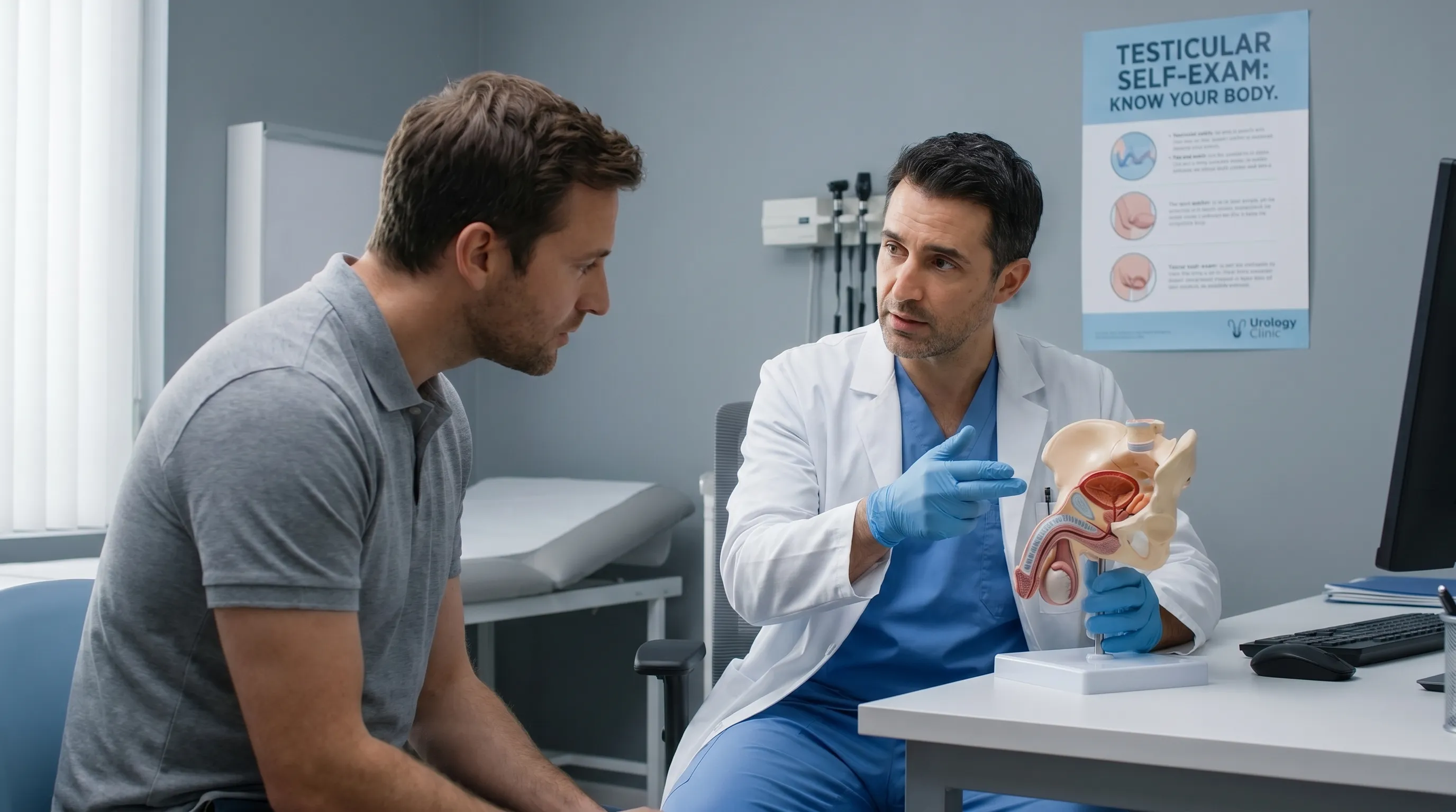
A structured testicular self-examination takes less than three minutes and should be integrated into monthly personal health routines. Begin during or immediately after a warm shower when scrotal muscles are relaxed. Stand in front of a mirror and visually inspect for swelling, redness, or asymmetry. Using both hands, gently roll each testicle between the thumbs and index fingers, feeling for irregularities, firm nodules, or cystic structures. The epididymis normally feels like a soft, rope-like band along the posterior border; cysts typically feel distinct, smooth, and slightly compressible. Note any tenderness, heaviness, or changes in size. Document findings and compare monthly. If you detect a new lump, experience sudden pain, or notice rapid enlargement, schedule a urological consultation promptly. Regular self-exams transform uncertainty into awareness and ensure that concerning symptoms receive professional evaluation before complications arise.
Lifestyle Adjustments for Scrotal Health
Beyond routine examination, several evidence-based lifestyle modifications support optimal scrotal and testicular health. Maintaining a healthy body weight reduces abdominal pressure and improves pelvic circulation. Avoid prolonged heat exposure from hot tubs, saunas, or tight clothing, as elevated scrotal temperatures can affect sperm production and increase fluid accumulation risks. Incorporate antioxidant-rich foods, omega-3 fatty acids, and adequate hydration to support tissue repair and inflammatory regulation. Practice safe sexual health protocols to prevent sexually transmitted infections that can cause epididymitis and secondary cystic changes. Engage in regular cardiovascular exercise to optimize systemic blood flow and lymphatic drainage. If you work in environments requiring heavy lifting or prolonged standing, utilize ergonomic supports and take regular movement breaks. These preventative strategies create a resilient physiological environment, reducing the likelihood of complications and ensuring that any benign findings remain manageable through conservative monitoring. When searching for cysts on testicles pictures, remember that visual documentation represents a single moment in a dynamic biological system that responds positively to consistent, informed self-care.
Comparing Scrotal Conditions: A Diagnostic Reference
Differentiating between various scrotal abnormalities requires understanding distinct clinical presentations, imaging characteristics, and management protocols. The following comparison table outlines key differences between epididymal cysts and other common scrotal conditions, providing a clear reference for patients navigating diagnostic information.
| Condition | Primary Location | Typical Size | Palpation Feel | Transillumination | Ultrasound Appearance | Primary Management |
|---|---|---|---|---|---|---|
| Epididymal Cyst | Epididymal head | 1-5 cm | Smooth, compressible, mobile | Positive | Anechoic, well-defined, no flow | Observation or excision |
| Spermatocele | Epididymis | 1-3 cm | Soft, cystic, slightly tender | Positive | Anechoic or low-level echoes | Conservative or surgery |
| Hydrocele | Tunica vaginalis | Variable | Tense, fluctuant, surrounds testicle | Positive | Fluid around testis, thin walls | Aspiration or hydrocelectomy |
| Varicocele | Spermatic cord veins | Enlarged veins | "Bag of worms" texture | Negative | Dilated veins >2mm, reflux on Valsalva | Embolization or ligation |
| Testicular Tumor | Testicular parenchyma | Variable | Firm, fixed, irregular, heavy | Negative | Heterogeneous, internal vascularity | Radical inguinal orchiectomy |
Understanding these distinctions prevents misdiagnosis and ensures appropriate referral pathways. While benign cysts dominate clinical presentations, the table highlights why professional evaluation remains non-negotiable when unfamiliar masses appear. Patients reviewing cysts on testicles pictures should recognize that accurate diagnosis integrates anatomical location, acoustic properties, and clinical correlation rather than superficial resemblance. For authoritative clinical guidelines and condition-specific information, resources such as the American Urological Association and Mayo Clinic provide comprehensive, peer-reviewed reference materials that support informed patient decision-making.
Frequently Asked Questions
Are epididymal cysts cancerous?
No. Epididymal cysts and spermatoceles are completely benign, fluid-filled sacs that do not transform into cancer. They arise from blocked ductules rather than malignant cellular proliferation. However, any new scrotal lump should be evaluated by a physician using ultrasound and clinical examination to definitively rule out malignancy, as testicular cancer remains highly treatable when detected early.
Can I safely drain a testicular cyst at home?
Absolutely not. Attempting to drain, puncture, or squeeze a cyst at home introduces significant infection risks, can cause internal bleeding, damages delicate epididymal structures, and may lead to chronic scarring or infertility. Only a licensed urologist should perform cyst aspiration or surgical excision under sterile, controlled conditions using appropriate imaging guidance.
When do testicular cysts require surgical removal?
Surgical intervention is typically reserved for cysts that cause persistent or worsening pain, significantly enlarge beyond three centimeters, interfere with sexual function or daily activities, or create substantial cosmetic concerns. Procedures like spermatocelectomy are highly effective, preserve reproductive anatomy when performed carefully, and are only recommended after conservative management fails to provide adequate symptom relief.
Will a cyst affect my fertility?
In the vast majority of cases, simple epididymal cysts do not impact fertility or sperm quality. However, exceptionally large cysts that compress the epididymal tubules or surgical interventions near the vas deferens may theoretically influence reproductive pathways. If you are actively planning for conception, discuss cyst management timelines with both a urologist and fertility specialist to optimize outcomes.
How often should I perform testicular self-exams?
Urologists recommend performing a comprehensive testicular self-examination once a month, ideally during or immediately after a warm shower when scrotal tissue is relaxed and blood vessels are dilated. This consistent routine helps you establish familiarity with normal anatomy, quickly recognize subtle changes, and seek prompt medical evaluation when necessary, aligning with early detection best practices.
Conclusion
Navigating concerns about scrotal abnormalities requires a balanced approach that combines medical knowledge, professional evaluation, and proactive self-care. While searching for cysts on testicles pictures can provide preliminary visual context, accurate diagnosis depends on ultrasound imaging, clinical palpation, and symptom correlation. The overwhelming majority of epididymal cysts and spermatoceles are benign, manageable through observation or conservative measures, and rarely impact long-term reproductive health. Understanding anatomical structures, recognizing red-flag symptoms, performing monthly self-examinations, and adhering to evidence-based treatment protocols empower individuals to maintain optimal scrotal wellness. Always prioritize consultation with a board-certified urologist when discovering new lumps, experiencing persistent discomfort, or noticing rapid changes in size or consistency. With informed awareness and timely medical guidance, you can confidently manage benign cystic conditions while safeguarding your overall reproductive vitality and long-term health outcomes.

About the author
Carlos Ruiz, MD, FACS, is a board-certified urologist specializing in minimally invasive and robotic surgery for urologic cancers. He is a senior partner at a large urology group in Houston, Texas, and is involved in clinical trials for new prostate cancer treatments.