Grover's Disease Food to Avoid: Evidence-Based Dietary Triggers and Skin Healing Strategies

Living with persistent skin inflammation can feel overwhelming, especially when standard topical treatments provide only temporary relief. Grover's disease, clinically known as transient acantholytic dermatosis, frequently disrupts daily life through intense pruritus, erythematous papules, and unpredictable flare-ups. While environmental factors like heat, humidity, and friction play well-documented roles, an often-overlooked contributor lies on our plates. Understanding the grover's disease food to avoid is a critical, evidence-based strategy that many patients and clinicians are beginning to integrate into comprehensive treatment plans. The connection between systemic inflammation, gut permeability, and cutaneous manifestation is no longer theoretical; modern dermatology recognizes that dietary patterns directly modulate immune responses, mast cell stability, and keratinocyte adhesion. When patients eliminate specific pro-inflammatory compounds, many experience significant reductions in lesion density and itching severity. This article provides a thorough, research-backed exploration of how nutrition influences this dermatological condition, outlines the primary dietary triggers, and delivers actionable protocols for sustainable skin healing.
Understanding Grover’s Disease and the Diet Connection
What Is Transient Acantholytic Dermatosis?
Grover’s disease is an acquired, non-hereditary dermatosis characterized by the sudden onset of small, intensely itchy, red or pink papules typically localized to the central chest and upper back. Histologically, it features focal acantholysis, meaning keratinocytes lose their intercellular attachments and separate within the epidermis. This structural breakdown compromises the skin barrier, allowing irritants, microbes, and inflammatory mediators to penetrate more deeply. Although the exact etiology remains multifactorial, established triggers include excessive sweating, prolonged heat exposure, ultraviolet radiation, and certain medications. Importantly, transient acantholytic dermatosis frequently coexists with other inflammatory or autoimmune conditions, suggesting a systemic predisposition rather than an isolated cutaneous anomaly.
Free calculatorSun Protection GuideSPF guide and sun protection recommendationsOpen the calculatorThe clinical presentation often mimics other papular dermatoses such as pityriasis rosea, dermatitis herpetiformis, or drug eruptions, making differential diagnosis essential. However, once confirmed through skin biopsy or clinical correlation, management shifts toward symptom control and trigger mitigation. Topical corticosteroids, phototherapy, and systemic retinoids constitute conventional therapy, but long-term reliance on pharmacological interventions without addressing underlying metabolic drivers often leads to relapse. This is where dietary intervention becomes clinically relevant. By identifying and removing specific nutritional compounds that exacerbate keratinocyte fragility and histamine release, patients can achieve more durable remission periods.
How Systemic Inflammation Drives Cutaneous Symptoms
The skin operates as a highly responsive immune organ. When systemic inflammation rises, cytokines such as interleukin-4, interleukin-13, and tumor necrosis factor-alpha circulate through the bloodstream and directly interact with dermal and epidermal cells. In Grover’s disease, this inflammatory cascade appears to weaken desmosomal junctions, which normally anchor neighboring keratinocytes together. Research published in the Journal of Clinical and Aesthetic Dermatology indicates that dietary-induced inflammation can accelerate acantholysis, particularly when combined with external stressors like sweating or friction.
Furthermore, the gastrointestinal tract houses approximately seventy percent of the body’s immune tissue. A compromised intestinal barrier, commonly referred to as leaky gut, permits partially digested proteins, bacterial endotoxins, and dietary antigens to enter systemic circulation. These circulating immune complexes can deposit in the dermis, triggering localized mast cell degranulation and subsequent histamine-mediated pruritus. Recognizing this gut-skin axis pathway clarifies why dietary modifications often yield profound improvements in Grover’s disease management. Patients who prioritize low-inflammatory eating patterns frequently report faster resolution of active lesions, reduced reliance on topical steroids, and improved overall quality of life.

The Top grover's disease food to avoid
Dairy Products and Lactose Sensitivity
Dairy ranks among the most frequently reported grover's disease food to avoid in clinical dermatology literature. The primary concern extends beyond lactose itself to casein and whey proteins, which are highly antigenic and can stimulate IgE-mediated and non-IgE-mediated inflammatory pathways. Beta-casein A1 variant, found in many commercial cow’s milk products, breaks down into beta-casomorphin-7 during digestion. This opioid-like peptide increases intestinal permeability and promotes systemic immune activation. For individuals with underlying mast cell reactivity, dairy consumption can rapidly elevate circulating histamine levels, intensifying cutaneous itching and erythema.
Additionally, many dairy products undergo fermentation or aging processes that naturally increase histamine concentration. Hard cheeses, aged yogurts, and cultured buttermilk frequently contain biogenic amines that directly trigger pruritus in histamine-sensitive patients. Even lactose-free dairy retains these inflammatory proteins, meaning switching to lactose-free variants does not automatically eliminate the grover's disease food to avoid profile. Patients are advised to replace conventional dairy with unsweetened almond milk, coconut yogurt, or oat-based alternatives fortified with calcium and vitamin D. Monitoring skin response over a four-week elimination period typically clarifies personal tolerance thresholds.
Refined Sugars and High-Glycemic Carbohydrates
Refined carbohydrates and added sugars represent another major dietary trigger. When consumed in excess, high-glycemic foods cause rapid spikes in blood glucose and subsequent insulin surges. Elevated insulin upregulates insulin-like growth factor-1 (IGF-1), which directly stimulates sebum production and accelerates keratinocyte proliferation. In the context of Grover’s disease, this hyperproliferative state disrupts normal epidermal maturation, weakening cell-to-cell adhesion and predisposing the skin to acantholytic separation.
Sugar also serves as a potent fuel for pathogenic gut bacteria. An overgrowth of dysbiotic microbes produces lipopolysaccharides that breach the intestinal barrier and initiate systemic inflammation. Clinical observations consistently show that patients who eliminate processed sweets, sugary beverages, white bread, and refined cereals experience marked reductions in lesion activity within weeks. Aligning with WHO guidelines on healthy diets, replacing high-glycemic items with low-glycemic alternatives such as quinoa, sweet potatoes, legumes, and steel-cut oats stabilizes blood sugar, modulates IGF-1 expression, and supports epidermal homeostasis. The evidence strongly supports minimizing added sugars to below ten percent of total daily caloric intake for optimal dermatological outcomes.
Alcohol and Fermented Beverages
Alcohol functions as a dual threat in inflammatory dermatoses. First, ethanol metabolism generates acetaldehyde, a highly reactive compound that depletes glutathione reserves and increases oxidative stress throughout the epidermal and dermal layers. Oxidative damage impairs collagen synthesis, compromises barrier lipid production, and directly activates sensory nerve endings, amplifying itch sensation. Second, alcohol acts as a potent vasodilator, increasing cutaneous blood flow and temperature, which are well-established exacerbating factors for Grover’s disease. Understanding the broader health effects of alcohol is crucial when managing inflammatory skin conditions.
Beyond pure ethanol, many alcoholic beverages undergo fermentation or aging processes that dramatically increase histamine and sulfite content. Red wine, craft beers, champagne, and kombucha frequently contain biogenic amines that bypass diamine oxidase (DAO) metabolism in susceptible individuals. When histamine accumulates systemically, it binds to H1 and H4 receptors on cutaneous mast cells and sensory neurons, triggering intense pruritus and vasodilation. For patients seeking lasting symptom control, treating alcohol as a primary grover's disease food to avoid is clinically justified. Hydration with electrolyte-enhanced water, herbal infusions, or sparkling mineral water provides safer alternatives that support detoxification pathways without triggering inflammatory cascades.
Nightshade Vegetables and Solanine
The Solanaceae family includes tomatoes, bell peppers, potatoes, eggplants, and chili peppers. These vegetables contain naturally occurring alkaloids, primarily solanine and chaconine, which evolved as plant defense mechanisms against pests. In humans, solanine can interfere with calcium channel function, disrupt cellular membrane integrity, and stimulate inflammatory eicosanoid production. While nightshades offer substantial nutritional benefits for the general population, patients with autoimmune dermatoses or mast cell activation syndromes frequently report flare-ups following consumption.
Solanine’s impact on keratinocyte adhesion remains an area of active investigation, but clinical case series consistently note symptom improvement when nightshades are temporarily removed from the diet. The grover's disease food to avoid list should therefore include a trial elimination of nightshade derivatives, particularly concentrated tomato sauces, smoked paprika, and heavily spiced processed foods. Many patients discover they tolerate cooked potatoes or small portions of bell peppers without issue, while others react to trace amounts in condiments. Reintroduction must proceed cautiously, one variety at a time, with careful symptom documentation over a seventy-two-hour window.
Ultra-Processed Foods and Additives
Modern food processing introduces numerous synthetic compounds rarely encountered by human metabolism throughout evolutionary history. Emulsifiers, artificial sweeteners, preservatives, and flavor enhancers disrupt gut microbiota composition, alter mucosal barrier function, and directly stimulate toll-like receptors on immune cells. Studies published by the National Institutes of Health demonstrate that dietary emulsifiers such as polysorbate-80 and carboxymethylcellulose degrade the mucus layer, enabling bacterial translocation and systemic inflammation.
For individuals managing Grover’s disease, ultra-processed snacks, packaged meals, and artificially flavored beverages represent a significant grover's disease food to avoid category. These products often contain hidden sources of histamine, glutamate, and inflammatory omega-6 fatty acids derived from refined seed oils. Transitioning to a whole-food, minimally processed diet dramatically reduces exposure to these immunogenic compounds. Patients who prioritize home-cooked meals using fresh ingredients, cold-pressed oils, and natural seasonings consistently report improved barrier resilience and fewer unpredictable flare cycles.
The Scientific Rationale: Histamine and Mast Cells
Understanding Histamine Intolerance
Histamine serves as a crucial neurotransmitter and immune mediator, but excessive accumulation leads to profound dermatological and systemic symptoms. The body normally regulates histamine through enzymatic degradation primarily via diamine oxidase (DAO) and histamine N-methyltransferase (HNMT). Genetic variations, gastrointestinal inflammation, nutrient deficiencies (particularly vitamin B6, zinc, and copper), and certain medications can impair DAO activity. When degradation pathways falter, dietary histamine enters circulation and binds extensively to cutaneous receptors.
Mast cell activation syndrome (MCAS) frequently overlaps with transient acantholytic dermatosis. Activated mast cells release histamine, prostaglandins, leukotrienes, and tryptase, all of which exacerbate pruritus, erythema, and epidermal fragility. Recognizing histamine intolerance as a physiological driver rather than a mere allergy shifts treatment paradigms toward dietary modulation, enzyme support, and gut healing. Patients who identify and eliminate high-histamine foods often experience rapid symptomatic improvement, validating histamine as a primary grover's disease food to avoid in susceptible populations.
Mast Cell Activation and Skin Barrier Disruption
Mast cells reside abundantly in the dermis and superficial blood vessels. Upon activation by dietary antigens, heat, friction, or stress, they degranulate, releasing inflammatory mediators that increase vascular permeability and stimulate keratinocyte dysfunction. In Grover’s disease, this process directly compromises desmosomal integrity, facilitating acantholysis. Chronic mast cell stimulation also upregulates matrix metalloproteinases, which degrade extracellular matrix proteins and further weaken structural support.
Research from dermatology journals emphasizes that stabilizing mast cell activity requires a multifaceted approach combining dietary histamine restriction, antioxidant intake, and temperature regulation. Quercetin-rich foods, omega-3 supplementation, and consistent sleep hygiene collectively reduce mast cell degranulation frequency. When patients integrate these strategies alongside targeted elimination of the grover's disease food to avoid categories, they frequently achieve prolonged remission phases previously unattainable with topical therapies alone.
Implementing a Targeted Elimination Protocol
Phase 1: Removal and Baseline Assessment
An effective elimination diet requires structured planning rather than arbitrary restriction. Following established Mayo Clinic guidelines for elimination diets, phase one involves removing all identified trigger categories simultaneously for a minimum of twenty-one days. During this period, patients should document daily food intake, symptom severity using a standardized numerical rating scale, sleep quality, and environmental exposures. Maintaining consistency prevents confounding variables from masking true dietary responses.
Baseline laboratory testing, including complete blood count, comprehensive metabolic panel, vitamin D levels, and inflammatory markers such as C-reactive protein and erythrocyte sedimentation rate, provides objective data to track systemic changes. Hydration should be optimized with two to three liters of purified water daily, and meals should center around lean proteins, low-glycemic vegetables, healthy fats, and herbal teas. This phase is not about starvation or nutrient deprivation but rather strategic removal to allow physiological reset.
Phase 2: Strategic Reintroduction and Tracking
After symptoms stabilize for at least one week, phase two begins. Reintroduction occurs systematically, introducing a single food group every three to four days while maintaining all other restrictions. For example, dairy products are reintroduced on day twenty-two, followed by careful monitoring for pruritus, lesion appearance, sleep disruption, or digestive changes. If no adverse reactions occur within seventy-two hours, that food remains in the diet, and the next category is introduced.
This methodical approach prevents false attribution of symptoms and clarifies individual tolerance thresholds. Patients frequently discover they can safely consume certain trigger categories in moderation while strictly avoiding others. The reintroduction phase transforms dietary restriction into personalized nutritional mapping, empowering individuals with precise knowledge of their grover's disease food to avoid profile without imposing unnecessary long-term limitations.

Nutrient-Rich Foods That Support Skin Healing
Omega-3 Fatty Acids and Anti-Inflammatory Lipids
While identifying what to eliminate remains foundational, understanding what to incorporate ensures long-term epidermal resilience. Omega-3 fatty acids, particularly eicosapentaenoic acid (EPA) and docosahexaenoic acid (DHA), directly compete with pro-inflammatory omega-6 derivatives for enzymatic conversion pathways. This competition downregulates leukotriene and prostaglandin synthesis, effectively calming systemic inflammation and reducing mast cell reactivity.
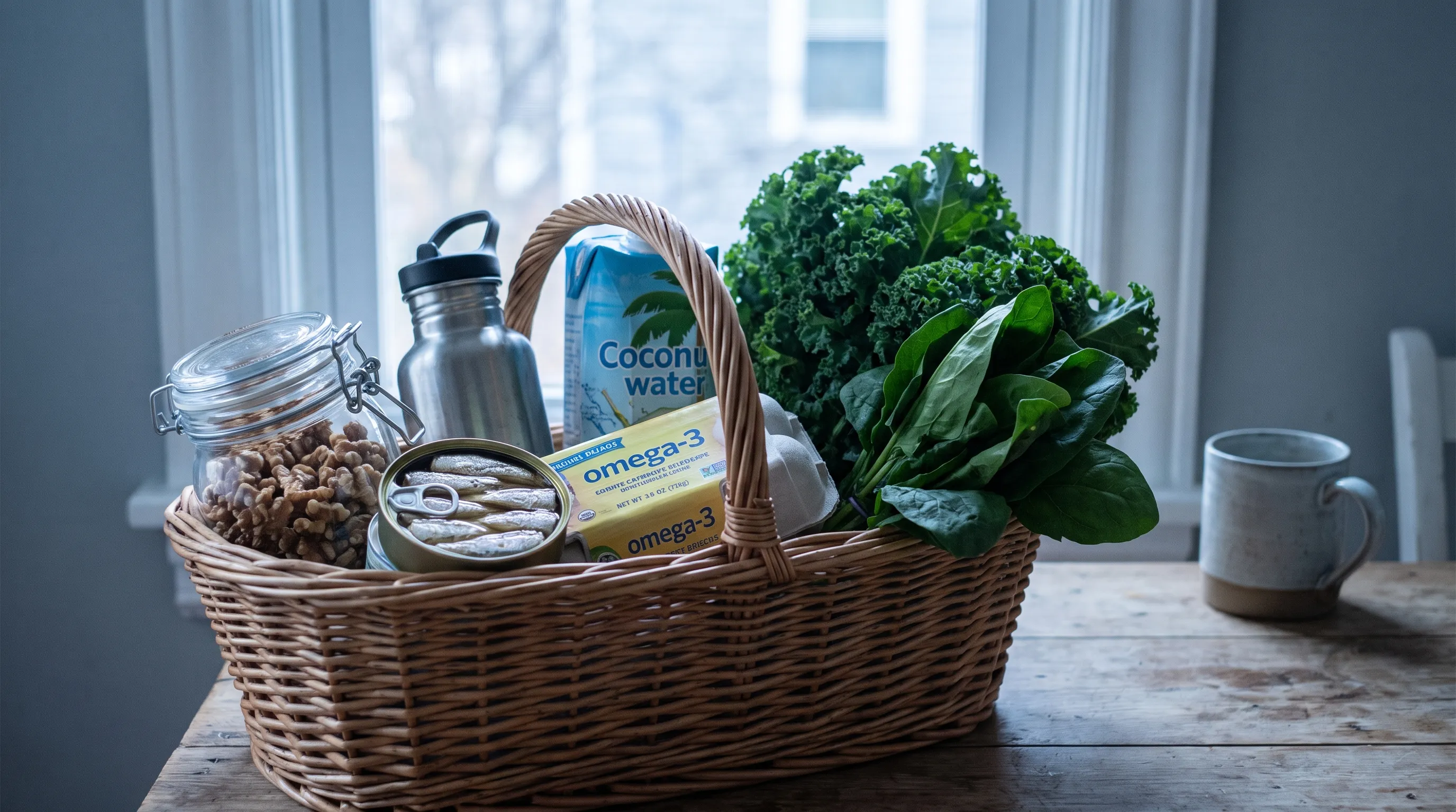
Cold-water fish such as wild-caught salmon, mackerel, and sardines provide the most bioavailable omega-3 sources. For plant-based patients, chia seeds, flaxseeds, walnuts, and algae-derived supplements offer viable alternatives. Consuming two to three servings weekly or supplementing with high-quality fish oil containing at least one thousand milligrams of combined EPA and DHA daily supports lipid bilayer integrity in keratinocytes. Strengthening cell membranes directly counters the acantholytic process, making omega-3s an indispensable component of Grover’s disease dietary management.
Antioxidants, Polyphenols, and Barrier Repair
Chronic oxidative stress accelerates epidermal degradation and impairs wound healing. Antioxidant-rich foods neutralize reactive oxygen species, preserve collagen architecture, and enhance barrier lipid production. Deeply pigmented berries, cruciferous vegetables, leafy greens, green tea, and extra virgin olive oil deliver potent polyphenols including resveratrol, quercetin, and epigallocatechin gallate.
Zinc plays an equally critical role, serving as a cofactor for over three hundred enzymatic reactions involved in DNA synthesis, immune modulation, and epithelial cell division. Clinical guidelines recommend dietary zinc from pumpkin seeds, lentils, and grass-fed beef, or supplementation at fifteen to thirty milligrams daily for eight to twelve weeks under medical supervision. Vitamin C further amplifies antioxidant defense and stimulates ceramide synthesis, which forms the mortar between epidermal corneocytes. Together, these nutrients create a biochemical environment hostile to acantholytic dermatosis, transforming nutritional intake from passive sustenance into active therapeutic intervention.
| Dietary Category | Pro-Inflammatory Examples (Avoid) | Anti-Inflammatory Alternatives (Include) | Primary Dermatological Impact |
|---|---|---|---|
| Fats | Refined seed oils, margarine, trans fats | Extra virgin olive oil, avocado oil, wild salmon | Modulates cytokine production and strengthens cell membranes |
| Carbohydrates | White bread, pastries, sugary cereals | Quinoa, sweet potatoes, legumes, berries | Stabilizes insulin/IGF-1 and reduces keratinocyte hyperproliferation |
| Proteins | Processed deli meats, conventional dairy | Organic poultry, grass-fed beef, hemp seeds, lentils | Lowers antigenic load and supports tissue repair |
| Beverages | Alcohol, energy drinks, sweetened sodas | Green tea, herbal infusions, filtered water | Decreases vasodilation and supports hepatic detoxification |
| Condiments | Soy sauce, artificial flavorings, vinegar-based sauces | Fresh herbs, turmeric, apple cider vinegar (low-histamine) | Reduces biogenic amine intake and provides phytonutrients |
Integrating Dietary Changes with Medical Management
Topical Therapies and Temperature Regulation
Dietary intervention functions synergistically rather than as a replacement for medical therapy. Topical corticosteroids, calcineurin inhibitors, and barrier repair creams remain essential during active flare phases. However, when patients remove systemic triggers from their grover's disease food to avoid list, the required potency and frequency of topical medications often decrease substantially. Temperature regulation amplifies these benefits. Since sweating and friction directly provoke acantholysis, wearing loose, breathable cotton garments, maintaining cool indoor environments, and avoiding prolonged hot showers become non-negotiable lifestyle adjuncts.
Patients frequently underestimate the compounding effect of dietary histamine and heat exposure. A single high-sodium, high-histamine meal followed by strenuous exercise in warm conditions can trigger severe outbreaks within hours. Educating patients on this interaction prevents unnecessary frustration and reinforces adherence to both nutritional and environmental modifications.
When to Consult a Dermatologist or Dietitian
Self-directed dietary changes yield excellent results for many, but professional guidance remains invaluable. Board-certified dermatologists can confirm diagnosis through biopsy, prescribe phototherapy or systemic retinoids when indicated, and monitor for secondary infections. Registered dietitians specializing in medical nutrition therapy ensure elimination diets remain nutritionally complete, prevent micronutrient deficiencies, and provide structured reintroduction frameworks. Collaboration between patients, dermatologists, and dietitians establishes a comprehensive care model that addresses both cutaneous manifestation and metabolic foundation.
Individuals experiencing unexplained weight loss, severe gastrointestinal distress, or rapidly progressive lesions should seek immediate medical evaluation. While Grover’s disease is typically benign, overlapping conditions such as paraneoplastic pemphigus or drug-induced acantholysis require urgent diagnostic differentiation. Maintaining open communication with healthcare providers ensures dietary strategies complement rather than compromise conventional treatment pathways.
Frequently Asked Questions
Is dairy the most common grover's disease food to avoid?
While dairy is frequently implicated due to casein proteins and histamine content, it is not universally the primary trigger. Clinical data indicates that refined sugars, alcohol, and high-histamine foods often drive more severe and predictable flare-ups. Individual biochemistry dictates tolerance, making personalized elimination tracking more reliable than generalized avoidance lists.
How long should an elimination diet last for Grover's disease?
The standard elimination phase requires twenty-one to thirty days to allow circulating inflammatory mediators and histamine levels to normalize. Following this baseline period, systematic reintroduction over four to six weeks identifies specific triggers without compromising skin barrier recovery.
Can supplements replace dietary changes for managing symptoms?
Targeted supplements such as omega-3 fish oil, vitamin D3, zinc, and quercetin provide meaningful biochemical support but cannot compensate for a chronically pro-inflammatory diet. Whole-food nutritional strategies establish the metabolic foundation upon which supplements effectively operate.
Does stress exacerbate dietary triggers in Grover's disease?
Chronic psychological stress elevates cortisol, which increases intestinal permeability, amplifies mast cell degranulation, and heightens inflammatory responses to dietary antigens. Integrating stress-reduction techniques such as mindfulness, controlled breathing, and adequate sleep significantly improves dietary tolerance.
Are nightshade vegetables always a grover's disease food to avoid?
Nightshades contain solanine and alkaloids that may trigger inflammation in sensitive individuals, but many patients tolerate them without adverse effects. The only reliable method to determine necessity involves a structured elimination and reintroduction protocol with careful symptom documentation.
Conclusion
Navigating the complexities of transient acantholytic dermatosis requires patience, clinical insight, and a willingness to explore systemic contributors beyond topical treatments. By systematically identifying the grover's disease food to avoid, patients actively participate in modulating inflammation, stabilizing mast cell activity, and reinforcing epidermal integrity. The evidence consistently demonstrates that dietary elimination, combined with temperature regulation, barrier-supportive nutrients, and collaborative medical oversight, produces the most durable and predictable outcomes. Skin health is fundamentally metabolic health. When individuals align their nutritional intake with physiological healing requirements, they transform unpredictable flare cycles into manageable, well-informed routines. Commit to the elimination process, track your responses with precision, and partner with qualified healthcare professionals to build a sustainable, evidence-based management strategy that restores comfort, confidence, and long-term dermatological wellness.
How we work
Researched and drafted with AI assistance, then reviewed by a human editor against the cited sources.
This article is general health information, not medical advice. Consult a qualified healthcare professional about your own situation.