Are Spider Veins Dangerous? Medical Insights, Risks, and Treatment Options

Millions of people notice thin, web-like lines appearing on their thighs, calves, or ankles and immediately wonder whether these markings indicate a serious health problem. The question are spider veins dangerous surfaces constantly in dermatology and vascular medicine clinics worldwide, reflecting a widespread concern that extends far beyond aesthetics. While these visible vessels are extremely common and largely benign, understanding their physiological origins, progression pathways, and potential associations with underlying circulatory dysfunction is essential for making informed healthcare decisions. Modern vascular science has clarified that spider veins represent a specific stage in venous capillary dilation, often driven by localized valve incompetence, hormonal fluctuations, prolonged standing, or genetic predisposition. For the vast majority of individuals, they remain a superficial cosmetic concern that does not interfere with systemic cardiovascular function. However, recognizing when spider veins transition from harmless vascular markings to indicators of progressive chronic venous disease requires a nuanced understanding of venous hemodynamics, clinical symptomatology, and evidence-based prevention strategies. This comprehensive guide explores the medical realities behind these visible vessels, outlines when professional intervention becomes necessary, and provides actionable, recommendations for maintaining optimal lower extremity circulation and preventing further vascular compromise.
Understanding Spider Veins: Anatomy and Physiology
To accurately assess whether these visible vessels pose a health risk, it is essential to examine the underlying vascular architecture that allows them to develop. The circulatory system relies on a complex network of arteries, capillaries, and veins to transport oxygenated blood from the heart to peripheral tissues and return deoxygenated blood against gravitational forces. Spider veins, medically termed telangiectasias (NIH), specifically involve the smallest caliber vessels located within the superficial dermal layer of the skin. When you ask are spider veins dangerous from a structural standpoint, the answer lies in how these microvessels respond to sustained hemodynamic stress and endothelial fatigue. Unlike larger muscular veins that rely on skeletal muscle contraction and deep fascial support, telangiectasias lack robust smooth muscle walls and instead depend on fragile connective tissue scaffolding. Over time, repeated pressure fluctuations cause the endothelial lining to stretch, the basement membrane to thin, and the vessel to permanently dilate, creating the characteristic branching patterns visible to the naked eye.
Free calculatorSun Protection GuideSPF guide and sun protection recommendationsOpen the calculatorHow Blood Circulation Works in the Lower Extremities
The venous return system in the legs operates under constant gravitational opposition. As blood travels downward through arterial networks, it perfuses muscle tissue, joints, and skin before collecting into progressively larger venules and superficial veins. A sophisticated series of bicuspid valves positioned at regular intervals along the venous pathway ensures unidirectional flow toward the heart. When these valves function correctly, they snap shut during muscle relaxation, preventing retrograde blood pooling. However, when microvascular pressure consistently exceeds structural tolerance limits, the smallest superficial venules begin to bulge outward. This is precisely why many individuals notice spider veins after extended periods of standing, pregnancy, or significant weight fluctuations (Mayo Clinic). The capillary beds in the lower dermis are particularly vulnerable to hydrostatic overload, especially in the calves and medial ankles where gravitational forces are strongest. Understanding this physiological cascade clarifies why spider veins rarely appear on the upper body or face unless specific dermatological or systemic conditions are present.
The Role of Venous Valves and Capillary Networks
Venous valve incompetence represents the primary mechanistic driver behind visible microvascular dilation. Each valve functions as a one-way gate, closing immediately after blood propulsion to eliminate backflow. When microscopic valve leaflets become stretched, inflamed, or structurally compromised, blood refluxes downward, accumulating in the superficial venous plexus. This chronic low-grade venous hypertension triggers endothelial cell remodeling, inflammatory cytokine release, and matrix metalloproteinase activation, all of which degrade collagen support and accelerate vessel dilation. Clinical research published in vascular medicine journals consistently demonstrates that even minor valve dysfunction can manifest as visible telangiectasias long before larger varicose veins develop. Patients frequently report that these markings appear gradually over months or years, often coinciding with lifestyle changes, hormonal transitions, or prolonged occupational standing. Recognizing this pathophysiology helps clinicians differentiate between isolated cosmetic concerns and early warning signs of progressive venous insufficiency.
Primary Causes and Risk Factors
While genetics play a substantial role, spider veins develop through multifactorial interactions between biological predisposition and environmental exposures. Evaluating are spider veins dangerous requires examining the cumulative impact of these risk elements on venous wall integrity and microcirculatory health. Familial inheritance accounts for approximately 60 to 80 percent of cases, suggesting a strong hereditary component in venous wall elasticity and valve development (Cleveland Clinic). Beyond genetics, hormonal fluctuations significantly influence venous compliance. Estrogen and progesterone receptors located in vascular smooth muscle tissue respond to hormonal shifts by altering connective tissue synthesis, collagen cross-linking, and endothelial permeability. This explains why pregnancy, oral contraceptive use, and perimenopause frequently correlate with accelerated spider vein formation. Additionally, occupational demands involving prolonged static postures disrupt the calf muscle pump mechanism, allowing venous pressure to accumulate in the lower extremities. When combined with systemic inflammation, sedentary habits, or poor dietary patterns, these factors create a perfect environment for microvascular dilation.
Genetic Predisposition and Familial Patterns
Chromosomal variations affecting collagen type IV synthesis, elastin fiber organization, and venous valve morphogenesis strongly influence an individual's susceptibility to superficial venous dilation. Family history remains the most reliable predictor of spider vein development, with first-degree relatives showing a three to fourfold increased likelihood of exhibiting visible telangiectasias by middle age. Specific genetic polymorphisms related to matrix metalloproteinase expression and venous endothelial growth factor pathways have been identified in genome-wide association studies, providing molecular evidence for hereditary venous vulnerability. Clinicians routinely document multi-generational vascular patterns during patient consultations, recognizing that early genetic screening and proactive lifestyle interventions can delay onset and reduce progression severity.
Hormonal Fluctuations and Gender-Specific Factors
Women represent approximately 75 percent of individuals seeking treatment for spider veins, a disparity largely driven by reproductive hormone dynamics. During pregnancy, circulating relaxin and progesterone levels induce widespread vascular smooth muscle relaxation, reducing venous wall tension while simultaneously increasing blood volume by up to 50 percent (WHO). The expanding uterus further compresses pelvic veins, elevating lower extremity venous pressure and promoting capillary engorgement. Similarly, synthetic hormone therapies, menopause-related estrogen decline, and thyroid dysfunction can alter endothelial homeostasis and accelerate telangiectasia formation. While men also develop spider veins, the absence of cyclical hormonal shifts and generally higher muscle mass in the lower extremities provides greater mechanical support to superficial venous networks.
Lifestyle, Occupational, and Environmental Triggers
Prolonged standing occupations, including healthcare, education, retail, and hospitality roles, subject the venous system to continuous gravitational stress without adequate calf muscle activation to facilitate upward blood flow. Conversely, extended sedentary periods involving desk work or long-haul travel impair venous return and promote blood stasis. Environmental heat exposure, including frequent sauna use, hot tub immersion, or intense lower-body workouts in high temperatures, temporarily dilates superficial vessels and exacerbates existing capillary fragility. Chronic constipation and habitual heavy lifting increase intra-abdominal pressure, directly transmitting mechanical force to the iliac and femoral venous segments, which compromises lower extremity drainage and accelerates microvascular dilation.
Signs, Symptoms, and Progression Patterns
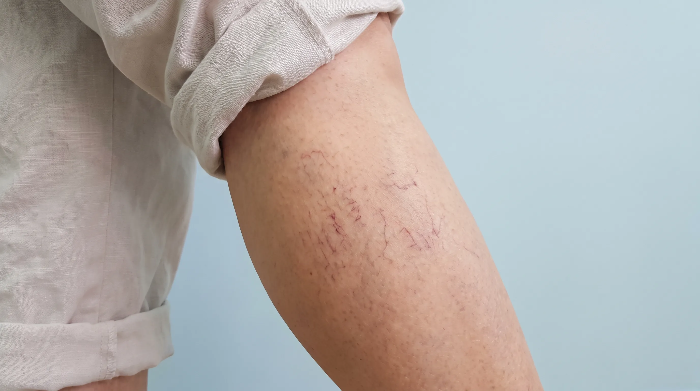
Spider veins typically manifest as fine, branching networks ranging from 0.1 to 1 millimeter in diameter, displaying vivid red, blue, purple, or occasionally black pigmentation depending on vessel depth, oxygenation levels, and dermal melanin content. While many individuals report no physical sensations, others experience localized warmth, mild itching, nighttime cramping, or a persistent feeling of leg heaviness after prolonged activity. Tracking these progression patterns helps patients and clinicians determine whether isolated cosmetic markings are evolving into broader circulatory dysfunction.
Visual Characteristics and Color Variations
The chromatic presentation of spider veins directly correlates with anatomical depth and hemoglobin oxygenation states. Superficial capillaries near the epidermal boundary typically appear bright red due to high oxygen content and minimal light absorption through thin overlying tissue. Deeper venules positioned closer to the dermal-hypodermal junction exhibit blue or purple hues because longer light wavelengths penetrate further, preferentially scattering shorter wavelengths while reflecting oxygen-depleted blood signatures. Linear, arborizing, and starburst configurations represent distinct morphological subtypes, each suggesting different hemodynamic stress patterns and underlying valve incompetence locations.
Associated Sensations and Physical Discomfort
Though primarily visual, spider veins occasionally produce tactile symptoms that impact daily comfort. Venous endothelial inflammation triggers localized histamine and cytokine release, causing intermittent pruritus, mild burning, or tingling sensations, particularly during temperature fluctuations or after extended upright positioning. Some patients report subtle aching that intensifies toward the evening, reflecting cumulative venous pooling throughout the day. When these sensations become frequent or interfere with sleep, mobility, or occupational performance, clinical evaluation becomes essential to rule out early chronic venous insufficiency or superficial thrombophlebitis.
When Are Spider Veins Dangerous? Medical Complications Explained
Addressing the core question directly, most spider veins remain entirely harmless and do not progress to life-threatening conditions. However, understanding when these visible vessels transition from cosmetic concerns to clinical warning signs requires recognizing the continuum of chronic venous disease. Venous hypertension rarely stays isolated indefinitely. When microvascular reflux remains unaddressed, hydrostatic pressure gradually transfers to larger superficial veins, reticular vessels, and eventually deep venous networks. This cascade initiates a series of structural and inflammatory changes that can compromise tissue perfusion, delay wound healing, and increase susceptibility to localized complications.
Chronic Venous Insufficiency (CVI) and Hemodynamic Stress
Chronic venous insufficiency develops when sustained venous hypertension impairs endothelial barrier function, allowing fibrinogen, leukocytes, and macromolecules to leak into perivascular tissue. This inflammatory response triggers fibroblast proliferation, collagen deposition, and eventual dermal thickening known as lipodermatosclerosis. Early indicators include persistent lower leg edema, hemosiderin deposition causing rust-colored skin staining, and eczematous dermatitis overlying dilated capillary beds. When patients ask are spider veins dangerous in the context of CVI, the clinical reality is that isolated telangiectasias serve as early phenotypic markers of underlying venous valve failure. Prompt intervention at this stage significantly reduces the likelihood of progression to advanced venous stasis syndromes.
Blood Clots and Superficial Thrombophlebitis Risks
Stagnant venous flow creates an environment conducive to localized coagulation activation. Superficial thrombophlebitis occurs when a spider vein or small reticular vessel develops a microthrombus, triggering intense local inflammation, palpable cord formation, erythema, and sharp tenderness along the affected tract. While these superficial clots rarely embolize to the pulmonary circulation, they frequently coexist with undiagnosed deep venous abnormalities. Ultrasound studies reveal that up to 15 percent of patients presenting with isolated superficial thrombophlebitis harbor concurrent asymptomatic deep vein thrombosis, emphasizing the importance of comprehensive vascular assessment rather than isolated symptom management.
Skin Changes, Pigmentation, and Ulceration Pathways
Prolonged venous hypertension ultimately compromises cutaneous microcirculation, depriving the epidermis and upper dermis of adequate oxygen and nutrient delivery. This hypoxic environment impairs keratinocyte migration, reduces collagen synthesis, and weakens barrier integrity, creating conditions where minor trauma or friction can initiate non-healing ulceration. Venous stasis ulcers typically develop over the medial malleolus or anterior lower third of the leg, displaying irregular borders, shallow depth, and copious serous exudate. Once ulceration occurs, the risk of secondary bacterial infection, delayed epithelialization, and functional impairment increases substantially. Preventing this endpoint requires recognizing early warning signs and implementing targeted venous decompression strategies before irreversible tissue remodeling occurs.
Diagnostic Approaches and Clinical Evaluation
Accurate diagnosis extends far beyond visual inspection. Modern vascular medicine employs systematic clinical frameworks combining patient history, physical examination, and objective imaging to determine whether visible telangiectasias represent isolated findings or early manifestations of progressive venous disease. Understanding are spider veins dangerous from a diagnostic standpoint involves evaluating hemodynamic parameters, anatomical mapping, and functional valve competence rather than relying solely on surface appearance.
Comprehensive Physical Examination and Medical History
Vascular specialists begin with detailed questioning regarding symptom onset, progression timeline, aggravating and relieving factors, family history, and occupational exposure patterns. Physical assessment includes systematic inspection with patients standing to visualize venous engorgement under gravitational stress, followed by palpation to detect warmth, tenderness, induration, or palpable cords. The tap test, manual compression maneuvers, and dependent positioning help identify reflux pathways and differentiate superficial telangiectasias from incompetent reticular veins or saphenous trunk abnormalities.
Duplex Ultrasound and Advanced Hemodynamic Testing
Duplex ultrasonography remains the gold standard for evaluating venous architecture, providing real-time B-mode anatomical imaging combined with color and spectral Doppler flow assessment. This noninvasive modality measures valve closure times, reverse flow duration, and pressure gradients across venous segments, establishing objective thresholds for pathological reflux. Extended durations exceeding 0.5 seconds in superficial veins confirm hemodynamic incompetence, guiding treatment selection and prognosis. When determining are spider veins dangerous, ultrasound findings revealing widespread reflux, deep venous anomalies, or perforator incompetence immediately elevate clinical concern and warrant structured intervention protocols.
Treatment Options and Medical Interventions
Once diagnostic evaluation confirms the need for intervention, patients benefit from a range of clinically validated treatment modalities tailored to vessel caliber, anatomical location, reflux severity, and individual cosmetic or symptomatic goals. Modern vascular medicine prioritizes minimally invasive techniques that deliver precise endothelial destruction, promote natural fibrotic absorption, and preserve surrounding tissue integrity.
Sclerotherapy: Mechanisms and Clinical Applications
Sclerotherapy remains the most extensively researched and widely utilized intervention for telangiectasia management. Physicians inject precisely measured volumes of detergent-based or osmotic sclerosants directly into targeted capillaries, triggering immediate endothelial desquamation, vessel spasm, and controlled fibrotic closure. Liquid formulations work effectively for vessels under 1 millimeter, while foam sclerosants provide greater endothelial contact area and prolonged contact time for slightly larger reticular networks. Multiple treatment sessions spaced four to six weeks apart yield cumulative clearance rates exceeding 70 to 85 percent, with most patients reporting significant aesthetic improvement and symptom reduction within three to six months. Post-procedural compression, ambulatory exercise, and sun avoidance optimize healing and prevent recanalization.
Laser and Light-Based Therapies
Non-invasive vascular lasers, including pulsed dye, Nd:YAG, and intense pulsed light systems, deliver selective photothermolysis to targeted chromophores within hemoglobin. The absorbed light energy converts to thermal heat, coagulating the vessel lumen without damaging overlying epidermal tissue. Laser therapy proves particularly effective for fine facial telangiectasias, vessels resistant to sclerotherapy, or patients preferring needle-free approaches. Treatment protocols typically require two to four sessions, with gradual fading occurring as the lymphatic system clears coagulated vascular debris. Combining laser modalities with compression therapy and topical vascular support enhances long-term outcomes and reduces recurrence rates.
Endovenous Procedures and Minimally Invasive Techniques
When diagnostic imaging reveals significant saphenous trunk reflux contributing to downstream spider vein formation, endovenous ablation techniques address the underlying hemodynamic source. Radiofrequency ablation, endovenous laser therapy, and mechanochemical occlusion deliver controlled thermal or chemical energy within incompetent superficial veins, inducing immediate contraction and permanent fibrotic closure. By eliminating the proximal reflux source, downstream telangiectasias frequently stabilize, preventing further recruitment of microvascular networks. Patients typically resume normal activities within 24 hours, with compression stocking use and structured ambulation accelerating recovery and minimizing post-procedural inflammation.
Prevention Strategies and Daily Management
Proactive vascular care significantly reduces the incidence and progression of spider veins, particularly for individuals with strong familial predisposition or high-risk occupational demands. Implementing evidence-based lifestyle modifications, optimizing daily movement patterns, and utilizing targeted compression strategies create a protective hemodynamic environment that preserves venous wall integrity and promotes efficient lower extremity circulation.
Targeted Exercise and Movement Routines
Regular calf muscle activation serves as the most effective physiological pump for venous return. Low-impact aerobic activities, including brisk walking, cycling, swimming, and elliptical training, stimulate rhythmic muscle contraction and relaxation cycles that actively propel blood upward against gravitational forces. Incorporating ankle alphabet exercises, heel raises, and seated foot flexion movements throughout prolonged standing or desk work interrupts venous stasis and maintains endothelial shear stress within optimal physiological ranges. Patients consistently following structured movement protocols report reduced evening swelling, fewer new telangiectasia formations, and improved overall leg comfort.
Compression Therapy and Proper Footwear Selection
Graduated medical compression stockings apply calculated pressure gradients, with maximum tension at the ankle gradually decreasing toward the upper calf, directly counteracting hydrostatic venous pressure. Clinical trials demonstrate that consistent daily wear of compression garments significantly reduces venous distension, minimizes endothelial stretch, and delays capillary recruitment in susceptible individuals. Proper footwear selection further supports venous mechanics; low-to-moderate heel heights maintain optimal ankle joint alignment and gastrocnemius pump efficiency, while avoiding completely flat soles or excessively elevated heels prevents altered gait mechanics and disproportionate calf muscle fatigue.
| Feature | Spider Veins (Telangiectasias) | Varicose Veins | Chronic Venous Insufficiency |
|---|---|---|---|
| Vessel Diameter | Less than 1 mm | Greater than 3 mm, raised | Variable, often involves deep system |
| Primary Cause | Superficial capillary dilation | Valve incompetence in larger veins | Prolonged venous hypertension |
| Typical Symptoms | Cosmetic web patterns, mild warmth | Aching, heaviness, visible bulging | Edema, skin changes, ulceration risk |
| Treatment Approach | Sclerotherapy, laser therapy | Endovenous ablation, phlebectomy | Compression, reflux management, wound care |


Frequently Asked Questions
Are spider veins dangerous or purely cosmetic?
Spider veins are typically benign and primarily cosmetic, but they can occasionally signal underlying venous valve dysfunction or chronic venous insufficiency. While most cases cause no serious harm, persistent swelling, skin changes, or pain warrant clinical evaluation to rule out progressive circulatory conditions.
What causes spider veins to suddenly appear in large numbers?
Rapid onset often correlates with acute hormonal shifts, pregnancy, significant weight changes, prolonged immobilization, or sudden increases in intra-abdominal pressure. These factors temporarily overwhelm venous compensatory mechanisms, triggering rapid endothelial dilation and visible telangiectasia clustering.
Can exercise eliminate existing spider veins completely?
While targeted movement improves overall venous return and prevents new vessel formation, exercise alone cannot reverse structurally dilated capillaries that have already undergone permanent wall remodeling. Clinical interventions such as sclerotherapy or laser treatment remain necessary for definitive clearance.
Is it safe to treat spider veins during pregnancy?
Most vascular specialists recommend postponing elective vein procedures until after delivery and breastfeeding completion. Hormonal fluctuations and increased blood volume during pregnancy often cause temporary telangiectasias that may spontaneously improve postpartum. Conservative management with compression garments and elevation remains the preferred approach.
How often should I follow up after spider vein treatment?
Patients typically require evaluation at four to six week intervals during active treatment phases, followed by annual vascular assessments to monitor hemodynamic stability, detect early recurrence, and adjust compression protocols as needed. Consistent follow-up ensures optimal long-term outcomes and prevents disease progression.
Conclusion
Evaluating whether visible telangiectasias represent a medical threat requires balancing cosmetic concerns with objective hemodynamic assessment. For the overwhelming majority of individuals, these delicate vascular patterns remain isolated, benign findings that respond effectively to modern minimally invasive treatments. However, recognizing when microvascular dilation reflects broader venous valve dysfunction, chronic hydrostatic overload, or impending chronic venous insufficiency fundamentally changes the clinical management approach. Proactive screening, consistent lifestyle modifications, appropriate compression utilization, and timely intervention when warning symptoms emerge collectively form a comprehensive vascular protection strategy. Patients who address early signs before progressive tissue remodeling occurs experience significantly better long-term circulatory health, reduced treatment complexity, and sustained lower extremity comfort. Consulting a board-certified vascular specialist ensures accurate diagnosis, personalized treatment planning, and ongoing hemodynamic monitoring, ultimately transforming a common cosmetic concern into an opportunity for comprehensive venous wellness optimization.
How we work
Researched and drafted with AI assistance, then reviewed by a human editor against the cited sources.
This article is general health information, not medical advice. Consult a qualified healthcare professional about your own situation.