Why Did My Toenail Fall Off Without Pain? Medical Causes, Care & Recovery Guide

Discovering that a toenail has detached from the underlying skin can be a startling experience, especially when it happens entirely without discomfort. Many individuals find themselves asking the same perplexing question: why did my toenail fall off without pain? The sudden absence of a toenail, whether noticed after stepping out of a hot shower or while pulling on a favorite pair of socks, often triggers immediate concern about underlying health issues or hidden injuries. However, the completely painless nature of this detachment is actually a well-documented medical phenomenon rooted in human anatomy. The nail plate itself is composed of hardened, keratinized cells that fundamentally lack both nerve endings and a direct blood supply. Once the structural connection between the nail and the vascular nail bed weakens, the separation occurs gradually, often going completely unnoticed until the exact moment of detachment. Understanding the precise physiological mechanisms, common clinical triggers, and proper care protocols behind this condition can help you navigate recovery safely, prevent secondary infections, and determine when professional medical evaluation is absolutely necessary.
Understanding the Anatomy of a Painless Toenail Detachment
To comprehend why nail separation occurs without discomfort, it is essential to first examine the fundamental architecture of the toenail. The human nail is a sophisticated biological structure designed to protect the distal phalanx while providing mechanical support during locomotion and daily activities.
Free calculatorA1C CalculatorA1C to average blood glucoseOpen the calculatorThe Structure of the Nail Plate
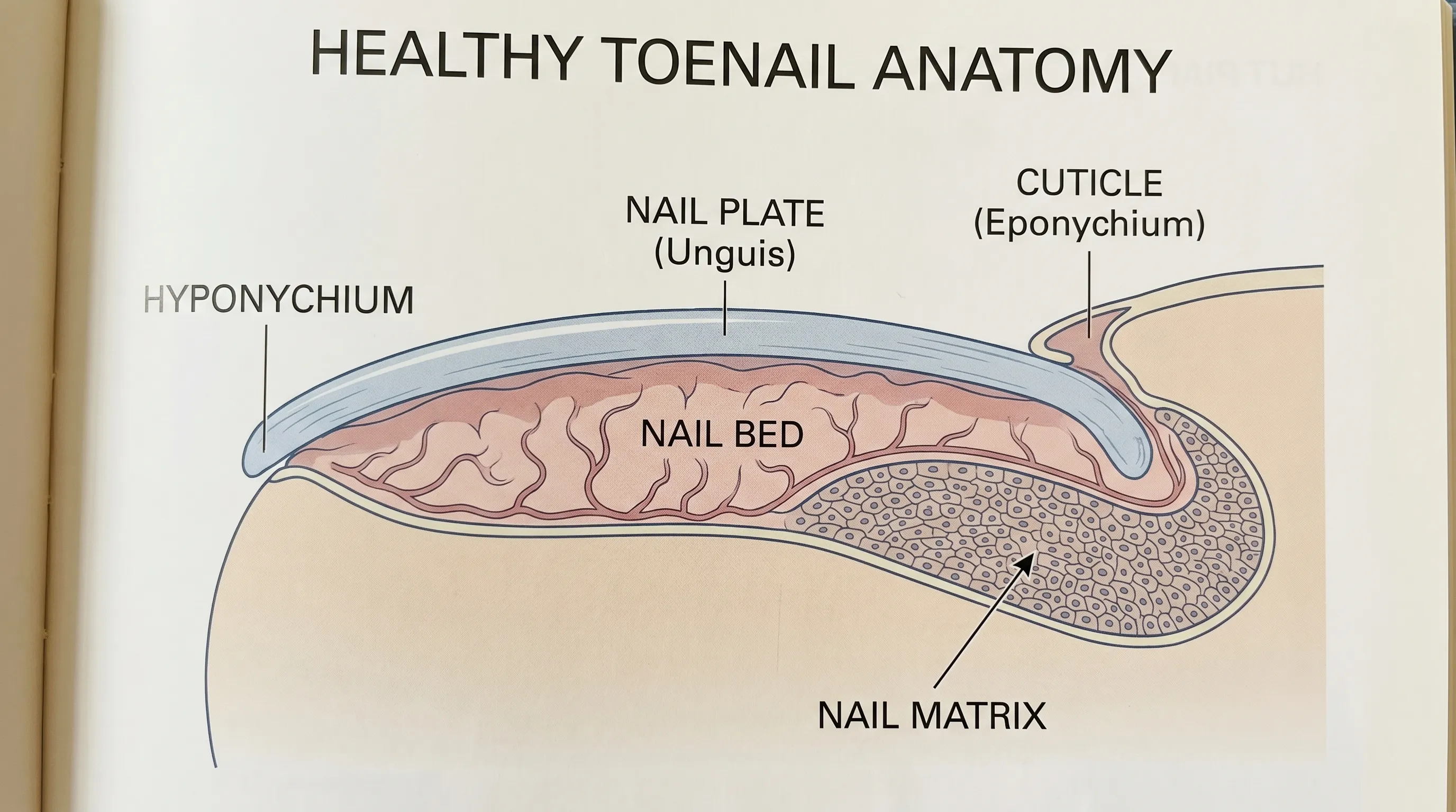
The visible portion of the nail, known clinically as the nail plate, is primarily composed of densely packed, non-living keratinocytes. These specialized epithelial cells undergo a continuous cycle of hardening, cross-linking, and compaction as they are pushed outward from their origin. Because the mature nail plate consists entirely of dead cellular material that has undergone complete keratinization, it possesses absolutely no nerve fibers or sensory receptors. This anatomical reality explains why cutting, filing, or even losing a toenail does not register as painful under normal physiological circumstances. The rigidity of the nail provides structural protection to the sensitive distal toe, acting as a natural shield against external impacts, abrasion, and mechanical stress. However, this protective barrier relies on a complex anchoring system to remain securely attached to the underlying tissue. When that anchoring system is compromised by moisture, infection, or metabolic disruption, the nail plate simply detaches and floats away without triggering nociceptive pathways in the central nervous system. Many patients initially panic when asking why did my toenail fall off without pain, but anatomical science provides clear reassurance that the absence of pain often reflects normal tissue turnover rather than acute injury.
The Role of the Nail Matrix and Bed
Beneath the visible nail plate lies the nail bed, a highly vascularized layer of specialized skin richly supplied with capillaries, sensory nerves, and squamous epithelial cells. The nail bed is fundamentally responsible for nourishing the overlying nail as it advances and maintaining its physical adherence through microscopic transverse ridges that interlock with the underside of the plate. At the proximal end of the nail, hidden beneath the protective proximal nail fold or cuticle, sits the nail matrix. This specialized germinative tissue acts as the primary growth center, continuously producing new keratinized cells that gradually push the nail forward over a period of twelve to eighteen months. When the nail matrix experiences temporary disruption or when the microscopic adhesion points between the plate and bed deteriorate, the structural integrity of the toenail begins to systematically fail. Because the pain-sensing nerve endings are heavily concentrated in the living tissue of the bed and surrounding dermis rather than the keratinized plate itself, the detachment process remains fundamentally painless unless the underlying tissue becomes acutely inflamed, infected, or mechanically sheared. This elegant biological arrangement allows the body to efficiently shed compromised keratin structures while actively protecting the sensitive vascularized tissue from further environmental harm.
Onycholysis: The Leading Cause of Painless Toenail Separation
Onycholysis represents the most frequently diagnosed medical condition responsible for painless nail detachment in clinical dermatology. Defined precisely as the gradual separation of the nail plate from the underlying nail bed, this condition typically begins at the distal free edge or lateral margins and progresses proximally toward the cuticle. As the separation advances, the affected portion often turns opaque white, pale yellow, or occasionally greenish due to trapped atmospheric air, accumulated keratin debris, or secondary microbial colonization beneath the lifted plate. Because the separation occurs incrementally over several weeks or even months, the nervous system gradually adapts to the changing pressure distribution, preventing acute pain signals from developing. The slow progression means that patients frequently wonder why did my toenail fall off without pain, completely unaware of the underlying pathological or mechanical activity that has been quietly dissolving the structural adhesion over time.
How Fungal Infections Gradually Weaken Nail Adhesion
Fungal pathogens, particularly dermatophytes such as Trichophyton rubrum, Trichophyton mentagrophytes, and Epidermophyton floccosum, are among the primary culprits behind chronic onycholysis. These microscopic organisms thrive in warm, humid, and dark environments, making the space beneath a toenail inside a closed shoe an ideal ecological niche. As the fungi proliferate and establish deep mycelial networks within the keratin matrix, they secrete specialized keratinase enzymes that systematically break down the tough protein bonds that give nails their structural strength and adhesive properties. Over a period of several months, this enzymatic degradation progressively weakens the microscopic anchoring bond between the nail plate and the underlying bed. According to comprehensive clinical data from the Mayo Clinic, chronic onychomycosis can silently compromise nail integrity long before any visible lifting or thickening becomes apparent to the patient. The exceptionally slow progression means that acute discomfort rarely accompanies the early or middle stages of detachment. The absence of significant inflammatory response in many chronic fungal cases further ensures that pain remains completely absent until structural failure occurs and the nail detaches entirely.
The Impact of Repetitive Microtrauma from Footwear and Exercise
While major acute trauma like dropping a heavy object directly onto the toe typically causes immediate bruising, subungual hematoma, and sharp pain, repetitive microtrauma operates as a far stealthier contributor to painless detachment. Runners, distance hikers, tennis players, and military personnel who regularly engage in high-impact activities or wear poorly fitted shoes with inadequate toe boxes subject their distal phalanges to constant, low-grade friction, shear stress, and compressive force. Each time the toe jams against the interior lining of a shoe during forward propulsion or rapid deceleration, the nail plate experiences a subtle mechanical shearing force against the nail bed. Over thousands of repetitions during training cycles or long work shifts, these micro-injuries cumulatively disrupt the microscopic epithelial adhesion points that secure the nail to the dermis. Moisture accumulation from excessive sweat further exacerbates the problem by softening the keratin structure and weakening the hydrophobic bonds that maintain adhesion. Since each individual micro-injury is physiologically too minor to trigger significant pain receptors, the cumulative structural damage progresses entirely unnoticed until the compromised nail eventually lifts and detaches. When individuals ask why did my toenail fall off without pain after months of intense training, clinicians typically identify repetitive shear trauma combined with moisture exposure as the primary mechanical drivers.
Psoriasis and Chemical Triggers
Autoimmune and chronic inflammatory dermatological conditions frequently manifest in the nails long before affecting visible cutaneous surfaces. Psoriatic nail disease affects up to 50% of individuals with cutaneous psoriasis and can present clinically with characteristic pitting, oil-drop discoloration, crumbling, and progressive onycholysis driven by autoimmune-mediated keratinocyte dysregulation. The inflammatory cytokines and accelerated epidermal turnover alter normal nail formation cycles, resulting in poorly adherent, structurally compromised plates that separate easily from the bed. Additionally, prolonged or repeated exposure to certain harsh chemicals—including strong acetone-based nail polish removers, formaldehyde-releasing hardeners, industrial cleaning agents, and extended immersion in detergent solutions—can systematically strip natural lipids and degrade the keratin matrix. For more detailed clinical insights into systemic and environmental triggers, the Cleveland Clinic provides an extensive overview of onycholysis and its multifaceted origins, emphasizing the importance of identifying underlying autoimmune or occupational factors.
Onychomadesis: Complete Nail Shedding After Systemic Stress
Unlike onycholysis, which involves gradual distal or lateral lifting from the bed, onychomadesis refers to the complete, proximal shedding of the entire nail plate following a temporary but absolute cessation of cellular growth in the nail matrix. This distinct clinical condition is characteristically preceded by the appearance of a deep transverse depression or groove, medically known as a Beau's line, that progressively migrates distally as new nail tissue eventually resumes formation. Eventually, this structural groove becomes deep enough to completely sever the proximal attachment of the nail from the regenerating matrix, resulting in the entire old nail plate falling off as a single unit or large fragment without tearing the underlying tissue.
The Mechanism of Temporary Nail Matrix Arrest
The nail matrix operates as a highly specialized epithelial factory, requiring precise cellular division, protein synthesis, and terminal differentiation to continuously produce healthy nail tissue. When the human body experiences severe physiological stress, acute illness, or major systemic inflammation, it instinctively prioritizes vital organ perfusion, central immune responses, and core metabolic functions over peripheral tissue maintenance like keratin production. This profound metabolic shift temporarily arrests the mitotic activity in the germinative matrix cells. Without continuous cellular production, a clear structural discontinuity forms horizontally across the nail plate. As the physiological stress resolves, homeostasis returns, and matrix activity successfully resumes, the newly generated nail tissue gradually pushes the old, detached segment distally toward the free edge. A comprehensive peer-reviewed analysis published in PubMed details exactly how this arrest-and-recovery biological cycle naturally produces onychomadesis. Because the eventual separation occurs entirely between dead keratin structures rather than involving live tissue trauma, vascular disruption, or nerve exposure, the shedding process remains fundamentally painless.
Post-Viral Illness and Medication-Induced Shedding
Viral infections, particularly coxsackievirus serotypes responsible for hand-foot-and-mouth disease, are exceptionally well-documented clinical triggers of onychomadesis in both pediatric and adult populations. The intense systemic inflammatory response, widespread cytokine release, and prolonged high fevers associated with acute viral illnesses create overwhelming metabolic demands that temporarily halt peripheral growth cycles. Similarly, severe systemic bacterial infections, major surgical procedures requiring extended anesthesia, and extreme physiological stressors like severe dehydration or acute nutritional deficiencies can produce identical matrix arrest effects. Certain pharmacological agents, including cytotoxic chemotherapy drugs, systemic isotretinoin and other high-dose retinoids, and specific immunosuppressant therapies, deliberately or inadvertently suppress rapidly dividing cells in the nail matrix. When patients successfully recover from these intensive medical challenges and inquire why did my toenail fall off without pain weeks or months after a serious illness or aggressive treatment course, board-certified clinicians typically identify onychomadesis as the expected physiological recovery outcome. The compromised nail detaches seamlessly and without discomfort as the renewed matrix steadily pushes forward a fresh, structurally sound replacement plate.
| Feature | Onycholysis | Onychomadesis | Acute Traumatic Injury |
|---|---|---|---|
| Primary Mechanism | Gradual loss of microscopic adhesion between plate and vascular bed | Temporary complete arrest of nail matrix cellular growth with proximal separation | |
| Pain Level | Typically entirely painless unless secondary infection occurs | Completely painless detachment due to dead tissue separation | |
| Onset Timeline | Progressive over weeks to months | ||
| Common Triggers | Fungal infections, psoriasis, repetitive microtrauma, harsh chemicals | ||
| Recovery Process | New nail grows outward from bed; requires careful bed protection |
Other Underlying Medical Conditions That Trigger Painless Nail Loss
Beyond localized nail disorders and acute systemic stress responses, several chronic endocrine and progressive neurological conditions can fundamentally alter peripheral nail biology, leading to unexpected, completely painless detachment. Recognizing and diagnosing these underlying metabolic factors is absolutely crucial for addressing true root causes rather than merely managing superficial symptoms repeatedly.
Thyroid Disorders and Hormonal Imbalances
Thyroid hormones play a critical regulatory role in keratinocyte metabolism, cellular turnover rates, and overall peripheral tissue integrity throughout the body. Both hyperthyroidism and hypothyroidism can significantly disrupt the delicate physiological equilibrium required for healthy nail growth, structural reinforcement, and sustained adhesion to the nail bed. In hyperthyroid states, dramatically accelerated basal metabolism leads to excessively rapid keratinization, often resulting in unusually brittle, fragile, and thin nails that separate easily from the underlying tissue—a clinical presentation historically termed Plummer's nails. Conversely, hypothyroidism drastically slows cellular turnover and reduces peripheral circulation, causing chronically dry, abnormally thickened, and poorly adherent nails that gradually detach over extended periods. These profound hormonal fluctuations directly affect microvascular supply to the distal extremities and alter the biochemical bonding between the nail plate and the underlying epithelial structures. When thyroid dysfunction remains clinically unmanaged, patients frequently experience multiple, unexplained instances of nail loss across several digits simultaneously.
Peripheral Neuropathy and the Diabetes Connection
Diabetes mellitus represents one of the most significant and clinically relevant risk factors for painless toenail complications and delayed structural healing. Chronic, poorly controlled hyperglycemia gradually damages the delicate peripheral nerves and small blood vessels supplying the distal lower extremities, a debilitating condition clinically known as diabetic peripheral neuropathy. This progressive nerve deterioration severely impairs the accurate transmission of pain, temperature, and pressure signals from the toes back to the central nervous system. Consequently, diabetic individuals may experience repetitive mechanical microtrauma, aggressive fungal colonization, or gradual structural nail separation without ever feeling the typical protective warning discomfort that would normally prompt behavioral changes or medical consultation. The American Academy of Dermatology emphasizes the critical importance of routine, scheduled foot examinations in diabetic and neuropathic populations, precisely because severely compromised sensory feedback can mask progressive structural damage until advanced clinical stages are reached. In these medically complex cases, the question why did my toenail fall off without pain is most often definitively answered by identifying underlying neuropathic masking combined with chronic microvascular changes that simultaneously impair tissue healing, structural resilience, and natural pain signaling pathways.
When to Seek Professional Medical Evaluation
While painless toenail detachment is frequently a benign, self-resolving physiological process, certain specific clinical presentations and patient demographics absolutely warrant prompt, thorough professional medical assessment. Early dermatological or podiatric intervention effectively prevents dangerous secondary complications and ensures appropriate, targeted management of underlying systemic conditions.
Red Flags Indicating Infection or Complications
The initial structural separation may begin completely painlessly, but the newly exposed nail bed creates a highly vulnerable, raw epithelial environment that is exceptionally susceptible to opportunistic bacterial and fungal invasion. If the affected area rapidly develops pronounced localized redness, significant swelling, noticeable warmth to the touch, purulent or serous drainage, or a distinct foul odor, these clinical signs strongly indicate a developing secondary infection such as acute paronychia, cellulitis, or localized bacterial overgrowth. Diabetic patients, elderly individuals with compromised immune systems, and patients with diagnosed peripheral arterial disease must exercise extreme clinical caution, as even minor nail bed exposure can rapidly progress to serious soft tissue infections, chronic non-healing ulcers, or dangerously delayed wound healing. Additionally, if the spontaneous detachment involves multiple toenails simultaneously, it often signals a widespread medication adverse reaction, aggressive systemic fungal colonization, or a poorly managed autoimmune condition requiring comprehensive dermatological or internal medicine evaluation.
Monitoring for Malignancy and Severe Dystrophy
In relatively rare but clinically serious instances, abnormal or unexplained nail changes can potentially indicate underlying malignancies or premalignant conditions. Subungual melanoma, an aggressive form of acral lentiginous skin cancer that develops beneath the nail plate, may initially present clinically as irregular dark longitudinal streaking, progressive structural deformity, or completely unexplained nail detachment that lacks any history of typical mechanical trauma. Persistent, severe discoloration that refuses to resolve with proper conservative hygiene, chronic unprovoked bleeding without known injury, or pronounced, asymmetric nail bed distortion require immediate specialist consultation. Board-certified dermatologists typically perform high-magnification dermoscopy or targeted nail matrix biopsy to definitively rule out malignant cellular transformation. Patients should never ignore progressive, unexplained structural abnormalities under the false assumption that they are harmless or purely cosmetic. Regular, attentive monitoring ensures that benign onycholysis or typical onychomadesis is never mistaken for serious oncological or degenerative conditions requiring urgent surgical or oncological intervention.
Step-by-Step Care and Management Strategies
Implementing proper wound management during and immediately after painless nail detachment is absolutely essential to prevent dangerous complications, protect the highly vulnerable exposed nail bed, and actively support healthy, unimpeded regrowth. Following established, evidence-based podiatric care protocols significantly reduces total recovery time, minimizes the risk of secondary infections, and prevents permanent structural scarring of the matrix.
Safe Detachment and Exposure Protection
When a toenail has partially lifted or completely separated, it is clinically crucial to strictly resist the natural urge to forcibly rip, pull, or twist it away from the toe. Premature, aggressive removal can easily tear delicate epithelial tissue in the exposed nail bed, trigger unnecessary localized bleeding, and create significantly larger microscopic entry points for environmental pathogens. Instead, patiently allow the compromised nail to detach entirely on its own as the new keratin plate gradually pushes the old tissue forward. Carefully and precisely trim any elevated, unstable edges using properly sterilized nail clippers to absolutely prevent the loose keratin from catching on thick socks, tight shoes, or bed linens. Once the nail bed is fully exposed, gently cleanse the area with mild, fragrance-free soap and lukewarm water, then pat it completely dry using a clean, lint-free towel or sterile medical gauze. Moisture control remains the single most important factor during healing; prolonged dampness aggressively encourages opportunistic microbial proliferation and delays epithelialization. Apply a thin, even layer of broad-spectrum over-the-counter antibiotic ointment to create an effective protective moisture barrier, then securely cover the area with a breathable, non-adhesive sterile bandage or medical gauze pad. Secure the dressing loosely enough to maintain healthy peripheral circulation but snugly enough to prevent constant friction during normal daily walking and standing.
Promoting Healthy Regrowth and Foot Hygiene
Toenail regeneration is a remarkably slow but highly predictable physiological process, typically advancing at a consistent biological rate of approximately one to one and a half millimeters per month in healthy adults. Complete structural regrowth from the proximal cuticle to the distal free edge strictly requires twelve to eighteen full months of consistent mechanical protection, optimal environmental conditions, and adequate nutritional support. During this extended recovery period, deliberately select footwear with generous, reinforced toe box room and adequate structural arch support to consistently prevent repetitive pressure or impact on the regenerating area. Switch daily to moisture-wicking socks manufactured from merino wool or specialized athletic synthetic blends to actively maintain a completely dry microenvironment around the toe. Absolutely avoid exposing the delicate healing tissue to harsh topical chemicals, including strong acetone-based polish removers, artificial acrylic nail products, or heavily pigmented cosmetic polishes, until the new plate has fully hardened and securely reattached. Consistently maintain a balanced diet exceptionally rich in lean protein, bioavailable zinc, dietary iron, and biotin to continuously provide essential amino acid building blocks required for optimal keratin synthesis. If you consistently find yourself asking why did my toenail fall off without pain despite diligently implementing careful foot care routines and protective strategies, schedule an appointment with a board-certified dermatologist to thoroughly evaluate potential persistent fungal colonization, irreversible matrix scarring, or underlying unmanaged metabolic factors.
Frequently Asked Questions
Why does my toenail separate from the bed without hurting?
Toenails are primarily composed of hardened, dead keratin cells that completely lack nerve endings and blood vessels. Pain only registers when living tissue, such as the underlying nail bed or surrounding skin, is injured or inflamed. Gradual separation allows the sensitive tissue to adapt to the changing structure, making the detachment process entirely painless in most cases.
How long does it take for a fallen toenail to grow back?
Toenails regenerate significantly slower than fingernails due to reduced peripheral circulation. Under optimal conditions, a new toenail grows at approximately 1 to 1.5 mm monthly. Complete replacement from the cuticle to the distal tip typically requires 12 to 18 months of consistent protection and proper nutrition.
Should I pull off a loose toenail that is hanging by a thread?
No, forcibly removing a partially attached toenail can tear the delicate vascularized nail bed, trigger bleeding, and create an open wound vulnerable to bacterial invasion. Trim carefully along the elevated edge to prevent snagging, apply a breathable protective dressing, and allow the nail to detach naturally as the new plate pushes it forward.
Can painless nail loss indicate a serious underlying illness?
While frequently caused by localized factors like fungal infections or microtrauma, widespread or recurrent painless shedding can sometimes signal systemic conditions. Thyroid disorders, uncontrolled diabetes, autoimmune diseases like psoriasis, and certain medication side effects can disrupt nail matrix function. Multiple affected nails warrant comprehensive medical evaluation.
What is the best way to prevent fungal reinfection during regrowth?
Maintain strict foot hygiene by washing and thoroughly drying your feet daily. Wear breathable, moisture-wicking socks and rotate footwear to ensure complete drying between uses. Apply antifungal powder to shoes and feet if you have a history of onychomycosis, and never share nail clippers or pedicure tools to prevent cross-contamination.
Conclusion
Discovering that a toenail has detached completely without accompanying discomfort is an exceptionally common clinical experience that typically points toward benign, well-understood physiological processes like gradual onycholysis or predictable post-stress onychomadesis. The human body is remarkably efficient at managing routine keratinized tissue turnover, frequently prioritizing quiet, gradual shedding over acute inflammatory responses to protect underlying structures. Whether the detachment was initially triggered by chronic low-grade fungal exposure, years of repetitive mechanical microtrauma, recovery from a severe systemic viral illness, or an unmanaged endocrine shift, the complete absence of pain usually indicates that vital living tissue and vascular networks remain fundamentally uncompromised. Implementing meticulous, evidence-based wound management, consistently protecting the exposed bed from further environmental trauma, and maintaining rigorous, daily foot hygiene collectively create the optimal biological environment for steady, healthy regeneration. However, actively recognizing clinical warning signs like persistent secondary infection, widespread systemic involvement, or significantly delayed epithelial healing ensures that entirely benign conditions are never mistakenly ignored or improperly self-treated. By thoroughly understanding why did my toenail fall off without pain and diligently following structured, conservative recovery protocols, you can confidently navigate the entire healing process, minimize complication risks, and actively support long-term structural nail resilience for years to come.
How we work
Researched and drafted with AI assistance, then reviewed by a human editor against the cited sources.
This article is general health information, not medical advice. Consult a qualified healthcare professional about your own situation.