Brown Spots at Bottom of Feet: Causes, Diagnosis, and Evidence-Based Care
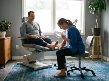
Noticing an unexpected pigmented mark on the underside of your foot can understandably trigger concern. The skin on the soles is uniquely thick, highly keratinized, and constantly subjected to mechanical stress, making it prone to distinct dermatological presentations. When you observe brown spots at bottom of feet, it is essential to understand the spectrum of potential causes, ranging from harmless physiological responses to conditions requiring professional medical intervention. Many individuals dismiss these marks as dirt or minor bruises, yet persistent or evolving lesions warrant careful assessment. Plantar skin lacks sebaceous glands and relies heavily on eccrine sweat glands, creating a specialized microenvironment that influences pigmentation patterns. By exploring the underlying mechanisms, clinical evaluation standards, and evidence-based management strategies, you can make informed decisions about your foot health. This comprehensive guide integrates current dermatological research, clinical guidelines, and practical self-care protocols to help you navigate this common yet frequently misunderstood symptom. Understanding when to monitor, when to treat, and when to seek specialized care ensures optimal outcomes and long-term dermatological wellness.
Understanding the Anatomy and Physiology of Plantar Skin
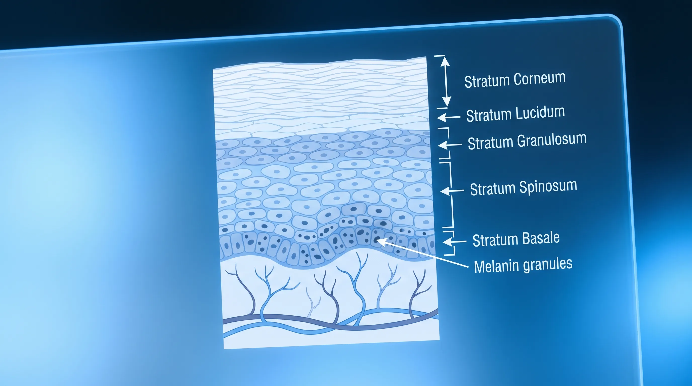
To accurately assess any dermatological change, it is crucial to first understand how the skin on your soles differs from skin elsewhere on your body. The plantar surface is classified as glabrous skin, meaning it is hairless and contains a significantly thicker stratum corneum, the outermost protective layer of the epidermis. This structural adaptation evolved to withstand continuous friction, shear forces, and weight-bearing pressure throughout your lifespan. The thick keratinocyte matrix naturally alters how pigmentation manifests and how clinicians interpret visual findings. Unlike thinner skin where melanin distribution appears uniform and readily visible through the epidermis, the dense keratin layer on the soles can obscure or distort pigment patterns, sometimes making benign lesions appear darker or more diffuse than they actually are.
Free calculatorSun Protection GuideSPF guide and sun protection recommendationsOpen the calculatorMelanocytes, the pigment-producing cells located at the dermoepidermal junction, function differently in acral areas. They are generally less numerous but produce melanin that is packaged into larger melanosomes, which transfer to surrounding keratinocytes more slowly. This unique physiology explains why brown spots at bottom of feet often develop gradually and may exhibit distinct dermatoscopic patterns compared to lesions on the trunk or extremities. Additionally, the absence of hair follicles and sweat glands in high-pressure zones creates a localized environment where post-inflammatory changes can linger for extended periods. Minor microtraumas, ill-fitting footwear, or even subtle gait abnormalities can trigger localized melanocyte activation, resulting in hyperpigmented areas that mimic more serious conditions.
The dermal layer beneath the epidermis on the plantar surface is densely packed with collagen fibers and elastic tissue designed for shock absorption. When trauma occurs, inflammatory mediators such as interleukins and tumor necrosis factor-alpha stimulate melanogenic pathways. This biological response is protective but can leave lasting pigmentation. Recognizing these physiological foundations helps separate normal adaptive responses from pathological processes. Clinicians rely on this anatomical knowledge when interpreting visual examinations, dermoscopic findings, and histological samples. Patients who understand the unique characteristics of plantar skin are better equipped to communicate effectively with healthcare providers and participate actively in their diagnostic journey.
Primary Causes of Brown Spots at the Bottom of Feet
Identifying the underlying etiology requires systematic evaluation of clinical presentation, patient history, and environmental factors. Pigmented lesions on the soles encompass a broad differential diagnosis, each with distinct pathophysiological mechanisms. Recognizing these categories prevents unnecessary anxiety while ensuring timely intervention when required.
Acral Nevi and Benign Pigmented Lesions
Acral nevi, commonly referred to as moles, represent the most frequent cause of localized brown pigmentation on weight-bearing surfaces. These benign melanocytic proliferations typically appear as well-circumscribed, uniformly pigmented macules or slightly elevated papules. Research published in dermatological literature indicates that acral nevi are particularly common in individuals with darker phototypes, though they occur across all demographic groups. The parallel furrow pattern, visible under dermoscopy, characterizes benign acral nevi, where pigment aligns along the skin's natural ridges rather than disrupting them. These lesions generally remain stable in size, shape, and coloration over decades. Regular self-examination remains the cornerstone of management, focusing on monitoring for any deviation from baseline appearance.
Post-Inflammatory Hyperpigmentation from Trauma or Friction
Mechanical stress is a primary driver of localized pigmentation changes. Repetitive pressure from footwear, athletic activities, or occupational standing can induce microvascular damage and localized inflammation. As the inflammatory cascade resolves, melanocytes often overproduce melanin in response to cytokine signaling, leaving behind flat, brown macules. This phenomenon, known as frictional melanosis, frequently presents in areas of maximal pressure such as the heel, ball of the foot, or lateral borders. Improving footwear ergonomics, utilizing custom orthotics, and incorporating silicone-based protective padding can significantly mitigate ongoing friction. Topical antioxidants and gentle keratolytic agents may assist in gradually fading residual pigmentation over several months.
Tinea Nigra and Superficial Fungal Presentations
Tinea nigra represents a distinct superficial mycosis caused by the dimorphic fungus Hortaea werneckii. This organism thrives in warm, humid environments and commonly colonizes the soles, particularly between toes or in sweat-retaining footwear crevices. Clinically, it presents as irregular, asymptomatic brown to black patches that may resemble superficial bruising or benign moles. Unlike deeper dermatophyte infections, tinea nigra does not penetrate living tissue and remains confined to the stratum corneum. Diagnosis is typically confirmed through potassium hydroxide preparation or fungal culture showing characteristic hyphae. Treatment involves consistent application of topical antifungal agents such as clotrimazole or miconazole for two to four weeks, alongside strict foot hygiene practices including daily washing, thorough drying, and moisture-wicking socks.
Early Melanocytic Changes and Acral Lentiginous Melanoma
While statistically less common than benign causes, acral lentiginous melanoma (ALM) demands heightened clinical awareness. This aggressive melanoma subtype accounts for a disproportionate percentage of melanoma cases in individuals with darker skin phototypes and typically arises on non-sun-exposed surfaces, including the soles. Early lesions may mimic benign nevi or bruising, making early recognition critical. Dermatologists utilize the ABCDE framework alongside the 'ugly duckling' sign, which identifies lesions that stand out from a patient's other pigmented marks. Advanced dermoscopy reveals parallel ridge patterns, atypical vascular networks, and blue-white structures that raise suspicion for malignancy. Early surgical excision remains the gold standard treatment, with prognosis heavily dependent on Breslow thickness at diagnosis. Public awareness campaigns emphasize that melanoma does not exclusively occur on sun-damaged skin.
Vascular and Systemic Pigmentary Disorders
Certain systemic conditions manifest with plantar pigmentation changes. Peripheral vascular disease, chronic venous insufficiency, and diabetic microangiopathy can alter local tissue oxygenation, leading to hemosiderin deposition that appears as brownish discoloration. Nutritional deficiencies, particularly vitamin B12 and folate, occasionally contribute to generalized hyperpigmentation that becomes more noticeable on thick plantar skin. Endocrine disorders such as Addison's disease or hemochromatosis produce widespread melanocyte stimulation, often accentuated in pressure points and creases. Comprehensive medical evaluation, including complete blood counts, metabolic panels, and vascular assessments, helps distinguish isolated dermatological findings from systemic manifestations. Addressing the underlying physiological imbalance typically results in gradual resolution of the pigmentation over time.
Clinical Evaluation and Diagnostic Approaches
Professional assessment begins with a comprehensive patient history, documenting onset, duration, symptomatology, footwear habits, occupational exposures, and personal or family dermatological history. Physical examination under adequate illumination allows clinicians to assess symmetry, border definition, color homogeneity, and elevation. The following diagnostic modalities represent current clinical standards.
Initial Physical Examination and Dermoscopy
Dermoscopy has revolutionized non-invasive dermatological diagnostics by magnifying subsurface structures invisible to the naked eye. A polarized dermoscope eliminates surface reflection, revealing pigment distribution within epidermal ridges and furrows. On acral skin, dermatologists evaluate the parallel furrow, lattice, or fibrillar patterns typical of benign lesions versus the parallel ridge pattern indicative of early melanoma. This technology significantly reduces unnecessary biopsies while improving diagnostic accuracy. Studies demonstrate that dermoscopic evaluation increases early melanoma detection rates by over thirty percent compared to clinical examination alone. Routine integration of dermoscopy into podiatric and dermatological practice has become a standard of care for evaluating brown spots at bottom of feet.
Biopsy Protocols and Histopathological Assessment
When clinical or dermoscopic findings raise suspicion, tissue sampling becomes necessary. Excisional biopsy is preferred when feasible, removing the entire lesion with narrow margins to preserve architectural context for histopathological evaluation. Incisional or punch biopsies may be utilized for larger or anatomically constrained areas. Pathologists examine hematoxylin and eosin-stained sections, assessing melanocyte proliferation, atypia, mitotic activity, and dermal invasion depth. Special immunohistochemical stains such as S100, Melan-A, and HMB-45 enhance diagnostic precision. Accurate histological grading guides subsequent management, including the need for wider surgical margins, sentinel lymph node mapping, or adjuvant therapies. Patients receive detailed counseling regarding procedure recovery, scar management, and follow-up surveillance protocols.
Differential Diagnosis Matrix
The following table summarizes key distinguishing features to aid in clinical differentiation and patient education.
| Feature | Acral Nevus | Post-Inflammatory Hyperpigmentation | Tinea Nigra | Acral Lentiginous Melanoma |
|---|---|---|---|---|
| Appearance | Well-defined, uniform brown macule | Flat, irregular patch matching friction zones | Superficial, non-scaly brown/black patch | Asymmetric, multicolored, evolving lesion |
| Borders | Sharp, symmetrical | Ill-defined, gradual fading | Slightly irregular but superficial | Jagged, blurred, or notched |
| Dermoscopy | Parallel furrow pattern | No pigment network, homogeneous structure | Focal black dots, absent vascular changes | Parallel ridge pattern, atypical vessels |
| Symptoms | Typically asymptomatic | May have mild tenderness initially | Asymptomatic, no scaling | Itching, ulceration, bleeding, or pain |
| Management | Observation or elective excision | Friction reduction, topical lightening | Topical antifungals, hygiene optimization | Wide excision, oncology referral |
Evidence-Based Treatment and Management Strategies
Therapeutic interventions must align with precise diagnosis, lesion characteristics, and patient preferences. Conservative approaches remain first-line for benign presentations, while malignant or rapidly progressive conditions require definitive intervention.
Conservative Monitoring and Watchful Waiting
For clinically benign acral nevi with stable dermoscopic features, active surveillance represents the standard of care. Patients receive documentation including standardized photographs, measurement charts, and scheduled follow-up intervals typically ranging from six to twelve months. Digital tracking applications facilitate longitudinal monitoring, enabling patients to upload sequential images for remote assessment by dermatology teams. Education on self-examination techniques empowers individuals to identify subtle changes promptly. Lifestyle modifications including moisture management, sun protection for exposed foot areas, and avoidance of repetitive trauma support long-term skin health without invasive procedures.
Topical Therapies and Pharmacological Interventions
When hyperpigmentation results from post-inflammatory pathways or superficial fungal colonization, targeted pharmacological treatments demonstrate efficacy. Hydroquinone, kojic acid, and azelaic acid inhibit tyrosinase activity, gradually reducing melanin synthesis in localized areas. Combination regimens often incorporate mild corticosteroids to suppress residual inflammation and retinoids to enhance epidermal turnover. Treatment courses typically span twelve to sixteen weeks, requiring consistent application and strict sun protection to prevent paradoxical repigmentation. Fungal-related pigmentation responds to allylamines and azoles that disrupt ergosterol synthesis. Patients with sensitive skin or contraindications to conventional lightening agents benefit from natural alternatives such as licorice root extract or niacinamide formulations, which offer moderate depigmenting effects with improved tolerability.
Surgical Excision, Cryotherapy, and Laser Modalities
Lesions demonstrating atypical features, symptomatic presentation, or persistent discomfort warrant procedural intervention. Surgical excision under local anesthesia ensures complete removal and definitive histological diagnosis. Wound closure follows tension-relieving techniques appropriate for the high-stress plantar environment, with post-operative instructions emphasizing off-loading protocols to prevent dehiscence. Cryotherapy utilizing liquid nitrogen effectively treats superficial tinea nigra and benign hyperkeratotic pigmented lesions, though it carries risk of hypopigmentation in darker skin types. Pulsed dye lasers and Q-switched Nd:YAG systems target melanin granules selectively, fragmenting pigment particles that are subsequently cleared by macrophages. Laser treatments require multiple sessions and strict post-procedural care to optimize outcomes and minimize scarring. Board-certified specialists determine the most appropriate modality based on lesion depth, skin phototype, and anatomical location.
Preventive Foot Care and Daily Self-Management
Long-term dermatological wellness relies on proactive maintenance, environmental modification, and consistent self-assessment. Implementing evidence-based foot care protocols significantly reduces the incidence of traumatic hyperpigmentation, fungal colonization, and delayed diagnosis of concerning lesions.
Optimizing Footwear and Reducing Plantar Pressure
Footwear selection profoundly influences plantar skin integrity. Shoes with inadequate cushioning, narrow toe boxes, or rigid soles concentrate pressure onto specific acral zones, triggering melanocyte activation. Invest in footwear featuring memory foam insoles, seamless interiors, and breathable mesh construction. Custom orthotics prescribed by a podiatrist correct biomechanical imbalances, redistributing weight across broader surface areas. Rotate shoes daily to allow moisture evaporation and material recovery. Replace athletic footwear every three hundred to five hundred miles to maintain shock-absorbing properties. These mechanical adjustments prevent recurrent microtrauma and subsequent post-inflammatory hyperpigmentation.
Establishing a Routine Inspection Protocol
Monthly self-examination remains the most effective strategy for early lesion detection. Conduct assessments in a well-lit environment after bathing, when the stratum corneum is softened and pigmentation is more visible. Utilize a full-length mirror or handheld device to inspect the soles, lateral borders, and interdigital spaces systematically. Document baseline findings using smartphone photography with consistent lighting and measurement references. Compare sequential images quarterly to identify asymmetry, border irregularity, or color shifts. Individuals with diabetes, immunosuppression, or personal melanoma history should perform weekly inspections and maintain scheduled dermatological appointments.

Dermatological Follow-Up and Professional Guidance
Routine dermatological visits complement home monitoring. Annual full-body skin examinations allow clinicians to assess total nevus burden, evaluate suspicious lesions, and reinforce patient education. High-risk individuals may require semi-annual surveillance with digital dermoscopic mapping. Maintain open communication regarding new lesions, changes in existing marks, or systemic symptoms. Consult the American Academy of Dermatology's guidelines on acral melanoma for updated risk stratification and monitoring protocols. Professional evaluation ensures that benign presentations remain monitored appropriately while concerning findings receive expedited diagnostic workup and treatment planning. Integrating expert care with disciplined self-management creates a comprehensive defense against preventable complications.
Frequently Asked Questions
Are brown spots at the bottom of feet usually a sign of skin cancer?
Not necessarily. The majority of pigmented lesions on the plantar surface are benign, including common acral nevi, post-inflammatory hyperpigmentation, or fungal conditions. However, acral lentiginous melanoma can present as a new or changing spot on the sole. Any irregular, asymmetrical, or rapidly evolving lesion should be evaluated by a dermatologist. Early detection through professional screening dramatically improves outcomes.
Can pressure from walking cause brown spots to appear?
Yes. Repetitive friction and pressure stimulate melanocytes and can lead to frictional melanosis or post-inflammatory hyperpigmentation. Proper footwear with adequate cushioning, arch support, and moisture-wicking properties significantly reduces this risk. Orthotic inserts and rotational shoe use further minimize localized stress on plantar skin.
How do doctors differentiate between a benign mole and melanoma on the foot?
Dermatologists use the ABCDE criteria combined with dermoscopy. Acral melanoma often displays parallel ridge patterns or atypical vascular structures under magnification, whereas benign lesions show parallel furrow patterns. Biopsy provides definitive diagnosis when visual assessment remains inconclusive. Advanced imaging and histopathological analysis guide appropriate management pathways.
Is tinea nigra contagious or dangerous?
Tinea nigra is a superficial fungal infection caused by Hortaea werneckii. It is mildly contagious through direct contact or contaminated surfaces but is entirely benign. It typically resolves with topical antifungal treatments and improved foot hygiene. Maintaining dry footwear and avoiding shared towels prevents reinfection and transmission to other household members.
Should I attempt to remove a dark spot on my sole at home?
No. Over-the-counter removal products can cause chemical burns, scarring, or alter the lesion's appearance, complicating future clinical diagnosis. Professional evaluation is essential to rule out malignancy before any removal or treatment is initiated. Licensed clinicians utilize sterile techniques, appropriate anesthesia, and precise wound closure to optimize healing and diagnostic accuracy.
Conclusion
Observing brown spots at bottom of feet warrants thoughtful attention rather than alarm. The plantar surface represents a unique dermatological environment shaped by constant mechanical stress, specialized cellular architecture, and distinct microbial ecosystems. Most pigmented lesions result from benign physiological responses, including acral nevi, frictional hyperpigmentation, or superficial fungal activity. However, recognizing the subtle warning signs of acral lentiginous melanoma remains critical for early intervention. Integrating regular self-examination, optimal footwear selection, and professional dermatological guidance forms the foundation of comprehensive foot health management. Avoiding DIY removal attempts and seeking evidence-based clinical evaluation ensures accurate diagnosis and appropriate treatment. By applying the diagnostic frameworks, preventive strategies, and care protocols outlined in this guide, you can confidently monitor your plantar skin and maintain long-term dermatological wellness. Consistent attention to these details, combined with timely professional consultation, transforms uncertainty into proactive health stewardship. Prioritize your foot health with the same diligence you apply to other vital aspects of your wellbeing, and consult licensed healthcare providers for personalized assessment and management plans tailored to your specific clinical presentation.

How we work
Researched and drafted with AI assistance, then reviewed by a human editor against the cited sources.
This article is general health information, not medical advice. Consult a qualified healthcare professional about your own situation.