Black Dot Under Toenail: Causes, Diagnosis, and Treatment Guide

Noticing a sudden black dot under toenail can immediately trigger concern, especially when the cause is unclear. Whether it appears after a long run, emerges gradually over weeks, or develops without any obvious trigger, understanding what lies beneath the nail plate is essential for both peace of mind and proper medical intervention. While most cases stem from minor trauma or harmless pigmentation changes, some may signal underlying infections or require specialized dermatological evaluation. Navigating the differences between benign and concerning nail changes can feel overwhelming without guidance. This comprehensive guide explores the anatomy, causes, diagnostic pathways, and evidence-based treatment strategies for a black dot under toenail, empowering you to make informed decisions about your foot health. By recognizing key warning signs, practicing safe home care, and knowing exactly when to consult a healthcare professional, you can protect your nails and maintain long-term lower extremity wellness.
Understanding the Anatomy of a Black Dot Under the Toenail
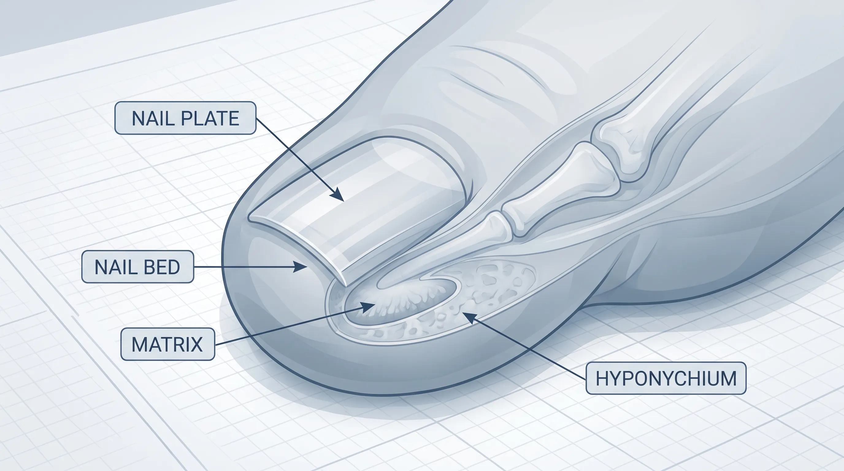
The toenail is a highly specialized appendage designed to protect the sensitive distal phalanx while providing mechanical support during walking and running. To fully grasp why a black dot under toenail appears and how it evolves, it is crucial to understand the complex architecture of the nail unit as outlined by the Cleveland Clinic. The visible portion, known as the nail plate, is composed of tightly packed, keratinized cells that grow continuously from the underlying nail matrix. Beneath this plate lies the nail bed, a highly vascularized tissue rich in capillaries that gives the nail its natural pinkish hue. Any disruption to this delicate ecosystem can result in visible discoloration, structural changes, or localized pigment deposits.
Free calculatorSun Protection GuideSPF guide and sun protection recommendationsOpen the calculatorThe Structure of the Toenail Unit
The nail matrix, located beneath the proximal nail fold, acts as the growth center. When melanocytes (pigment-producing cells) in the matrix become stimulated or damaged, they release melanin that becomes trapped in the forming nail plate, appearing as linear streaks or discrete dots. The nail bed adheres tightly to the plate through a thin layer of epithelial cells. Blood vessels run parallel to the direction of nail growth, meaning any trauma that ruptures capillaries will leave behind pooled blood that oxidizes from red to dark brown or black over time. The hyponychium, situated beneath the free edge of the nail, forms a critical waterproof seal that prevents pathogens from entering the deeper nail unit. Compromise to any of these structures can manifest visually as localized pigmentation or structural deformity.
How Pigmentation and Trauma Affect Nail Beds
When mechanical stress occurs, such as repetitive microtrauma from ill-fitting shoes or acute injury from stubbing a toe, capillaries in the nail bed rupture. Since the nail plate is rigid and non-compressible, the blood becomes trapped in a confined space, forming a subungual hematoma that appears as a black dot under toenail. Over time, enzymatic breakdown of hemoglobin causes the color to shift from bright red to dark purple, then black. Conversely, benign pigmentation arises when melanocytes produce excess melanin due to hormonal fluctuations, sun exposure, or genetic predisposition. In rare instances, dysregulated melanocyte proliferation can lead to malignant transformation. Understanding these pathways helps differentiate between transient trauma and conditions requiring clinical intervention.
Common Causes of a Black Dot Under Toenail
Identifying the underlying etiology is the cornerstone of appropriate management. While the visual presentation may appear similar across different conditions, the clinical history, growth pattern, and associated symptoms provide vital diagnostic clues. Below are the most prevalent medical explanations for this presentation.
Subungual Hematoma (Trauma)
A subungual hematoma is by far the most common reason patients notice a black dot under toenail. It typically occurs after repetitive microtrauma, such as prolonged hiking, marathon running, or sudden impact injury. The pooled blood creates immediate pressure, often accompanied by throbbing pain. As the nail grows forward at approximately one millimeter per month, the discolored area gradually migrates toward the distal edge. In cases where the hematoma covers more than twenty-five to forty percent of the nail surface or causes severe pain, medical drainage may be indicated to relieve pressure and prevent permanent matrix damage. Without intervention, small hematomas resolve spontaneously over six to twelve months, consistent with clinical guidance from the Mayo Clinic.
Fungal Infections (Onychomycosis)
Dermatophytes and nondermatophyte molds can invade the nail plate and bed, leading to discoloration that sometimes presents as a localized black dot under toenail. Fungal pigmentation occurs when certain organisms produce melanin as a metabolic byproduct or when secondary bacterial colonization occurs alongside the primary infection. Unlike trauma-related spots, fungal lesions often accompany thickening, crumbling edges, yellowish-brown streaking, or subungual debris. According to the CDC, diagnosis requires a potassium hydroxide preparation or fungal culture. Treatment involves prolonged topical antifungals, oral terbinafine, or laser therapy, depending on the extent of nail involvement and patient medical history.
Pigmented Lesions and Nevi (Moles)
Benign melanocytic nevi frequently appear on the digits and may present as a solitary black dot under toenail. These moles originate from nests of melanocytes within the nail matrix or bed. They typically exhibit uniform color, regular borders, and stable size over months or years. Dermoscopic evaluation usually reveals parallel banding or homogenous pigmentation patterns that distinguish them from malignant transformations. Routine monitoring is generally sufficient unless changes in shape, color intensity, or symmetry occur. Patients with darker skin tones are more prone to benign nail pigmentation due to higher baseline melanocyte activity.
Melanoma and Serious Conditions
Subungual melanoma accounts for one to three percent of all melanoma cases globally but carries significant morbidity if diagnosed late. A black dot under toenail may represent the early manifestation of acral lentiginous melanoma, which commonly affects the great toe. Warning signs include irregular borders, color variegation, involvement of the nail fold (Hutchinson sign), nail splitting, or failure of the lesion to grow out with the nail plate, as detailed by the National Institutes of Health (NIH). Early biopsy and wide local excision are critical for optimal outcomes. Delayed recognition often leads to subungual bone invasion or metastasis, underscoring the importance of prompt dermatological assessment for atypical presentations.
Other Rare Causes
Less frequently, a black dot under toenail may result from foreign body retention, such as a wood splinter or glass fragment embedded in the nail bed. Pseudomonas aeruginosa infection can also cause dark greenish-black discoloration due to pyocyanin production, particularly in chronically moist or damaged nails. Certain medications, including chemotherapy agents, antimalarials, and minocycline, induce diffuse or focal nail pigmentation as an adverse effect. Systemic conditions like Addison disease or hemochromatosis rarely present with isolated nail changes. Accurate differentiation requires comprehensive clinical correlation and targeted diagnostic testing.
Recognizing Symptoms and Warning Signs
Careful observation of accompanying clinical features provides essential diagnostic information. While isolated discoloration is often benign, certain symptom clusters warrant immediate professional evaluation.
Distinguishing Benign vs. Concerning Features
Benign lesions typically demonstrate uniform pigmentation, smooth borders, and gradual migration toward the free nail edge. They rarely cause pain beyond the initial trauma period and maintain stable dimensions over time. Conversely, concerning features include asymmetry, jagged or blurred margins, multiple colors within the same spot, rapid enlargement, or extension into the periungual skin. Nail plate dystrophy, such as splitting, lifting, or crumbling, alongside a black dot under toenail, raises suspicion for invasive pathology. The presence of bleeding, ulceration, or persistent tenderness without recent injury should never be ignored.
Associated Symptoms to Monitor
Patients should track pain intensity, swelling, warmth, discharge, and changes in nail texture. Localized erythema, purulent drainage, or fever may indicate secondary bacterial infection requiring antibiotic therapy. Lymphadenopathy in the popliteal or inguinal regions suggests possible malignancy spread. Functional impairment, such as difficulty wearing shoes or altered gait mechanics, can result from chronic pressure or structural nail changes. Documenting these symptoms with dated photographs assists clinicians in tracking progression and determining appropriate intervention timelines.
Diagnostic Approaches and Medical Evaluation
Accurate diagnosis relies on a systematic clinical approach that combines visual inspection, advanced imaging, and histopathological confirmation when indicated. Dermatologists and podiatrists utilize standardized protocols to differentiate between traumatic, infectious, and neoplastic etiologies.
Clinical Examination and Dermoscopy
The initial evaluation involves thorough inspection of all digits, assessment of nail thickness, and palpation of surrounding tissues. Transillumination with a bright penlight can reveal trapped fluid versus solid pigmentation. Dermoscopy, a noninvasive technique using polarized light and magnification, allows visualization of microvascular patterns and melanin distribution. Parallel pigmented bands, homogeneous brown-black coloration, and regular architecture suggest benign processes. Atypical networks, radial streaming, or irregular dots strongly indicate malignancy. This tool significantly reduces unnecessary biopsies while improving early cancer detection rates.
Biopsy and Laboratory Testing
When clinical suspicion remains high, a nail matrix or bed biopsy provides definitive histological diagnosis. Punch biopsy, longitudinal excision, or tangential shaving techniques are selected based on lesion location and size. Tissue samples undergo hematoxylin and eosin staining, immunohistochemistry, and sometimes molecular profiling to confirm melanoma subtype or identify fungal elements. As noted by the Mayo Clinic, pathologists evaluate mitotic rate, atypia grade, and depth of invasion to guide prognosis and surgical planning.
When Imaging Is Necessary
Radiographic imaging becomes essential if bone involvement is suspected. Plain X-rays can detect subungual osteomyelitis, foreign bodies, or underlying fractures. High-frequency ultrasound evaluates soft tissue thickness and fluid accumulation. Magnetic resonance imaging provides superior contrast resolution for assessing tumor depth, tendon involvement, and regional lymph node status. Advanced imaging modalities inform surgical margins, reconstructive planning, and staging protocols for malignant lesions.
Treatment Options and Management Strategies
Management strategies are tailored to the underlying cause, lesion characteristics, and patient preferences. Conservative monitoring, targeted pharmacotherapy, and surgical intervention represent the primary therapeutic pathways.
Conservative Home Care and Observation
For trauma-induced hematomas without significant pain, observation is the standard of care. Keeping the affected toe clean, dry, and protected from further impact promotes natural resolution. Elevation reduces swelling, while over-the-counter analgesics manage discomfort. Avoiding tight footwear and moisture-trapping socks prevents secondary complications. If a black dot under toenail remains stable and migrates distally over several months, no active intervention is required. Regular photographic documentation helps track natural nail growth and ensures early detection of any concerning changes.
Medical Interventions for Infections and Hematomas
Large, painful hematomas may require trephination, a minor procedure where a sterile needle or cautery device creates a small opening in the nail plate to release trapped blood. This provides immediate pain relief and preserves nail viability. Fungal infections respond to topical ciclopirox or efinaconazole applied daily for nine to twelve months. Oral terbinafine achieves higher cure rates but requires baseline liver function testing due to potential hepatotoxicity, per Cleveland Clinic guidelines. Bacterial superinfections are treated with targeted antibiotics based on culture sensitivity. Adjunctive debridement improves topical medication penetration and accelerates recovery.
Surgical and Procedural Options for Malignancy
Early-stage subungual melanoma typically undergoes wide local excision with two-centimeter margins, followed by histological verification of clear borders. Advanced cases may require amputation of the affected phalanx to ensure oncological clearance. Reconstructive techniques, including skin grafting or flap transfer, restore function and cosmesis. Sentinel lymph node biopsy determines metastatic spread, guiding adjuvant immunotherapy or targeted therapy regimens. Multidisciplinary coordination among dermatologists, surgical oncologists, and podiatric surgeons optimizes patient outcomes.
Pharmacological Treatments
Emerging therapeutic options include topical immunomodulators, intralesional injections, and systemic retinoids for refractory nail dystrophy. Antifungal combinations targeting both dermatophytes and yeast improve eradication rates. Pain management protocols incorporate neuropathic agents, NSAIDs, and localized nerve blocks for chronic discomfort. Nutritional supplementation with biotin, zinc, and omega-three fatty acids supports keratin synthesis and overall nail integrity during recovery phases.
| Condition | Primary Cause | Key Symptoms | Recommended Treatment | When to See a Doctor |
|---|---|---|---|---|
| Subungual Hematoma | Trauma/Micro-injury | Sudden onset, throbbing pain, migrates upward | Observation, ice, elevation, trephination if severe | Pain uncontrolled, covers >25% nail, or persists >3 months |
| Onychomycosis | Fungal infection | Thickening, crumbling, yellow-black discoloration, debris | Topical antifungals, oral terbinafine, laser therapy | Spreads, causes pain, diabetic patient, or unresponsive to OTC care |
| Melanocytic Nevus | Benign melanocyte cluster | Uniform dot, stable size, regular borders, asymptomatic | Routine monitoring, dermoscopic tracking | Changes shape/color, bleeds, or spreads to skin fold |
| Subungual Melanoma | Malignant melanocyte proliferation | Irregular borders, color variation, Hutchinson sign, nail destruction | Surgical excision, sentinel node biopsy, immunotherapy | Any suspicious pigmentation without trauma history |
| Foreign Body/Pseudomonas | Splinter, moisture, bacterial colonization | Localized black-green spot, swelling, tenderness | Extraction, topical/oral antibiotics, wound care | Signs of infection, embedded object, or delayed healing |
Prevention and Long-Term Foot Health
Proactive foot care significantly reduces the risk of developing a black dot under toenail and maintains optimal lower extremity function. Implementing evidence-based hygiene practices, selecting appropriate footwear, and scheduling routine evaluations create a sustainable foundation for nail health.
Proper Footwear and Nail Care
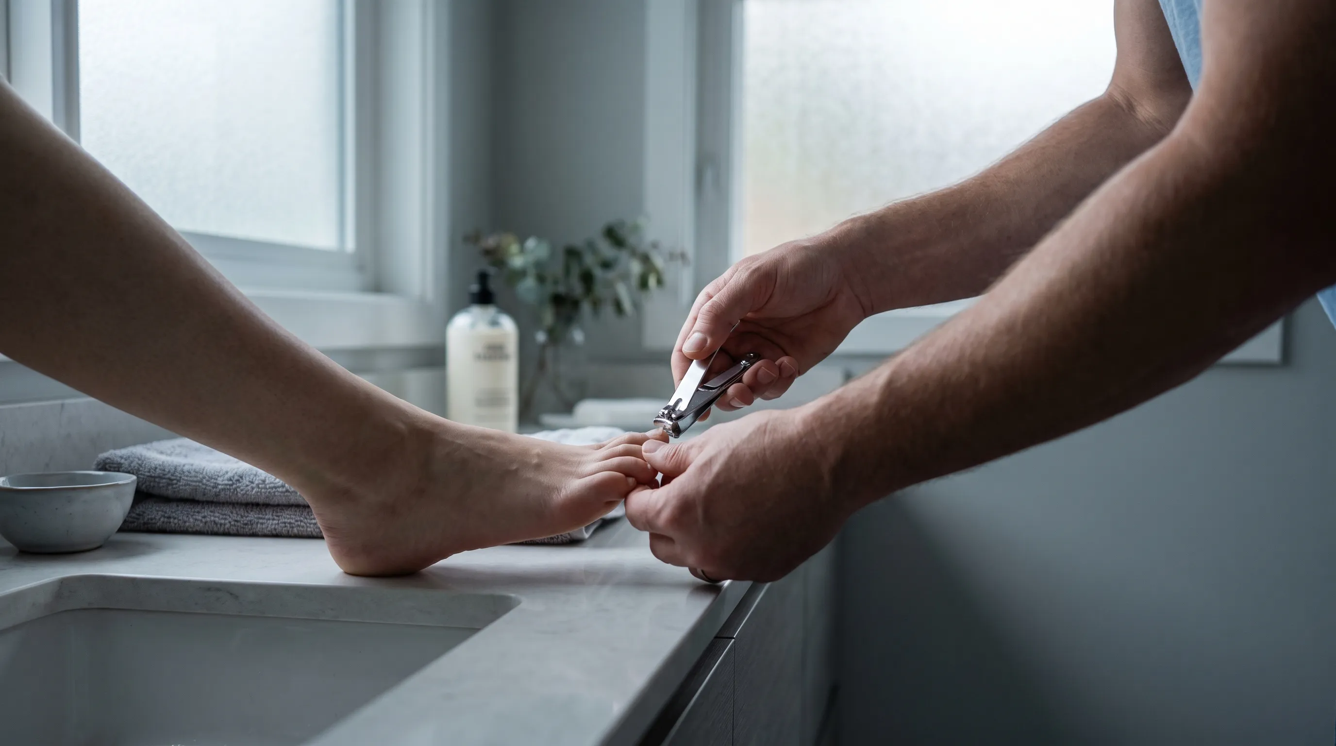
Shoes must provide adequate toe box width, arch support, and moisture-wicking properties to minimize repetitive friction. Athletic footwear should be replaced every three to five hundred miles as cushioning degrades. Toenails should be trimmed straight across using clean, sterilized clippers to prevent ingrown edges and microtrauma. Filing sharp corners eliminates pressure points that cause subungual bleeding. Avoid sharing pedicure tools and disinfect home foot spas regularly to prevent fungal and bacterial transmission. Applying a light, non-comedogenic moisturizer to cuticles maintains tissue elasticity without creating occlusive environments that promote microbial overgrowth. For comprehensive hygiene standards, refer to WHO foot care guidelines.
Sports Safety and Activity Modification
Runners and hikers should lace shoes securely to prevent forward toe slippage during downhill descents. Moisture-wicking double-layer socks reduce friction and blister formation. Gradually increasing mileage allows tissues to adapt to mechanical stress. Cross-training minimizes repetitive impact on specific digits. If a black dot under toenail appears after intense activity, temporarily reducing load-bearing exercises and switching to low-impact alternatives like swimming or cycling allows natural healing without compromising cardiovascular fitness.
Routine Monitoring and Early Intervention
Monthly self-examinations enable early detection of pigment changes, texture alterations, or structural abnormalities. Use natural lighting and a magnifying lens for precise inspection. Document findings with dated photographs to track progression. Schedule annual podiatric or dermatological screenings if you have a history of trauma, immunosuppression, diabetes, or family melanoma risk. Prompt evaluation of atypical lesions prevents unnecessary anxiety and ensures timely, appropriate management. Consistent foot care routines integrate seamlessly into overall wellness protocols, supporting mobility and independence throughout life. The CDC emphasizes that routine monitoring is critical for preventing secondary complications in active populations.
Frequently Asked Questions
What causes a sudden black dot under toenail?
A sudden black dot under toenail is most frequently the result of minor trauma that ruptures capillaries in the nail bed, leading to a subungual hematoma. Repetitive friction from tight footwear, stubbing injuries, or high-impact sports commonly trigger this response. Less frequently, it may indicate early fungal colonization, a benign mole, or, rarely, a malignant transformation. Tracking symptom onset and recent activity helps narrow the underlying cause.
Can a black dot under toenail go away on its own?
Yes, when caused by trauma, the pigment will naturally grow out as the nail plate advances. This process typically takes six to twelve months, depending on individual growth rates and nutritional status. Fungal-related spots may persist or worsen without targeted antifungal therapy. Any lesion that remains stationary, expands, or alters appearance should be professionally evaluated rather than ignored.
How can I tell if a black dot under toenail is serious?
Serious lesions exhibit irregular borders, multiple shades of brown or black, rapid growth, nail plate destruction, or pigment extension into the surrounding skin (Hutchinson sign). Persistent pain without trauma, bleeding, or failure to migrate upward are additional red flags. Benign spots generally maintain uniform color and smooth margins while progressing distally over time.
Should I try to drain or pop a black dot under toenail at home?
Absolutely not. Home drainage introduces high infection risk, can permanently damage the nail matrix, and often worsens pain or scarring. If pressure becomes unbearable or the hematoma covers a large portion of the nail, a licensed podiatrist can safely perform sterile trephination in a controlled clinical environment, ensuring proper aftercare and preventing complications.
Does a black dot under toenail always mean cancer?
No. The overwhelming majority of black dots are benign and stem from trauma, fungal activity, or harmless nevi. Subungual melanoma represents a small percentage of cases but requires careful monitoring and professional assessment when atypical features emerge. Early dermatological consultation provides definitive answers and peace of mind without unnecessary alarm.
Conclusion
A black dot under toenail is a common clinical presentation that spans a wide spectrum of causes, from benign trauma to rare malignancy. By understanding nail anatomy, recognizing distinguishing features, and applying evidence-based diagnostic criteria, patients and clinicians can effectively differentiate between self-resolving conditions and those requiring targeted intervention. Conservative observation, proper wound hygiene, and timely medical consultation form the cornerstone of optimal management. Implementing preventive foot care strategies, selecting appropriate footwear, and maintaining regular self-examination routines significantly reduce recurrence risk and promote long-term lower extremity health. When in doubt, seeking evaluation from a board-certified podiatrist or dermatologist ensures accurate diagnosis and personalized treatment planning. Prioritizing proactive foot wellness not only resolves immediate concerns but also safeguards mobility and overall quality of life for years to come.
How we work
Researched and drafted with AI assistance, then reviewed by a human editor against the cited sources.
This article is general health information, not medical advice. Consult a qualified healthcare professional about your own situation.