Sagging Breasts: Causes, Evidence-Based Solutions, and Long-Term Prevention

Understanding the natural evolution of breast shape over time is essential for developing realistic expectations, maintaining body confidence, and making informed healthcare decisions. While cultural narratives often frame changes in breast firmness as purely cosmetic concerns, the underlying physiological processes involve complex interactions between hormonal fluctuations, connective tissue integrity, and biomechanical stressors. For millions of individuals navigating these shifts, sagging breasts represent a common yet highly individualized experience influenced by genetics, lifestyle habits, reproductive history, and aging. By exploring the anatomical foundations, identifying modifiable risk factors, and evaluating both conservative and medical interventions, you can build a sustainable approach to breast wellness. This guide synthesizes current clinical evidence, dermatological research, and surgical best practices to deliver comprehensive, actionable strategies that prioritize health, functionality, and self-assurance at every stage of life.
Understanding Breast Ptosis: The Science Behind Sagging Breasts
Breast Anatomy and the Cooper’s Ligaments
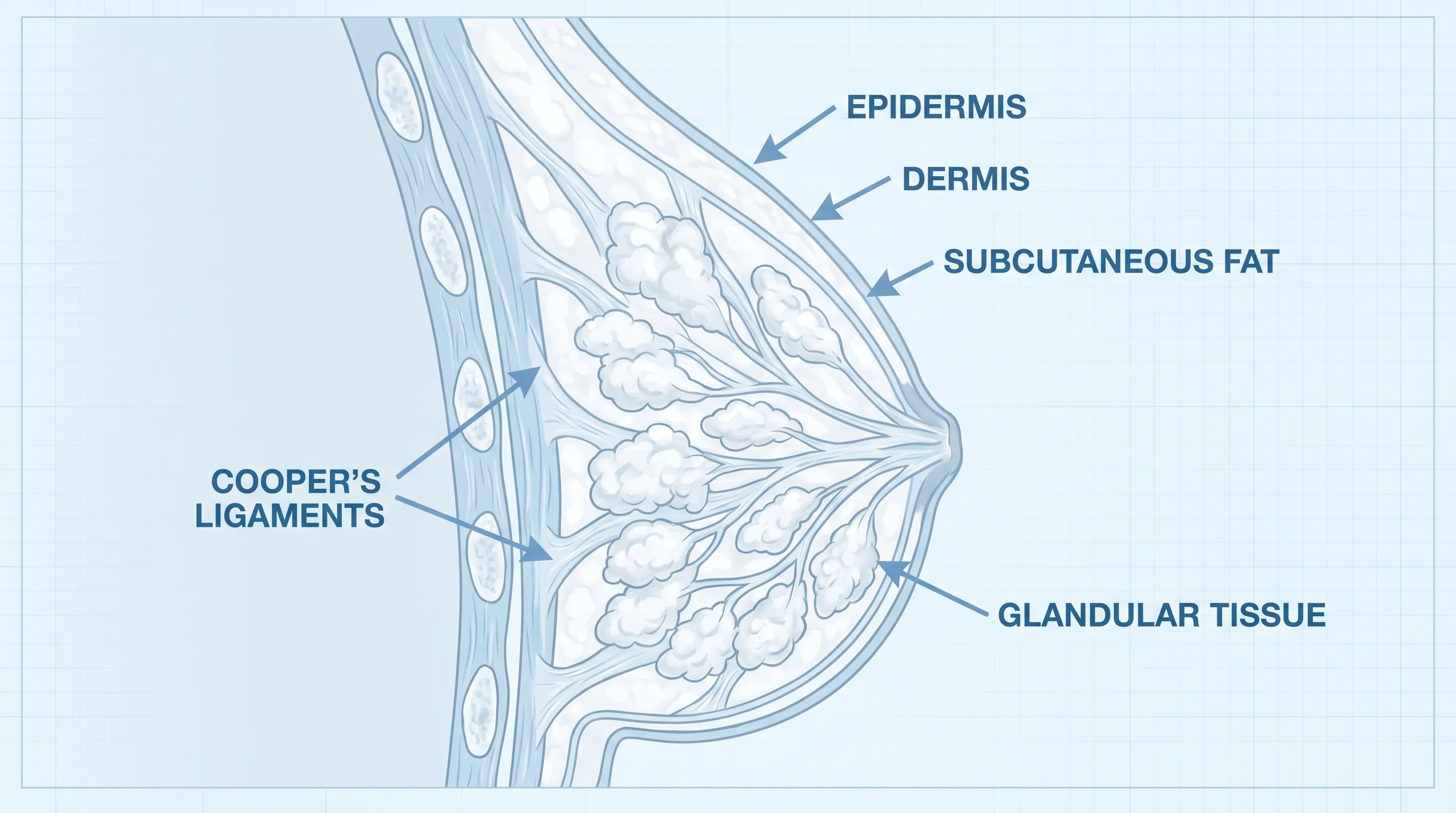
Breast tissue is fundamentally composed of glandular lobules, adipose tissue, fibrous connective structures, and an extensive network of blood vessels and nerves. What determines breast shape and positional stability is not primarily muscle, as breast tissue contains no intrinsic contractile fibers, but rather the architectural support provided by the suspensory ligaments of Cooper. These delicate, web-like connective bands originate from the deep fascia and extend through the breast parenchyma to attach to the dermis, functioning as a natural internal scaffolding system. Over time, mechanical tension, hormonal shifts, and environmental stressors gradually degrade collagen and elastin fibers within these ligaments. As the structural matrix weakens, gravitational forces exert continuous downward pull, resulting in tissue descent and skin redundancy.
Free calculatorWater Intake CalculatorCalculate daily water needsOpen the calculatorThe dermal layer itself undergoes parallel degenerative changes. Aging reduces fibroblast activity, slowing collagen synthesis by approximately one percent annually after the mid-twenties, as documented in NIH research on skin aging. Ultraviolet exposure accelerates this decline through photoaging pathways that generate matrix metalloproteinases, enzymes that break down extracellular matrix proteins. Consequently, skin loses tensile strength and recoil capacity, further contributing to the visual presentation of sagging breasts. Recognizing that ptosis originates from internal connective tissue changes rather than superficial skin laxity alone clarifies why topical applications have limited structural impact and why comprehensive approaches targeting both tissue support and skin health yield the most meaningful outcomes.
The Grading Scale of Ptosis
Clinicians utilize the Regnault classification system to objectively measure breast ptosis and guide treatment planning. This standardized framework evaluates the positional relationship between the nipple-areolar complex and the inframammary fold. Grade 0 describes a breast with the nipple positioned at or above the fold, representing optimal anatomical positioning. Grade I, or mild ptosis, indicates the nipple sits at the level of the fold while the lower breast tissue descends slightly below it. Grade II involves the nipple falling one to three centimeters beneath the fold, with noticeable lower pole elongation and reduced upper fullness. Grade III, or severe ptosis, occurs when the nipple rests more than three centimeters below the fold and points downward, often accompanied by significant skin stretching, tissue deflation, and altered chest wall contour.
Accurate grading enables healthcare providers to match patients with appropriate interventions. Conservative measures typically yield meaningful improvements for Grade 0 and I presentations, particularly when combined with lifestyle optimization. Grade II often benefits from a combination of non-invasive modalities and structured physical therapy, though surgical consultation becomes increasingly relevant as tissue descent progresses. Grade III ptosis generally requires anatomical reconstruction through mastopexy to restore functional positioning, alleviate dermatological complications such as intertrigo or skin crease irritation, and reestablish proportional chest aesthetics. Understanding this spectrum empowers individuals to set realistic goals and pursue interventions aligned with their specific anatomical profile.
Primary Causes and Contributing Factors
Aging and Hormonal Changes
The most pervasive driver of breast tissue remodeling is chronological aging, which intersects with profound endocrine transitions. During perimenopause and menopause, ovarian estrogen production declines significantly, directly impacting tissue hydration, collagen density, and glandular volume. Estrogen receptors densely populate breast stromal cells, and reduced signaling initiates apoptotic processes within fibroglandular elements. This involution replaces functional tissue with adipose depots that lack the structural rigidity of glandular networks, leading to softer consistency and reduced support. Simultaneously, progesterone fluctuations during menstrual cycles historically stimulate lobular proliferation and temporary swelling; as cycles become irregular, the tissue experiences inconsistent expansion and contraction, accelerating ligament fatigue.
Research, supported by Mayo Clinic guidelines on menopause and hormonal shifts, confirms that prolonged hypoestrogenic states correlate with measurable decreases in dermal thickness and microvascular density within breast tissue. This physiological reality underscores why individuals may notice accelerated changes in firmness during their late thirties through fifties. While chronological aging cannot be reversed, targeted interventions focusing on hormone-informed nutrition, consistent physical activity, and evidence-based dermatological protocols can significantly slow structural degradation and preserve tissue resilience.
Pregnancy, Breastfeeding, and Weight Fluctuations
Pregnancy induces dramatic breast transformation as progesterone and prolactin drive alveolar development and ductal expansion. The tissue can increase by one to three cup sizes, stretching the skin envelope and loading the Cooper’s ligaments with unprecedented mechanical stress. Following delivery, whether or not breastfeeding occurs, the glandular apparatus undergoes involution, shrinking back toward baseline dimensions. However, the stretched dermal matrix rarely returns to its original configuration, leaving behind excess skin and diminished upper pole volume.
Extensive epidemiological studies, including research highlighted by the Cleveland Clinic on breast changes during pregnancy, demonstrate that breastfeeding itself does not independently cause long-term ptosis. Instead, the primary determinants are the number of pregnancies, maternal age at conception, pre-pregnancy body mass index, and smoking status. Rapid postpartum weight loss further exacerbates tissue laxity by creating a mismatch between skin surface area and underlying volume. Gradual, nutritionally supported weight management during and after pregnancy, combined with progressive strength training, significantly mitigates structural compromise. Wearing appropriately fitted support garments during pregnancy and nursing also reduces mechanical strain on suspensory structures.
Genetics and Skin Elasticity
Hereditary factors establish the baseline architecture and biochemical composition of connective tissue. Variations in genes regulating collagen cross-linking, elastin fiber organization, and matrix protein turnover dictate inherent tissue resilience. Individuals with familial histories of premature skin laxity, joint hypermobility, or connective tissue disorders often experience earlier or more pronounced breast descent. Additionally, melanin concentration influences photoaging susceptibility, with lower baseline protection increasing susceptibility to UV-mediated elastosis in sun-exposed chest areas.
While genetic predisposition cannot be altered, understanding familial patterns enables proactive management. Early adoption of protective skincare, consistent postural alignment training, and avoidance of known tissue-degrading behaviors (such as smoking or extreme dieting) can offset hereditary vulnerabilities. Genetic counseling is rarely necessary for isolated ptosis, but individuals presenting with systemic symptoms like widespread skin hyperextensibility or joint instability should consult rheumatology or genetics specialists to rule out syndromic connective tissue conditions.
Lifestyle Habits: Smoking, Sun Exposure, and Bra Usage
Cigarette smoking represents one of the most preventable accelerators of breast tissue degeneration. Nicotine induces vasoconstriction, reducing blood flow to the dermis by up to forty percent and depriving fibroblasts of oxygen and essential nutrients required for matrix synthesis. Carbon monoxide further displaces oxygen at the hemoglobin level, compounding tissue hypoxia. The resulting environment favors collagen fragmentation, elastin clumping, and accelerated cellular senescence. CDC research on tobacco and premature aging consistently links heavy smoking to earlier onset of Grade II and III ptosis, independent of age or weight.
Chronic ultraviolet exposure degrades superficial connective networks through reactive oxygen species generation and inflammatory cytokine release. The décolleté receives disproportionate sun exposure compared to other covered body regions, making daily broad-spectrum SPF application non-negotiable for long-term skin preservation. WHO guidelines on ultraviolet radiation emphasize the cumulative impact of unshielded UV exposure on dermal integrity. Regarding undergarment usage, clinical evidence does not support the notion that wearing bras prevents ptosis; however, properly fitted support garments significantly reduce dynamic tissue movement during exercise, decreasing ligament fatigue and improving comfort. Conversely, consistently wearing ill-fitting, non-supportive bras during high-impact activities can contribute to cumulative microtrauma over years.
Non-Surgical Approaches and Lifestyle Modifications
Targeted Exercise and Posture Correction
While breast tissue itself cannot be toned through exercise, strengthening the underlying pectoralis major and minor, trapezius, rhomboids, and serratus anterior muscles creates a firmer anatomical foundation that visually lifts and stabilizes the chest contour. Progressive resistance training focusing on chest presses, push-up variations, dumbbell flyes, and resistance band rows improves muscular hypertrophy and neuromuscular recruitment. NIH recommendations for strength training highlight that a structured three-day weekly protocol incorporating compound movements and controlled eccentrics yields measurable improvements in posture, upper pole definition, and shoulder blade alignment within eight to twelve weeks.
Postural correction addresses a frequently overlooked contributor to breast ptosis. Anterior pelvic tilt, thoracic kyphosis, and forward head posture shift the center of gravity, increasing gravitational pull on the anterior chest wall and stretching anterior fascial chains. Implementing daily mobility routines that open the pectorals, strengthen the mid-back extensors, and activate the deep core stabilizers realigns the spine, reduces tissue strain, and enhances breathing mechanics. Integrating posture-aware habits during desk work, phone usage, and sleep positioning amplifies exercise benefits and supports sustainable tissue health.
Supportive Underwear and Bra Fitting
Proper bra selection serves as a mechanical intervention rather than a preventive miracle. Studies indicate that approximately eighty percent of individuals wear incorrect bra sizes, leading to inadequate lift, shoulder groove formation, and tissue migration toward the axillae. The Cleveland Clinic's bra fitting guidelines emphasize that professional fittings assess underband tension, cup volume, wire width, and strap distribution to optimize load-bearing efficiency. During physical activity, high-impact sports bras with encapsulated cups minimize oscillation by up to fifty-four percent, reducing microtrauma to suspensory structures.
Beyond exercise, strategic undergarment choices align with daily activities and physiological states. Seamless, breathable fabrics reduce friction and moisture retention, preventing dermatological irritation. For those experiencing significant volume changes postpartum or post-weight loss, adjustable multi-hook closures and adaptive materials accommodate ongoing tissue remodeling. Rotating between supportive daily wear and relaxed styles during rest periods ensures continuous circulation while maintaining structural guidance. Understanding that bras manage dynamics rather than reverse anatomy enables realistic expectations and informed purchasing decisions.
Nutrition, Hydration, and Skin Health
Dermal resilience depends on systemic nutritional status. Adequate protein intake provides amino acids necessary for collagen peptide synthesis, with studies recommending one to one point two grams per kilogram of body weight for optimal tissue repair. NIH Office of Dietary Supplements data notes that vitamin C acts as a mandatory cofactor for proline and lysine hydroxylation, essential cross-linking reactions that stabilize the collagen triple helix. Zinc facilitates matrix metalloproteinase regulation and cellular proliferation, while vitamin E and omega-3 fatty acids mitigate lipid peroxidation and inflammatory degradation.
Hydration directly influences dermal turgor and interstitial fluid balance. Chronic mild dehydration impairs keratinocyte function, reduces glycosaminoglycan synthesis, and compromises skin barrier integrity, making tissues more susceptible to creasing and sagging. Incorporating collagen-supportive foods such as bone broth, citrus fruits, leafy greens, nuts, seeds, and fatty fish establishes a biochemical environment conducive to connective tissue maintenance. Topical retinoids and peptide serums complement systemic nutrition by stimulating epidermal turnover and fibroblast activity, though results require consistent application over several months to manifest visible improvements.
Evidence-Based Treatments and Medical Interventions
Topical Creams and Skincare
The market abounds with firming lotions promising rapid reversal of sagging breasts, yet clinical evidence demonstrates that topical agents primarily influence epidermal and superficial dermal layers rather than deep structural networks. Ingredients such as retinol, niacinamide, peptides, and ceramides improve skin hydration, reduce fine lines, and enhance barrier function, creating a temporarily tighter appearance. However, the Mayo Clinic notes that no topical formulation can rebuild stretched Cooper’s ligaments or restore lost glandular volume.
For individuals with Grade 0 or I ptosis, consistent application of regimens can optimize skin quality, improve radiance, and slow photoaging progression. Products containing stabilized vitamin C, broad-spectrum SPF, and moisturizing humectants like hyaluronic acid deliver measurable benefits when paired with gentle massage techniques that promote localized circulation. Managing expectations is crucial; while skincare supports overall tissue health, it functions as an adjunct rather than a standalone solution for structural ptosis.
Non-Invasive Procedures: Radiofrequency and Ultrasound
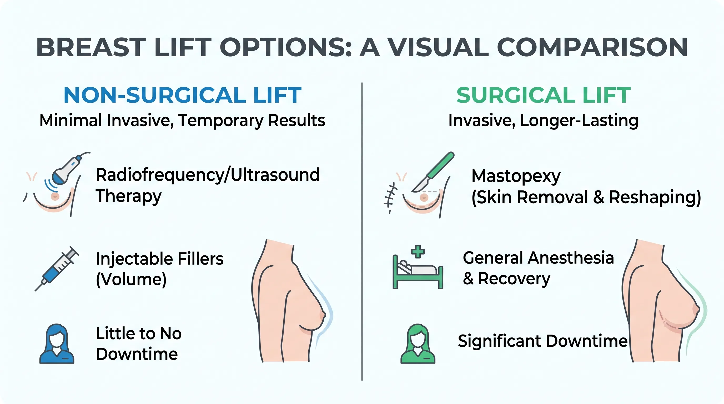
Technological advancements have expanded non-surgical options for mild to moderate tissue laxity. Monopolar and bipolar radiofrequency devices deliver controlled thermal energy to the deep dermis, triggering neocollagenesis and dermal remodeling over multiple treatment sessions. Micro-focused ultrasound platforms such as Ultherapy penetrate deeper into the superficial muscular aponeurotic system, stimulating collagen reorganization and producing gradual tightening over three to six months. Clinical trials, reviewed in Cleveland Clinic evaluations of radiofrequency skin tightening, report average satisfaction scores of seventy to eighty percent for early-stage ptosis, with effects typically lasting twelve to twenty-four months depending on lifestyle factors.
These modalities require realistic patient selection, optimal skin elasticity, and commitment to maintenance protocols. Combining energy-based devices with targeted nutrition, exercise, and skincare amplifies outcomes and prolongs longevity. While not equivalent to surgical tissue repositioning, non-invasive treatments provide a valuable middle ground for individuals seeking gradual improvement without operative recovery.
Surgical Mastopexy: What to Expect
Mastopexy, commonly referred to as a breast lift, directly addresses anatomical descent by excising excess skin, reshaping the parenchymal mound, and repositioning the nipple-areolar complex to a youthful projection. Surgical techniques vary based on severity: the periareolar (donut) method suits mild cases with minimal skin excess; the vertical (lollipop) approach corrects moderate ptosis with efficient scar placement; and the inverted-T (anchor) incision manages severe laxity with comprehensive tissue redistribution. Mayo Clinic surgical overviews detail that the procedure requires general anesthesia, typically lasting two to four hours, with recovery spanning four to six weeks.
Patients follow structured postoperative protocols including compression garments, activity restrictions, and scar management strategies such as silicone sheeting and massage. Complication rates remain low in board-certified hands, though risks include altered nipple sensation, delayed wound healing, asymmetry, and rare implant-related issues if augmentation is combined. Comprehensive preoperative counseling ensures patients understand realistic outcomes, maintenance requirements, and long-term care expectations.
Breast Implants vs. Natural Lifting
Choosing between volume restoration and native tissue repositioning depends on individual anatomy, aesthetic goals, and physiological history. Individuals experiencing significant glandular involution but maintaining adequate skin elasticity may benefit from implants alone, as restored volume often naturally lifts the tissue envelope. Conversely, those with pronounced skin laxity and inferior nipple positioning require mastopexy regardless of volume considerations. Augmentation-mastopexy combines both approaches, addressing dual deficits simultaneously.
Implant selection involves evaluating shape, projection, surface texture, and placement relative to the pectoral muscle. FDA safety and monitoring guidelines for breast implants emphasize that autologous fat grafting offers a natural alternative for mild volume deficits, utilizing purified adipose tissue harvested from other body regions. Both approaches demand careful surgical planning, with long-term monitoring recommended for implant integrity or fat retention. Open dialogue with a certified plastic surgeon ensures alignment between anatomical reality and aesthetic aspirations.
Psychological Impact and Body Image Considerations
Navigating Emotional Well-Being
Breast morphology intersects deeply with personal identity, cultural messaging, and self-perception. Media narratives frequently emphasize static, youthful aesthetics as the standard of attractiveness, fostering unnecessary anxiety around natural aging processes. For many, sagging breasts trigger feelings of diminished femininity, reduced confidence, or discomfort during social and intimate interactions. Acknowledging these emotional responses validates individual experiences while separating physiological reality from socially constructed ideals.
Cognitive restructuring, body neutrality practices, and community engagement significantly improve psychological resilience. Shifting focus from appearance-based validation to functional appreciation—recognizing breasts as organs capable of nourishment, sensory experience, and bodily integrity—cultivates sustainable self-acceptance. Integrating mindfulness techniques, limiting exposure to unrealistic imagery, and celebrating bodily autonomy reinforce mental wellness alongside physical care.
When to Seek Professional Counseling
Persistent distress, avoidance of intimacy, clothing restrictions, or depressive symptoms related to breast changes warrant professional intervention. NIH resources on body image and mental health recommend licensed therapists specializing in body image, somatic experiencing, or cognitive behavioral therapy to provide evidence-based frameworks for processing aesthetic concerns without pathologizing normal anatomy. Support groups, both in-person and virtual, offer shared narratives that normalize transitions and reduce isolation. When emotional distress impairs daily functioning or coexists with body dysmorphic tendencies, psychiatric evaluation ensures comprehensive, compassionate care tailored to individual needs.
| Treatment Modality | Best Suited For | Recovery Time | Longevity of Results | Invasiveness Level |
|---|---|---|---|---|
| Targeted Exercise & Posture Training | Grades 0-I, early laxity, muscle weakness | None | Sustained with consistency | Non-invasive |
| Radiofrequency & Ultrasound | Mild-moderate ptosis, fair skin elasticity | Minimal (1-3 days) | 12-24 months | Non-invasive |
| Mastopexy (Breast Lift) | Grades II-III, significant skin excess | 4-6 weeks | Long-term (10+ years) | Surgical |
| Augmentation-Mastopexy | Volume loss combined with tissue descent | 4-8 weeks | Long-term, implant monitoring required | Surgical |
| Fat Grafting | Mild volume deficit, natural tissue preference | 1-2 weeks | Partial retention (50-70%) | Minimally invasive |
Frequently Asked Questions
Can sagging breasts be fully reversed without surgery?
Non-surgical methods such as targeted strength training, posture improvement, optimized nutrition, and skin-care protocols can significantly improve firmness and appearance, especially in mild to moderate cases. However, severe ligament stretching and significant skin laxity typically require surgical intervention like mastopexy for a complete anatomical lift. Consulting a board-certified plastic surgeon or dermatologist can help determine the most appropriate approach based on individual tissue characteristics and goals.
Does breastfeeding actually cause breasts to sag?
Research consistently shows that breastfeeding itself does not cause long-term breast ptosis. The structural changes are primarily driven by the physiological expansion during pregnancy, followed by tissue involution postpartum, rather than the act of nursing. Maintaining gradual weight management, wearing supportive nursing bras, and practicing chest-strengthening exercises can help preserve tissue integrity during and after pregnancy.
How do I know if I need a surgical breast lift?
Candidates for mastopexy typically present with Grade II or III ptosis, where the nipple falls significantly below the inframammary fold and the majority of breast tissue sits lower on the chest wall. Additional indicators include stretched skin, loss of upper pole fullness, and persistent discomfort despite conservative measures. A thorough evaluation with a qualified surgeon will assess skin elasticity, tissue volume, and overall health to determine surgical candidacy.
Are there specific vitamins that improve breast skin elasticity?
Collagen synthesis and dermal resilience rely heavily on vitamin C, vitamin E, zinc, and adequate protein intake. Omega-3 fatty acids support cell membrane integrity and reduce inflammatory degradation of elastin fibers. While topical and dietary nutrients cannot reverse severe ligamentous laxity, consistent supplementation combined with a balanced diet supports overall skin quality, hydration, and tissue repair capacity over time.
Is it safe to combine a breast lift with implants?
Yes, augmentation-mastopexy is one of the most frequently performed combined procedures in aesthetic surgery. When executed by an experienced, board-certified plastic surgeon, combining tissue reshaping with volume restoration yields superior contour and long-term symmetry. The procedure requires careful preoperative planning to account for blood supply preservation, skin envelope dynamics, and implant positioning to minimize complications and optimize healing.
Conclusion
Navigating changes in breast morphology is a deeply personal journey that intersects anatomy, lifestyle, and self-perception. Understanding that sagging breasts result from natural connective tissue remodeling, hormonal shifts, and environmental stressors removes stigma and empowers proactive care. Whether pursuing conservative optimization through exercise and nutrition, exploring non-invasive technologies, or considering surgical restoration, informed decision-making remains the cornerstone of sustainable wellness. Prioritizing evidence-based interventions, maintaining realistic expectations, and embracing bodily evolution fosters lasting confidence. Always consult qualified healthcare professionals for personalized assessments, and remember that health, functionality, and self-acceptance form the true foundation of long-term well-being.

How we work
Researched and drafted with AI assistance, then reviewed by a human editor against the cited sources.
This article is general health information, not medical advice. Consult a qualified healthcare professional about your own situation.