Dissolvable Stitches Sticking Out: Causes, Care, and When to Worry
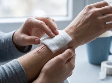
Discovering that your surgical closure has not seamlessly blended into your healing skin can be surprisingly unsettling. Many patients anticipate a clean, invisible transition as their incision knits together, only to find thin thread ends or knots visibly protruding from the wound margin. While this can initially trigger anxiety about infection or surgical complications, the reality is far more reassuring. Dissolvable sutures are engineered to break down gradually, but their absorption rate is heavily influenced by individual biology, wound location, suture material composition, and postoperative care routines. Understanding the physiological mechanisms behind why dissolvable stitches sticking out is actually a common occurrence will empower you to manage your recovery confidently. Instead of rushing to remove them or ignoring warning signs, you can follow evidence-based wound care protocols that support optimal tissue regeneration. This comprehensive guide explores exactly what happens beneath the surface during suture degradation, outlines safe home management strategies, highlights critical red flags, and provides actionable steps to minimize scarring while ensuring your body heals exactly as it should. Whether you are recovering from a minor dermatological procedure or a major abdominal surgery, navigating the dissolution phase with proper knowledge transforms uncertainty into controlled, informed recovery.
What Are Dissolvable Stitches and How Do They Work?
Dissolvable stitches, medically referred to as absorbable sutures, are specialized surgical threads designed to hold wound edges together until the underlying tissue regains sufficient tensile strength to remain closed independently. Unlike traditional non-absorbable materials that require clinical removal, absorbable sutures undergo a controlled breakdown process within the human body. The degradation mechanism primarily occurs through hydrolysis or enzymatic digestion, depending on the polymer used. Synthetic absorbable sutures like polyglycolic acid, polydioxanone, and polylactic acid break down via hydrolysis, a chemical reaction where water molecules penetrate the polymer chain and gradually cleave it into smaller, biocompatible fragments that the body can safely metabolize and excrete. Natural absorbable materials, such as plain or chromic catgut, degrade through phagocytic cellular activity, where immune cells enzymatically break down the collagen-based material.
Free calculatorWater Intake CalculatorCalculate daily water needsOpen the calculatorThe Science Behind Absorbable Sutures
The healing process dictates the necessity and timeline of suture absorption. When a surgical incision occurs, the body immediately initiates a complex, four-phase cascade: hemostasis, inflammation, proliferation, and remodeling. During the proliferation phase, fibroblasts migrate into the wound bed and synthesize new collagen fibers to bridge the gap between tissue edges. Dissolvable stitches provide mechanical support precisely during this vulnerable window, preventing wound dehiscence while allowing natural tissue regeneration. As collagen cross-linking matures and reaches approximately 20 to 30 percent of original tissue strength, the body no longer requires external reinforcement. This is precisely when hydrolysis or enzymatic breakdown accelerates, allowing the suture to lose structural integrity safely without compromising the wound.
The rate of absorption is meticulously engineered by medical manufacturers. Sutures intended for fast-healing mucosal tissues, like those inside the mouth or vaginal walls, are designed to dissolve within 7 to 14 days. Sutures used for fascial closure in abdominal surgeries or orthopedic applications must maintain tensile strength for weeks to months, with complete absorption occurring over 90 to 180 days. This material science ensures that the mechanical support timeline perfectly matches the biological healing timeline of the specific anatomical region.
Common Types and Absorption Timelines
Not all absorbable sutures behave identically. Polyglactin 910 (Vicryl) retains significant tensile strength for up to 21 days and typically absorbs completely within 56 to 70 days. Polydioxanone (PDS) is a monofilament suture prized for prolonged strength retention, maintaining integrity for up to 42 days and fully absorbing between 91 and 180 days. Poliglecaprone 25 (Monocryl) offers excellent handling characteristics but degrades faster, losing most strength by day 14 and absorbing entirely within 28 to 42 days. Chromic catgut, derived from bovine or ovine intestinal submucosa, undergoes rapid enzymatic breakdown and usually disappears within 9 to 14 days. Understanding which material your surgeon selected provides crucial context for managing dissolvable stitches sticking out and sets realistic expectations for the dissolution phase.
Why Are My Dissolvable Stitches Sticking Out?
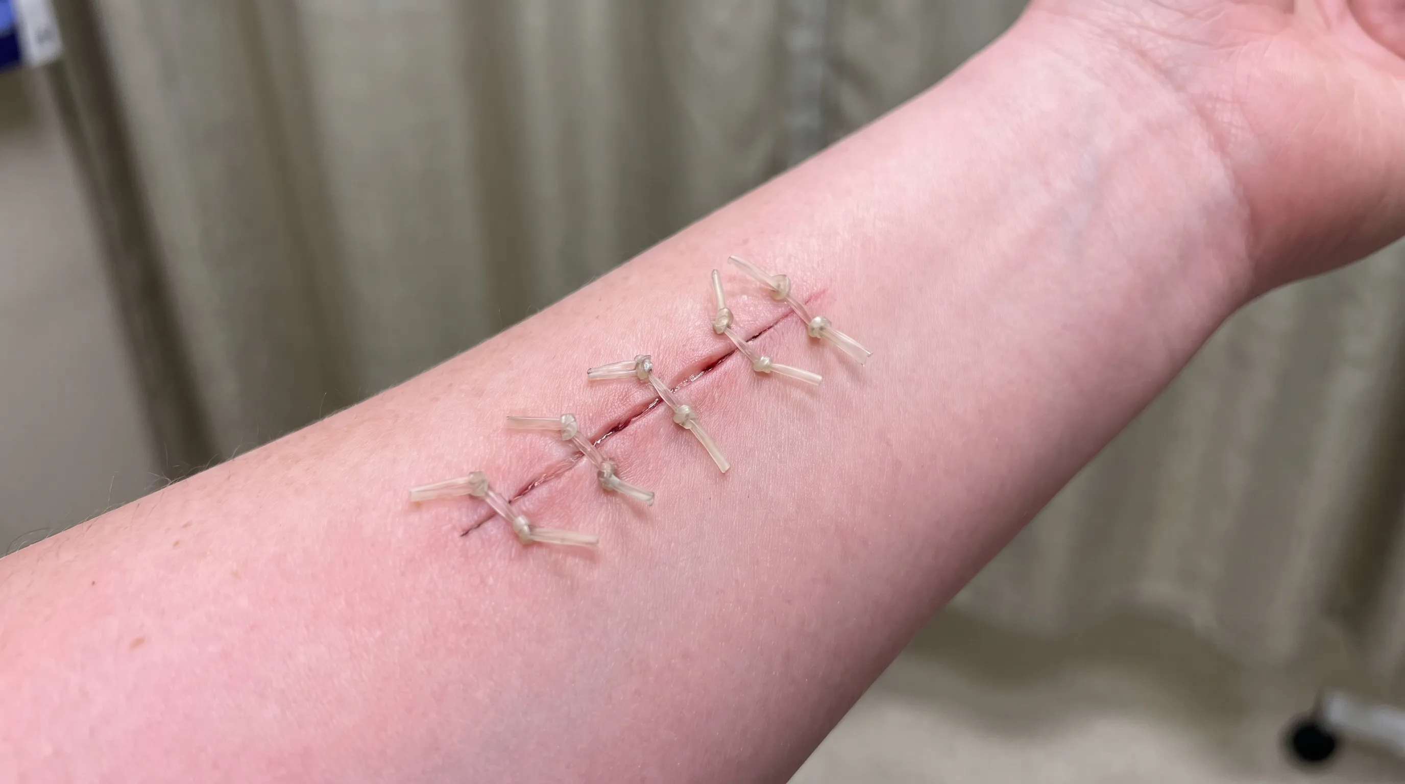
Encountering dissolvable stitches sticking out from a healing wound is surprisingly common and rarely indicates surgical error or impending complications. The phenomenon typically stems from normal physiological variations, localized tissue dynamics, or the physical properties of the suture material itself. As the underlying tissue regenerates and swells slightly during the inflammatory and proliferative phases, the wound margin can subtly push previously buried suture loops or knots toward the epidermal surface. Additionally, as the suture begins to hydrolyze, the material often becomes brittle, shrinks, or stiffens, causing what was once flush with the skin to gradually protrude.
Normal Healing Variations
Human biology does not follow a uniform template. Individual differences in skin thickness, subcutaneous fat distribution, metabolic rate, and local blood flow significantly influence how quickly and smoothly sutures integrate and dissolve. Areas with high tension or frequent movement, such as joints or the abdominal wall, experience constant micro-stresses that can cause suture ends to migrate upward as the tissue remodels. Furthermore, natural skin exfoliation and epidermal turnover gradually expose deeper layers, which can bring previously subdermal knot loops closer to the surface. In most cases, this outward migration is a completely benign part of the healing trajectory.
Signs of Delayed Absorption or Improper Placement
While minor protrusion is expected, certain patterns warrant closer attention. If the protruding material remains rigid, causes localized irritation, or fails to soften over several weeks, it may indicate delayed absorption. This can occur in patients with compromised microcirculation, uncontrolled diabetes, chronic inflammation, or specific nutritional deficiencies that slow hydrolytic breakdown. Occasionally, a surgeon may intentionally tie a suture knot slightly more superficially to prevent deep tissue irritation, which naturally results in a visible or palpable end as healing progresses. If the area feels excessively tight, or if the suture appears to be "spitting" through a tiny pinpoint opening surrounded by a small inflammatory bump, you may be experiencing a foreign body reaction where the immune system actively expels material it cannot rapidly metabolize.
When Spitting Out Sutures Is Expected
The term "spitting sutures" describes a benign but slightly uncomfortable phenomenon where the body pushes a dissolvable suture to the surface through a small opening. This typically happens when a deeper stitch begins to dissolve faster than the overlying tissue has fully sealed, or when the immune system recognizes the material as a persistent foreign entity and initiates localized inflammatory expulsion. You may notice a small red bump, a tiny pimple-like opening, or a visible thread end emerging. This is not an infection but rather a physiological clearance mechanism. Gentle cleansing and sterile gauze protection usually resolve the issue as the remaining fragment naturally detaches and falls away.
How to Safely Manage Dissolvable Stitches Sticking Out
Managing dissolvable stitches sticking out requires a delicate balance between patience, hygiene, and proactive wound observation. The golden rule is to avoid aggressive pulling, excessive scrubbing, or premature cutting. Every unnecessary touch introduces potential pathogens and risks disrupting fragile granulation tissue. Instead, adopt a structured, evidence-based care routine that prioritizes cleanliness, moisture balance, and tissue protection while allowing the body to complete its natural degradation process.
Cleaning and Protecting the Area
Proper cleansing prevents bacterial colonization around protruding suture material. Wash your hands thoroughly with antibacterial soap before touching the wound area or dressing. Gently cleanse the incision site using lukewarm water and a mild, fragrance-free cleanser. Avoid hydrogen peroxide, rubbing alcohol, or iodine solutions, as these harsh antiseptics can damage healthy regenerating cells, delay epithelialization, and paradoxically increase infection risk. Pat the area dry with a clean, lint-free towel using light dabbing motions rather than rubbing.
Once clean and dry, apply a thin layer of medical-grade ointment or petroleum jelly if recommended by your surgeon. This maintains a moist wound environment, which clinically accelerates healing by 50 percent compared to dry wound management. Cover the area with a sterile, breathable gauze pad or a hydrocolloid dressing, securing it with hypoallergenic medical tape. Change the dressing daily or whenever it becomes damp, soiled, or loose. Consistent dressing changes prevent maceration, reduce friction against clothing, and minimize the chance of the protruding stitch catching on fabrics.
What to Trim and What to Leave Alone
It is natural to feel tempted to cut dissolvable stitches sticking out for comfort or aesthetic reasons. If you must trim, only remove the portion that freely extends beyond the skin surface. Never pull on the thread to bring more material out, and never cut near or at the wound margin. Use sterile, medical-grade scissors cleaned with rubbing alcohol and allowed to air dry completely. Position the scissors parallel to the skin surface, snip only the dangling loop, and avoid touching the wound bed. If you feel resistance, if the thread is attached to a knot, or if trimming causes sharp pain, stop immediately and leave it alone. Professional evaluation is always safer than guesswork, especially when dealing with deep tissue sutures or complex surgical sites.
When to Seek Professional Medical Intervention
Home management has clear boundaries. Contact your surgeon or visit an urgent care clinic if the protruding suture is accompanied by spreading redness, warmth, purulent discharge, foul odor, fever, or escalating pain that does not respond to over-the-counter analgesics. Medical professionals can safely evaluate whether the stitch requires removal, assess for underlying suture abscesses, apply appropriate topical or oral antimicrobial therapy if needed, and reinforce wound edges if partial dehiscence has occurred. Do not attempt to extract deeply embedded knots or manipulate inflamed tissue at home, as this can convert a minor irritation into a significant complication.
Preventing Infection and Promoting Optimal Healing
The dissolution phase coincides with the body's transition from acute inflammation to mature tissue remodeling. Protecting this delicate window requires a comprehensive approach that extends beyond local wound care. Systemic health, nutritional status, hydration levels, and lifestyle habits profoundly influence how efficiently your immune system manages suture degradation and tissue regeneration.
Daily Wound Care Routine
Consistency is the cornerstone of effective wound management. Establish a predictable schedule for inspection, cleansing, and redressing. During each check, document changes in color, swelling, discharge consistency, and suture appearance using a simple journal or smartphone photographs. Early detection of adverse changes dramatically improves outcomes. Keep the wound elevated above heart level when resting, especially for extremity incisions, to reduce hydrostatic pressure and minimize edema. Avoid direct sunlight on healing tissue, as ultraviolet exposure can cause hyperpigmentation and delay collagen maturation, leaving the scarred area vulnerable to long-term cosmetic concerns.
Nutrition and Hydration for Tissue Repair
Your body requires specific building blocks to synthesize new collagen, form microvascular networks, and execute hydrolysis safely. Prioritize high-quality protein sources such as lean poultry, fish, eggs, legumes, and dairy, which supply essential amino acids like proline and lysine for collagen cross-linking. Incorporate vitamin C-rich foods like citrus fruits, bell peppers, and leafy greens to support enzymatic processes critical for tissue synthesis. Zinc, found in pumpkin seeds, beef, and whole grains, accelerates epithelial migration and immune cell function. Maintain hydration by consuming at least 64 ounces of water daily, as proper fluid balance ensures optimal blood viscosity, nutrient delivery, and waste removal from the wound microenvironment.
Lifestyle Adjustments During Recovery
Physical strain and environmental factors significantly impact healing velocity. Avoid heavy lifting, strenuous exercise, or repetitive movements that place direct tension on the incision for at least 4 to 6 weeks, or as specifically directed by your surgical team. Smoking and vaping introduce nicotine and carbon monoxide into the bloodstream, causing severe vasoconstriction that starves the wound bed of oxygen and essential nutrients, dramatically increasing the risk of delayed suture absorption and tissue necrosis. Similarly, excessive alcohol consumption impairs neutrophil function and disrupts sleep architecture, both of which are vital for tissue repair. Prioritize restorative sleep, manage stress through gentle breathing exercises or meditation, and maintain a stable, comfortable environment to allow your physiological systems to dedicate maximum resources to healing.
Understanding Suture Materials and Timelines
Not all absorbable sutures share identical degradation profiles, absorption rates, or clinical applications. Understanding the specific material used in your procedure provides essential context for interpreting why dissolvable stitches sticking out may occur and how long the process should reasonably take. The table below outlines the most common absorbable suture types, their primary composition, retention of tensile strength, complete absorption timelines, and typical clinical applications.
| Suture Type | Composition | Tensile Strength Retention | Complete Absorption Timeline | Common Applications |
|---|---|---|---|---|
| Polyglactin 910 (Vicryl) | Synthetic braided copolymer | 75% at 14 days, 50% at 21 days | 56 to 70 days | Soft tissue approximation, gastrointestinal surgery, subcuticular closure |
| Polydioxanone (PDS) | Synthetic monofilament polyester | 70% at 14 days, 50% at 28 days | 91 to 180 days | Fascial closure, abdominal wall reconstruction, pediatric cardiovascular surgery |
| Poliglecaprone 25 (Monocryl) | Synthetic monofilament glycolide | 60% at 10 days, negligible by day 21 | 28 to 42 days | Subcutaneous tissue closure, dermatological procedures, plastic surgery |
| Chromic Catgut | Chemically treated collagen | 50% at 7 to 10 days | 9 to 14 days | Mucosal surfaces, oral cavity procedures, gynecological applications |
| Polyglycolic Acid (Dexon) | Synthetic braided polymer | 65% at 14 days, negligible by day 28 | 60 to 90 days | Urinary tract surgery, skin closure in low-tension areas, general surgery |
This data demonstrates why your surgeon selected a specific thread based on anatomical location, tissue type, and expected healing velocity. When dissolvable stitches sticking out occurs, referencing your surgical documentation regarding suture material provides immediate clarity on whether the timeline aligns with expected physiological breakdown or warrants clinical reassessment.
When to See a Doctor: Red Flags You Should Never Ignore
While mild protrusion, occasional itching, and gradual softening of suture ends fall within normal recovery parameters, certain clinical indicators demand immediate professional evaluation. The human immune system efficiently manages controlled inflammation, but overwhelming that defense system with bacterial pathogens, severe mechanical stress, or systemic compromise requires medical intervention. Recognizing the difference between expected healing discomfort and pathological warning signs prevents minor irritation from escalating into serious complications.
Recognizing Signs of Infection
Surgical site infections represent one of the most common postoperative complications. Monitor your incision for erythema that spreads outward beyond 2 centimeters from the wound margin, escalating warmth, swelling that worsens rather than gradually improves, and purulent exudate ranging from yellow to greenish in color. A foul, foul-smelling discharge is a definitive clinical indicator of bacterial proliferation. Systemic signs include fever exceeding 100.4°F (38°C), chills, rapid heart rate, generalized malaise, and lymphadenopathy in nearby regions. If you observe these symptoms, contact your healthcare provider promptly, as untreated surgical infections can progress to cellulitis, abscess formation, or systemic sepsis.
Complications Like Dehiscence and Granulomas
Wound dehiscence occurs when the surgical incision partially or completely separates, exposing underlying tissue layers. This often presents as a sudden increase in drainage, visible gap formation, sharp tearing pain during movement, or sudden bleeding. Immediate medical attention is critical, as delayed management can lead to organ exposure, severe infection, or hernia formation in abdominal cases. Suture granulomas represent localized inflammatory nodules that develop when the immune system walls off undissolved suture material. These appear as firm, occasionally tender, red or purple bumps directly over the stitch path. While typically benign and self-resolving over weeks to months, persistent or painful granulomas may require minor in-office steroid injection or suture extraction by a qualified clinician.
Frequently Asked Questions
How long does it take for dissolvable stitches to disappear completely?
The timeline depends entirely on suture composition, anatomical location, and individual metabolic factors. Most absorbable sutures lose significant tensile strength within 7 to 21 days and undergo complete hydrolytic or enzymatic breakdown between 4 weeks and 3 months. Deep fascial sutures, particularly those made of polydioxanone, may take up to 6 months to fully absorb. Tracking your specific material type provides the most accurate expectation.
Can I safely cut dissolvable stitches that are sticking out at home?
You may carefully trim only the portion that freely protrudes beyond the skin using sterile scissors. Never pull on the thread, never cut at the knot or wound edge, and stop immediately if you feel resistance or experience pain. When in doubt, allow your surgeon or wound care specialist to evaluate the site. Improper cutting can introduce pathogens, damage fragile granulation tissue, or inadvertently disrupt wound closure.
Why do my dissolvable stitches feel hard or look black as they dissolve?
Hardening and darkening are normal physiological changes during hydrolysis. As water molecules cleave polymer chains and immune cells break down the material, the remaining fragments often stiffen, dry out, and oxidize, resulting in a darker, crusty appearance. This indicates active degradation. Monitor for accompanying symptoms like spreading redness or pus, which would warrant clinical evaluation.
What should I do if a dissolvable stitch accidentally pulls out early?
If a suture dislodges before your incision has adequately sealed (typically before 10 to 14 days), keep the wound clean, covered, and protected. Avoid stretching the area, monitor for bleeding or tissue separation, and contact your surgical team promptly. They will assess whether additional reinforcement, adhesive strips, or modified wound care protocols are necessary to prevent dehiscence or delayed healing.
Is mild itching and tingling around dissolvable stitches normal during the healing process?
Yes, mild to moderate itching, tingling, or localized tenderness is a positive indicator of active tissue remodeling. Nerve endings regenerate, histamine is released during the inflammatory phase, and epithelial cells migrate across the wound bed, all of which create characteristic sensations. Avoid scratching, use cool compresses instead of heat, and apply surgeon-approved moisturizers to soothe the area without compromising the sterile barrier.
Conclusion
Navigating the recovery phase when you notice dissolvable stitches sticking out can initially trigger unnecessary concern, but understanding the physiological mechanisms behind suture absorption transforms anxiety into informed vigilance. Your body is executing a precisely coordinated healing cascade, and occasional suture protrusion, mild hardening, or gradual exposure represents the natural progression of tissue remodeling and polymer breakdown. By adhering to strict hygiene protocols, avoiding aggressive manipulation, maintaining optimal nutrition and hydration, and recognizing definitive red flags, you actively participate in safeguarding your surgical outcome. Always reference your specific suture material and surgeon guidelines, maintain open communication with your healthcare provider, and trust the biological timeline your body requires to rebuild structural integrity. With patience, evidence-based wound management, and proactive monitoring, your incision will mature smoothly, minimizing scarring and restoring full function while ensuring that dissolvable stitches sticking out remains a manageable, temporary milestone in your complete recovery journey. For ongoing wound care guidance and postoperative resources, refer to authoritative medical platforms such as Mayo Clinic's Wound Care Guide and Cleveland Clinic's Surgical Recovery Resources.
How we work
Researched and drafted with AI assistance, then reviewed by a human editor against the cited sources.
This article is general health information, not medical advice. Consult a qualified healthcare professional about your own situation.