Enamel Decalcification: Causes, Symptoms, and Evidence-Based Treatments

Your teeth are remarkably resilient, yet their most visible outer layer operates in a constant state of biological negotiation between loss and repair. Every meal, every sip of water, and every brushing session shifts this delicate balance. When the scales tip toward mineral loss, a process known as enamel decalcification begins. This early stage of tooth structure breakdown often goes unnoticed until chalky white spots appear, signaling that the protective barrier has been compromised. Understanding this process is the first step toward preserving your smile for decades to come, a priority underscored by CDC oral health guidelines. By exploring the underlying science, identifying personal risk factors, and implementing clinically validated prevention strategies, you can actively defend against progressive damage and support your mouth's natural healing mechanisms.
What Is Enamel Decalcification?
Enamel decalcification represents the initial phase of tooth mineral loss, occurring when acids in the mouth dissolve the crystalline structure of hydroxyapatite. Tooth enamel is composed of approximately 96 percent inorganic minerals, primarily calcium and phosphate, which form tightly packed hexagonal prisms as outlined by the National Institute of Dental and Craniofacial Research. These minerals are not inert; they exist in a dynamic equilibrium with the surrounding oral environment. When bacterial metabolism, dietary acids, or gastric reflux lower the local pH below 5.5, the chemical balance shifts. Calcium and phosphate ions leach out of the enamel lattice, leaving behind microscopic pores and subsurface voids.
Free calculatorWater Intake CalculatorCalculate daily water needsOpen the calculatorThis process is entirely reversible in its earliest stages because the enamel's structural framework remains intact. The saliva in your mouth naturally delivers calcium, phosphate, and fluoride ions to the porous areas, acting as a biological repair system. However, when acid attacks outpace remineralization, the subsurface damage becomes visible as opaque white lesions. These spots are not merely cosmetic concerns; they indicate structural weakness that can rapidly progress to frank cavitation if left unaddressed. Recognizing enamel decalcification as an early warning system empowers patients to intervene before restorative dentistry becomes necessary.
The Biology of Tooth Enamel
Hydroxyapatite crystals arrange themselves in a highly organized pattern, interwoven with small amounts of protein and water to create a material harder than bone. Ameloblast cells form this structure during tooth development, but once they disappear after eruption, enamel loses its cellular regeneration capacity. This biological reality makes enamel entirely dependent on external mineral sources for maintenance. Saliva serves as the primary delivery mechanism, supersaturated with calcium and phosphate ions that can diffuse into early lesions. Fluoride dramatically accelerates this repair process by forming fluorapatite, a more acid-resistant crystalline compound that integrates into the existing matrix, a mechanism validated by clinical research at Mayo Clinic.
The pellicle, a thin protein film that forms on clean tooth surfaces within minutes, acts as a selective barrier. It facilitates ion exchange while protecting against extreme pH fluctuations. When plaque biofilm matures unchecked, however, the pellicle becomes compromised. Bacterial colonies embedded in the matrix produce organic acids as metabolic byproducts, creating localized environments where dissolution occurs far faster than natural repair. Understanding this microscopic battlefield explains why consistent disruption of plaque and strategic use of remineralizing agents are foundational to preventing enamel decalcification.
Decalcification vs. Dental Caries: Understanding the Difference
Many patients use these terms interchangeably, but they represent distinct phases of the same pathological continuum. Enamel decalcification is strictly a mineral loss event without bacterial penetration or structural collapse. The surface remains intact, and the lesion appears as a translucent or chalky area. Dental caries, commonly called cavities, occur when acid-producing bacteria infiltrate the porous subsurface, multiply, and destroy the organic collagen matrix that supports the remaining mineral structure. At this stage, the enamel fractures, creating a permanent void that cannot remineralize spontaneously.
Clinically, the transition from decalcification to caries hinges on the concept of critical pH duration. Brief acid exposures trigger temporary demineralization that saliva can reverse within an hour. Prolonged or frequent exposures prevent recovery, allowing the lesion front to advance toward the dentin. The distinction matters significantly for treatment planning, as emphasized in preventive dental protocols by the Cleveland Clinic. Early white spots respond beautifully to conservative therapies like professional fluoride varnish or resin infiltration. Once a cavity forms, intervention shifts to excavation and restorative materials. Early identification remains the most powerful tool in modern preventive dentistry.
Primary Causes and Risk Factors
Enamel decalcification rarely occurs from a single isolated event. Instead, it develops through the cumulative effect of multiple risk factors that consistently shift the oral environment toward acidity and plaque retention. Recognizing these contributors allows for targeted lifestyle modifications and clinical interventions. The primary drivers include inadequate plaque removal, dietary patterns rich in fermentable carbohydrates and acids, physiological conditions that reduce salivary protection, and mechanical factors that complicate cleaning.
Poor Oral Hygiene and Plaque Accumulation
Dental plaque is a structured, adherent biofilm composed of bacteria, salivary proteins, and extracellular polysaccharides. When left undisturbed, plaque matures and becomes increasingly acidogenic. Streptococcus mutans and Lactobacillus species dominate mature plaque and possess exceptional abilities to metabolize sugars into lactic acid. This acid diffuses through the enamel surface, creating a localized demineralization zone. Brushing and flossing physically disrupt this biofilm before it reaches pathogenic maturity.
Research consistently demonstrates that inadequate cleaning, particularly around the gingival margin and between proximal surfaces, creates sheltered environments where plaque calcifies into calculus. Once mineralized, calculus provides a rough substrate that accelerates new plaque formation. This cycle perpetuates chronic acid exposure, making enamel decalcification nearly inevitable without professional intervention and disciplined home care consistent with CDC recommendations. The frequency and technique of cleaning matter more than duration. A systematic approach covering all surfaces, including the distal aspects of back molars and the lingual surfaces of anterior teeth, prevents plaque from establishing stable colonies.
Dietary Triggers: Sugar and Acidic Foods
The relationship between diet and demineralization follows well-documented biochemical pathways. Fermentable carbohydrates serve as primary energy sources for acidogenic bacteria. Sucrose, glucose, and fructose rapidly lower plaque pH to levels that dissolve hydroxyapatite within minutes. More importantly, the frequency of carbohydrate exposure matters far more than total daily intake. Each eating event triggers a new acid attack, and the mouth requires thirty to sixty minutes of neutral pH exposure to remineralize. Snacking throughout the day keeps enamel in a perpetual state of demineralization.
Dietary acids present a direct threat independent of bacterial activity. Citric acid in citrus fruits and sodas, phosphoric acid in colas, and malic acid in apples directly chelate calcium ions from the enamel surface. This chemical erosion weakens the crystal lattice, making it more susceptible to subsequent bacterial acid attacks. The World Health Organization strongly recommends limiting frequent sugar exposure to prevent progressive enamel demineralization and systemic health risks. Alcohol consumption further complicates the scenario by drying oral tissues and reducing salivary buffering capacity. Patients who consume acidic beverages frequently, especially those who sip slowly or hold liquids in the mouth before swallowing, create ideal conditions for progressive enamel loss. Modifying consumption patterns represents one of the most effective non-clinical interventions for preventing enamel decalcification.
Medical Conditions and Medications
Systemic health profoundly influences oral mineral balance. Xerostomia, or chronic dry mouth, eliminates the natural protective mechanisms of saliva, a condition thoroughly documented by the NIDCR. Conditions such as Sjögren syndrome, diabetes, and salivary gland dysfunction drastically reduce flow rates and alter salivary chemistry. Without adequate buffering, calcium, and phosphate delivery, even minor dietary acids trigger significant demineralization. Gastroesophageal reflux disease (GERD) and eating disorders like bulimia introduce gastric hydrochloric acid into the oral cavity repeatedly, causing rapid and severe enamel dissolution, particularly on the palatal and occlusal surfaces.
Pharmaceutical interventions frequently exacerbate these risks. Antihistamines, antidepressants, diuretics, and antihypertensive medications commonly reduce salivary secretion. Inhalers used for asthma and allergy management deposit particulate matter that irritates oral tissues and disrupts microbial balance. Patients managing these conditions require modified preventive protocols, including prescription remineralizing gels, salivary substitutes, and more frequent professional monitoring. Collaboration between dental providers and physicians ensures that medical treatments do not inadvertently compromise tooth structure.
Orthodontic Appliances and Lifestyle Factors
Fixed orthodontic brackets and wires create numerous sheltered niches where plaque accumulates beyond the reach of standard brushing and flossing. The enamel surrounding brackets remains highly susceptible to demineralization because cleaning techniques must adapt to complex hardware. Without meticulous hygiene, patients often develop characteristic white spot lesions precisely at bracket margins. Removable appliances, retainers, and clear aligners can also contribute to decalcification if worn over teeth without adequate cleaning or if they create an enclosed, low-oxygen environment that favors acid-producing bacteria.
Tobacco use and recreational drugs alter oral microbiome composition, reduce vascularity to gingival tissues, and dry oral mucosa. Smoking impairs immune response in the gingival crevice, allowing pathogenic bacteria to thrive and produce acids more aggressively. Chronic stress elevates cortisol levels, which can suppress salivary flow and increase cravings for high-carbohydrate comfort foods. Lifestyle modifications that address appliance maintenance, substance avoidance, and stress management significantly reduce the cumulative risk factors driving enamel decalcification.
Recognizing the Early Warning Signs
Early detection dramatically changes the prognosis for compromised enamel. The initial stages of mineral loss produce subtle visual and sensory changes that patients and clinicians can identify with careful observation. Understanding these indicators transforms passive dental visits into proactive preservation strategies.
Visual Indicators: White Spots and Texture Changes
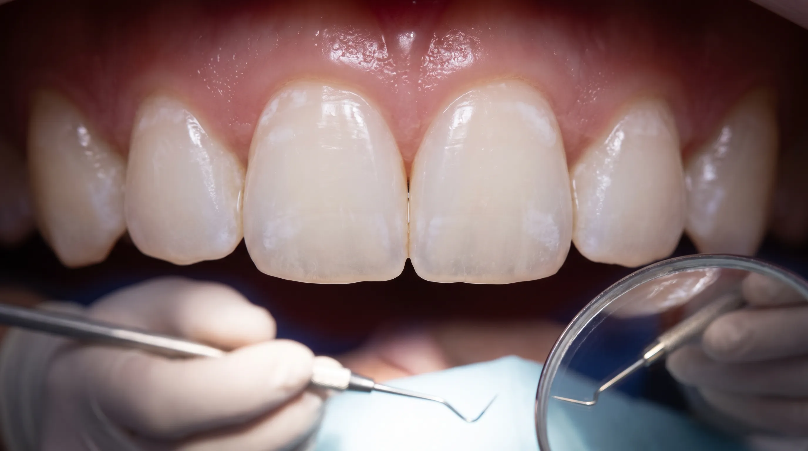
The hallmark of early enamel decalcification is the appearance of chalky, opaque white spots on otherwise translucent tooth surfaces. These lesions result from subsurface mineral loss that alters the refractive index of the enamel. When light penetrates the demineralized zone, it scatters differently, creating a milky appearance. Initially, these spots may only be visible when the tooth is dry, becoming more pronounced as mineral loss progresses. The lesions typically develop near the gingival margin, around orthodontic brackets, and along the cervical third of the anterior teeth.
Surface texture provides additional diagnostic clues. Healthy enamel feels remarkably smooth under a dental explorer. As decalcification advances, the surface may develop a chalky or rough texture that catches on instruments or feels different to the tongue. In advanced stages without cavitation, the enamel may exhibit slight translucency at the incisal edges while the cervical areas remain opaque. Regular self-examination with proper lighting helps patients identify changes early. Dentists use visual-tactile assessment combined with quantitative light-induced fluorescence devices to measure mineral density and track lesion progression objectively.
Sensitivity and Discomfort Progression
As mineral loss exposes the underlying dentinal tubule network, patients often report increased thermal sensitivity. Cold beverages or ambient air may trigger sharp, transient pain that subsides quickly. This occurs because hydrodynamic fluid shifts within exposed tubules stimulate nerve endings in the pulp. Sensitivity serves as a valuable biological feedback mechanism, signaling that the protective enamel barrier has thinned or fractured.
In the earliest stages of enamel decalcification, sensitivity may be absent because the lesion remains subsurface. As porosity increases, fluid exchange accelerates, and discomfort emerges. Some patients also notice mild soreness when chewing hard foods or experience a sticky feeling on tooth surfaces due to altered plaque accumulation patterns. Tracking symptom progression alongside visual changes helps clinicians determine whether conservative remineralization or more invasive interventions are warranted. Prompt attention to these signs prevents irreversible structural collapse.
Prevention Strategies You Can Start Today
Preventing enamel decalcification requires a multi-layered approach that combines meticulous hygiene, strategic nutrition, and targeted chemical interventions. The strategies outlined below are grounded in contemporary clinical research and designed for practical integration into daily routines.
Optimizing Your Daily Oral Hygiene Routine
Effective plaque removal depends on technique, consistency, and appropriate tools. A soft-bristled manual or powered toothbrush should be positioned at a forty-five-degree angle to the gumline, allowing filaments to penetrate the sulcus without traumatizing tissue. Gentle circular motions covering two to three teeth at a time ensure uniform cleaning. Brushing for two minutes, twice daily, remains the gold standard, but timing matters. Brushing immediately after consuming acidic foods or beverages can actually abrade softened enamel. Waiting thirty to forty minutes allows saliva to neutralize pH and re-harden the surface before mechanical cleaning resumes.
Interdental cleaning is non-negotiable for comprehensive plaque removal. Floss, interdental brushes, or water flossers disrupt biofilm between contact points where brushing cannot reach. Patients with tight contacts may benefit from waxed floss that glides smoothly without fraying. Those with wider spaces or orthodontic appliances should use specialized brushes designed for hardware navigation. Consistent interdental care prevents the formation of proximal lesions that remain invisible until radiographic examination reveals subsurface destruction. Establishing a nightly routine that includes thorough cleaning before sleep eliminates the prolonged acid production that occurs during resting hours.
Dietary Modifications for Stronger Enamel
Dietary adjustments represent the most powerful modifiable risk factor for enamel preservation. Limiting fermentable carbohydrate exposure reduces the fuel available for acid-producing bacteria. Patients should aim to consolidate sugar consumption into defined meal times rather than continuous grazing throughout the day. This clustering creates extended recovery periods where saliva can effectively remineralize early lesions. Rinsing with water immediately after meals accelerates acid clearance and reduces contact time.
Incorporating enamel-supporting nutrients strengthens resistance to future demineralization. Dairy products like cheese and yogurt provide bioavailable calcium and phosphate while stimulating salivary flow. Green leafy vegetables supply essential minerals and polyphenols that inhibit bacterial adhesion. Chewing fibrous foods like carrots and celery naturally scrubs tooth surfaces and stimulates glandular secretion. Conversely, limiting carbonated beverages, sports drinks, and highly acidic fruits minimizes direct chemical erosion. When consuming acidic items, using a straw directs liquid past anterior teeth, and following consumption with water or cheese helps neutralize residual acid. These simple modifications create an oral environment that actively opposes enamel decalcification.
Fluoride and Remineralization Products
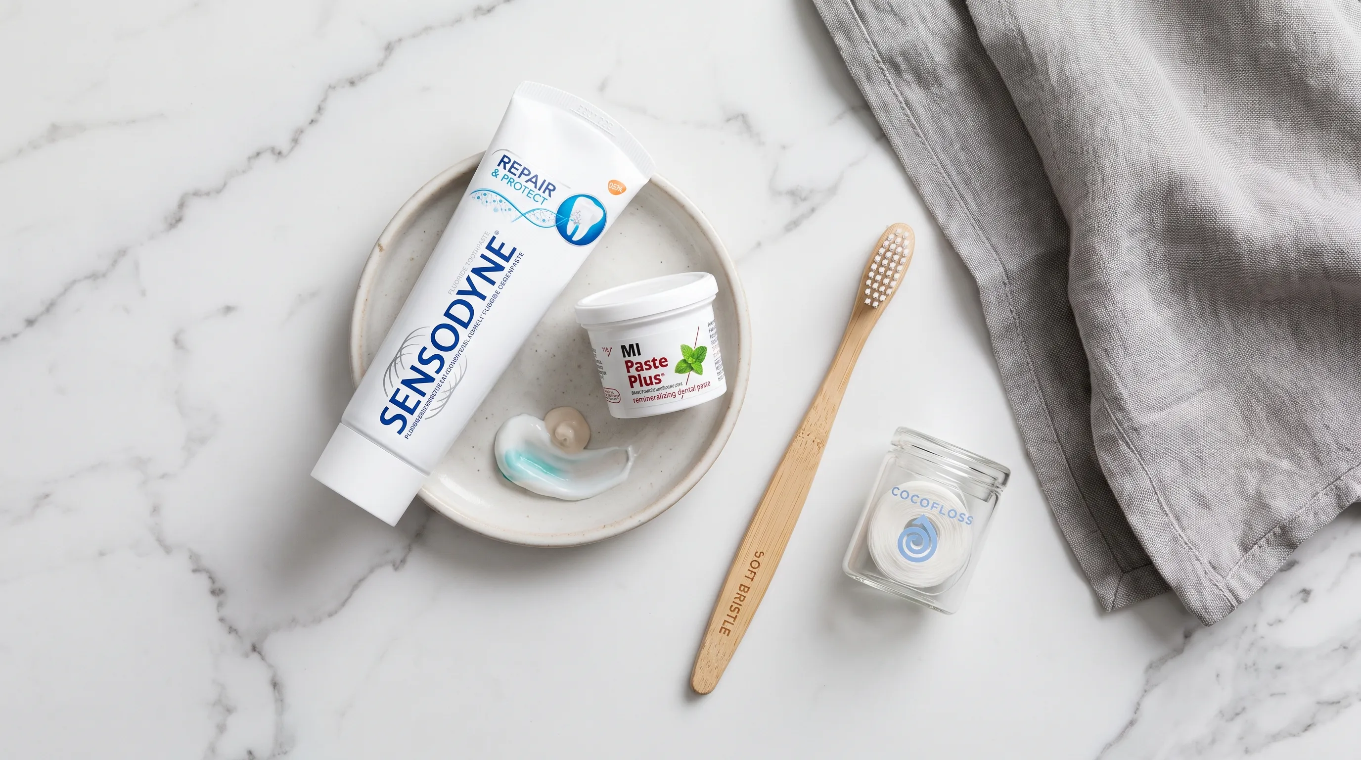
Fluoride remains the most extensively researched and clinically validated agent for preventing enamel mineral loss, with extensive safety and efficacy data compiled by the CDC. At optimal concentrations, fluoride integrates into the enamel lattice, replacing hydroxyl groups to form fluorapatite, which dissolves at a pH of approximately 4.5 rather than 5.5. This increased acid resistance dramatically slows demineralization rates while accelerating remineralization. Over-the-counter toothpaste typically contains 1000 to 1500 ppm fluoride, while prescription formulations reach 5000 ppm for high-risk patients.
Casein phosphopeptide-amorphous calcium phosphate (CPP-ACP) pastes offer an alternative or complementary approach. These products stabilize calcium and phosphate in a soluble form, delivering them directly to lesion sites. Clinical trials demonstrate significant improvements in white spot lesion regression when used consistently alongside fluoride therapy. Hydroxyapatite nanoparticle toothpastes provide direct mineral replacement without fluoride, appealing to patients seeking alternative formulations. Applying remineralizing gels before bedtime maximizes contact time during low-saliva hours. Patients should follow manufacturer instructions carefully, spitting excess product without rinsing to prolong active ingredient retention on tooth surfaces.
Professional Treatment Options
When home care proves insufficient or lesions advance despite prevention, professional interventions offer targeted solutions that address both cosmetic concerns and structural integrity. Modern dentistry provides a spectrum of treatments ranging from minimally invasive chemical therapies to restorative procedures.
In-Office Remineralization Therapies
Dental professionals apply concentrated fluoride varnishes, gels, or sealants during routine visits to deliver high-dose mineral therapy directly to at-risk surfaces. Varnish formulations adhere to enamel for hours, releasing fluoride gradually as saliva slowly dissolves the matrix. This sustained release maximizes uptake during periods of low home-care compliance or high acid exposure. Silver diamine fluoride (SDF) has gained traction for arresting active lesions, particularly in pediatric and special-needs populations, though it permanently stains carious tissue black, making it unsuitable for esthetic zones.
Professional prophylaxis removes calcified deposits that harbor acid-producing bacteria and obstruct natural remineralization pathways. Polishing smooths surface irregularities, reducing plaque retention sites. For patients with generalized demineralization, clinicians may recommend custom-fitted delivery trays loaded with prescription-strength remineralizing gels. Patients wear these overnight, ensuring prolonged contact with high-concentration minerals. Regular reapplication every three to six months maintains therapeutic levels while clinicians monitor lesion progression through standardized imaging and tactile evaluation.
Resin Infiltration and Microabrasion
Resin infiltration represents a revolutionary conservative treatment for white spot lesions. The procedure begins with surface etching to remove the hypermineralized outer layer that blocks penetration. Alcohol drying expands the porous subsurface network, allowing low-viscosity resin monomers to infiltrate the lesion body via capillary action. Once cured, the resin matches the refractive index of healthy enamel, rendering the white spot virtually invisible while physically blocking acid diffusion. This technique preserves tooth structure, requires no anesthesia, and demonstrates excellent longevity when indicated appropriately.
Microabrasion combines controlled enamel removal with chemical agents to eliminate superficial stains and minor demineralization. Dental professionals apply a slurry containing hydrochloric acid and pumice, followed by gentle polishing with specialized abrasives. The acid softens the outermost compromised layer, allowing precise removal without affecting underlying healthy structure. Microabrasion works best for isolated, shallow white spots or fluorosis-related discoloration. It cannot reverse subsurface mineral loss or address structural defects, but it effectively improves appearance when lesions are purely esthetic concerns. Combining microabrasion with professional bleaching often yields optimal results for patients seeking cosmetic enhancement.
Dental Veneers and Bonding for Advanced Cases
When enamel decalcification progresses to cavitation or severe structural compromise, restorative dentistry becomes necessary. Composite resin bonding allows clinicians to rebuild missing tooth structure directly in the mouth using tooth-colored materials cured with specialized light. The technique requires meticulous isolation, acid etching, and adhesive application to ensure durable retention. Bonding addresses both functional deficits and cosmetic concerns but may stain over time and requires periodic maintenance.
Porcelain veneers offer superior longevity and stain resistance for patients with extensive demineralization across multiple anterior teeth. These custom-fabricated ceramic shells require minimal tooth preparation, though irreversible enamel removal is necessary for proper adaptation. Veneers completely mask underlying white spots while providing exceptional strength and esthetics. Patients must weigh the benefits of immediate cosmetic transformation against the long-term commitment to restorative maintenance. Regardless of the chosen modality, successful outcomes depend on addressing the underlying risk factors that caused enamel decalcification initially.
The Role of Lifestyle and Systemic Health
Oral health never exists in isolation. Systemic conditions, daily habits, and psychological factors create an interconnected network that either supports or undermines enamel integrity. Addressing these broader influences ensures sustainable prevention and long-term success.
Hydration and Saliva Production
Water serves as the foundation of oral homeostasis. Adequate hydration maintains salivary flow rates necessary for natural buffering, mineral delivery, and bacterial clearance. Dehydration reduces volume, concentrates acids, and impairs the mouth's self-cleaning mechanisms. Patients who exercise vigorously, live in dry climates, or take medications that reduce fluid retention should consciously increase water intake throughout the day. Sugar-free gum containing xylitol stimulates salivary glands while inhibiting bacterial metabolism, providing dual benefits for dry mouth sufferers.
Saliva contains critical proteins like statherin and proline-rich peptides that prevent spontaneous calcium phosphate precipitation in ducts, ensuring minerals remain soluble for delivery to enamel. These proteins also form protective films that shield against acid penetration. Chronic dry mouth disrupts this sophisticated delivery system, leaving teeth vulnerable to rapid demineralization. Using salivary substitutes, avoiding alcohol-based mouthwashes, and consulting physicians about medication alternatives can restore natural protective mechanisms and significantly reduce the incidence of enamel decalcification.
Stress, Sleep, and Oral Microbiome Balance
Psychological stress triggers physiological cascades that directly impact oral health. Elevated cortisol levels suppress immune function in the gingival crevice, allowing pathogenic bacteria to proliferate and produce more aggressive acids. Stress also correlates with bruxism, where chronic grinding creates microfractures that compromise enamel integrity and increase susceptibility to chemical erosion. Furthermore, stress-induced dietary choices often favor high-sugar, low-nutrient foods that fuel demineralization.
Sleep quality influences immune regulation and tissue repair cycles. During deep sleep, cellular regeneration and systemic inflammation modulation occur. Chronic sleep deprivation impairs these processes, reducing the mouth's ability to recover from daily acid attacks. Poor sleep also increases mouth breathing, which dries oral tissues and shifts the microbiome toward acid-tolerant species. Implementing stress-reduction techniques like mindfulness meditation, regular exercise, and consistent sleep schedules supports systemic balance. A balanced oral microbiome, dominated by commensal rather than pathogenic bacteria, naturally maintains pH equilibrium and minimizes enamel decalcification without requiring aggressive interventions.
Navigating Orthodontic Treatment Safely
Orthodontic therapy fundamentally alters the cleaning landscape, creating predictable risk zones for plaque accumulation. Patients must adapt their hygiene protocols before treatment begins to prevent irreversible damage. Specialized orthodontic brushes with cut-away bristle designs access hardware efficiently, while floss threaders enable navigation beneath wires. Water flossers with targeted tips effectively flush debris from bracket bases and molar bands.
Dietary vigilance becomes paramount during orthodontic treatment. Sticky candies, hard snacks, and prolonged sugary beverage consumption dramatically increase decalcification risk around fixed appliances. Many orthodontists provide high-concentration fluoride mouth rinses for nightly use and schedule more frequent prophylaxis visits to monitor vulnerable surfaces. If white spots develop during treatment, prompt intervention with professional remineralization prevents progression to permanent cavities. Successful orthodontic outcomes depend entirely on maintaining enamel integrity throughout the mechanical adjustment process.
| Intervention | Primary Mechanism | Ideal Use Case | Longevity | Professional Requirement |
|---|---|---|---|---|
| Fluoride Varnish | Sustained ion release, fluorapatite formation | High-risk patients, post-prophylaxis application | 3-6 months | Yes |
| CPP-ACP Paste | Bioavailable calcium/phosphate delivery | Active white spots, dry mouth, orthodontic patients | Ongoing with daily use | Prescription available |
| Resin Infiltration | Capillary penetration, refractive index matching | Isolated white spots without cavitation | 5+ years | Yes |
| Microabrasion | Controlled superficial enamel removal | Shallow stains, mild fluorosis | Permanent for treated areas | Yes |
| Composite Bonding | Direct structural replacement and sealing | Cavitated lesions, structural defects | 5-10 years with maintenance | Yes |
Frequently Asked Questions
Is enamel decalcification reversible?
Early-stage enamel decalcification can often be reversed through remineralization using fluoride, calcium-phosphate products, and improved oral hygiene according to clinical standards from the NIH. However, once a cavity forms or the enamel surface breaks down, the damage becomes irreversible and requires professional restoration. Consistent application of evidence-based products and dietary modifications significantly improve the likelihood of complete lesion arrest.
How long does it take for enamel to remineralize after decalcification?
Remineralization is a gradual biological process. With consistent use of remineralizing agents like fluoride or CPP-ACP, visible improvements typically appear within 3 to 6 months. The timeline depends on lesion depth, salivary flow, and dietary habits. Regular clinical monitoring ensures that therapy remains effective and adjustments are made when necessary.
Can enamel decalcification occur without cavities?
Yes. Decalcification is the initial stage of demineralization where calcium and phosphate are lost, creating chalky white spots. This can happen long before bacterial acids penetrate deep enough to create a structural cavity. Identifying and treating lesions at this pre-cavitation stage prevents the need for drilling and filling procedures.
What is the best toothpaste for reversing decalcification?
Dental professionals recommend toothpaste containing at least 1000-1500 ppm fluoride, sodium monofluorophosphate, or stannous fluoride. Formulations with casein phosphopeptide-amorphous calcium phosphate (CPP-ACP) are also clinically proven to enhance remineralization. Patients with high demineralization risk may benefit from prescription-strength 5000 ppm fluoride pastes used nightly.
Does drinking acidic beverages directly cause enamel decalcification?
Yes. Acidic drinks lower the pH in the mouth below the critical threshold of 5.5, triggering immediate dissolution of hydroxyapatite crystals. Frequent sipping, rather than the volume consumed, significantly increases demineralization risk as detailed by the Mayo Clinic. Modifying consumption patterns, using straws, and rinsing with water afterward substantially reduces direct chemical erosion.
Key Takeaways
Enamel decalcification represents the critical warning phase where tooth structure begins to lose mineral density but has not yet suffered permanent structural collapse. The process is driven by acid-producing bacteria, dietary sugars, direct chemical exposure, and systemic factors like reduced salivary flow. Early identification of white spot lesions and surface texture changes allows for timely intervention before irreversible cavitation occurs. Preventing enamel decalcification requires a comprehensive strategy combining meticulous plaque removal, strategic dietary modifications, consistent use of remineralizing agents, and regular professional monitoring. When early lesions do form, conservative treatments like resin infiltration, microabrasion, and high-concentration fluoride therapies effectively halt progression and restore aesthetics without invasive procedures. Ultimately, preserving tooth enamel depends on maintaining the delicate balance between demineralization and natural repair. By understanding the science, implementing daily protective habits, and partnering with dental professionals for personalized care, you can ensure that your teeth remain strong, functional, and healthy for a lifetime. Proactive management of enamel health transforms potential crises into manageable routines, securing a confident smile well into the future.
How we work
Researched and drafted with AI assistance, then reviewed by a human editor against the cited sources.
This article is general health information, not medical advice. Consult a qualified healthcare professional about your own situation.