Fractured Thumb: Comprehensive Guide to Diagnosis, Treatment, and Recovery
A fractured thumb is far more than a minor inconvenience; it is a potentially debilitating hand injury that can dramatically alter your daily independence. The thumb serves as the cornerstone of human manual dexterity, contributing to roughly forty to fifty percent of overall hand function. When one of the three critical bones that comprise the thumb sustains a fracture, the immediate loss of pinch strength, grip stability, and fine motor coordination becomes glaringly apparent. From simple tasks like buttoning a shirt and typing on a keyboard to essential daily routines like opening jars, holding utensils, or driving a vehicle, the thumb is constantly engaged. Understanding the clinical reality of this injury is the first step toward preserving long-term function. Proper medical evaluation, accurate classification of the fracture pattern, adherence to prescribed immobilization protocols, and a structured rehabilitation program are all non-negotiable components of optimal recovery. Without timely intervention, a fractured thumb can lead to chronic pain, permanent joint stiffness, malunion, or early-onset post-traumatic arthritis. This comprehensive guide explores the anatomy, diagnostic pathways, evidence-based treatment options, and detailed rehabilitation strategies you need to navigate a fractured thumb safely and effectively, ensuring a return to your highest level of manual performance.
Understanding Thumb Anatomy and Its Critical Role
To fully grasp the clinical implications of a fractured thumb, it is essential to understand the intricate biomechanics that make this digit uniquely powerful and functionally dominant. The human hand relies heavily on the opposable thumb, which allows for precision grip and power grip. This capability stems from a complex arrangement of bones, joints, ligaments, and tendons that work in seamless coordination.
Free calculatorHigh Calcium Foods ChartChart of calcium-rich foods for bone healthOpen the calculatorThe Three Primary Bones
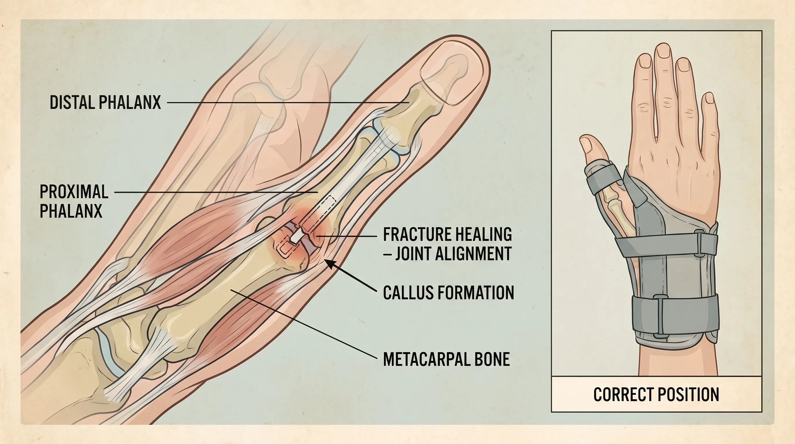
The thumb is structurally distinct from the other four digits, containing only three phalanges instead of four. The distal phalanx forms the tip of the thumb, supporting the nail bed and enabling fine tactile manipulation. The proximal phalanx sits in the middle segment, serving as the primary lever for flexion and extension. Connecting the thumb to the wrist and carpus is the first metacarpal, the longest and most robust of the three. This bone acts as the foundational anchor, transmitting forces generated during gripping, pushing, and pinching directly into the wrist and forearm.
Key Joints and Ligamentous Support
Three major synovial joints govern thumb mobility. The interphalangeal (IP) joint allows bending at the tip, while the metacarpophalangeal (MCP) joint facilitates wider flexion and extension. The carpometacarpal (CMC) joint at the base of the first metacarpal is a saddle joint that enables circumduction, opposition, and the remarkable rotational capacity that defines human hand function. Stability across these joints is maintained by a network of collateral ligaments, with the ulnar collateral ligament (UCL) at the MCP joint being particularly vulnerable to traumatic avulsion during falls or sports injuries. The flexor pollicis longus (FPL) and extensor pollicis longus (EPL) tendons run longitudinally, providing the muscular force required for movement.
Why Thumb Integrity Matters
Because the thumb accounts for nearly half of all hand function, even a minimally displaced fracture can cascade into significant disability. Compromised alignment alters joint mechanics, leading to uneven cartilage wear, tendon attrition, and compensatory muscle overuse in the wrist and forearm. Recognizing the anatomical complexity behind a fractured thumb underscores why specialized orthopedic assessment is crucial rather than relying on home remedies or generic splints.
Recognizing the Signs and Symptoms of a Fractured Thumb
Trauma to the hand can produce overlapping symptoms, making it difficult for laypersons to distinguish between a severe sprain, tendon rupture, or a true bone fracture. However, specific clinical indicators strongly suggest osseous disruption and warrant immediate medical imaging.
Acute Pain and Localized Tenderness
Sharp, throbbing pain immediately following trauma is the primary hallmark of a fractured thumb. Unlike soft tissue injuries that may produce dull, radiating discomfort, bone fractures typically elicit intense, pinpoint tenderness directly over the fracture site. Palpation often reproduces severe pain, particularly over the anatomical snuffbox at the base of the metacarpal or along the phalangeal shafts.
Visible Deformity and Structural Misalignment
In displaced fractures, the thumb may appear crooked, shortened, or rotated unnaturally. A step-off deformity along the bone shaft or a dorsal/volar angulation is unmistakable. Swelling typically develops within minutes, accompanied by rapid ecchymosis (bruising) that migrates toward the fingers and palm as gravity pulls extravasated blood downward.
Functional Impairment and Joint Instability
Patients often report an immediate inability to actively extend or flex the thumb against resistance. Attempts to pinch between the thumb and index finger feel weak or mechanically blocked. In cases involving ligamentous avulsion or severe comminution, the thumb joint may feel loose or abnormally mobile when gently manipulated by a clinician.
Neurovascular Warning Signs
While less common, severe swelling or bone fragment displacement can compress the digital nerves or radial artery branches. Numbness, tingling, coldness, or a pale bluish discoloration of the thumb tip indicates potential neurovascular compromise. This requires urgent orthopedic evaluation to prevent tissue ischemia or permanent nerve damage.
Types of Thumb Fractures: A Clinical Breakdown
Not all fractured thumb injuries are created equal. Orthopedic specialists classify these fractures based on location, fracture line pattern, joint involvement, and degree of displacement. Accurate classification dictates the entire treatment pathway.
Bennett and Rolando Fractures
Both injuries occur at the proximal base of the first metacarpal near the CMC joint. A Bennett fracture involves an intra-articular oblique break that leaves a large volar-ulnar fragment attached to the palmar ligaments while the metacarpal shaft subluxates proximally and radially due to unopposed muscle pull. Because it violates the CMC joint surface, restoring joint congruity to within one to two millimeters is critical to prevent degenerative arthritis. A Rolando fracture is essentially a comminuted Bennett fracture, where the base shatters into three or more fragments in a T or Y configuration. The increased fragmentation makes surgical reconstruction highly complex, often requiring meticulous internal fixation or staged external fixation.
Metacarpal Shaft Fractures
These fractures run longitudinally through the long, tubular portion of the first metacarpal. They frequently occur from direct blunt force trauma, such as striking a hard object or crushing injuries. Because they typically spare the articular surface, shaft fractures are often amenable to conservative management with closed reduction and casting, provided rotational alignment is preserved.
Proximal and Distal Phalangeal Fractures
Fractures of the phalanges usually result from axial loading, such as jamming the thumb against a ball or floor. Extra-articular phalangeal shaft fractures generally heal well with buddy taping or brief immobilization. Intra-articular phalangeal fractures involving the IP or MCP joints, however, risk joint incongruity and stiffness, sometimes necessitating surgical intervention to restore smooth articulation.
Avulsion Fractures
Often clinically grouped with ligament injuries, avulsion fractures occur when an extreme force causes a ligament to tear away, taking a cortical bone fragment with it. Gamekeeper’s or Skier’s thumb typically involves ulnar collateral ligament avulsion at the proximal phalanx base. While primarily a ligamentous injury, the bony component classifies it alongside fractures. Treatment depends on fragment size and displacement, ranging from splinting for small, stable avulsions to surgical repair for large fragments that destabilize the joint.
Diagnosis: How Medical Professionals Assess the Injury
Accurate diagnosis of a fractured thumb relies on a systematic clinical evaluation paired with advanced imaging modalities. Self-diagnosis is strongly discouraged, as subtle displacement or intra-articular involvement may only be visible on specialized radiographic views.
Physical Examination and Stress Testing
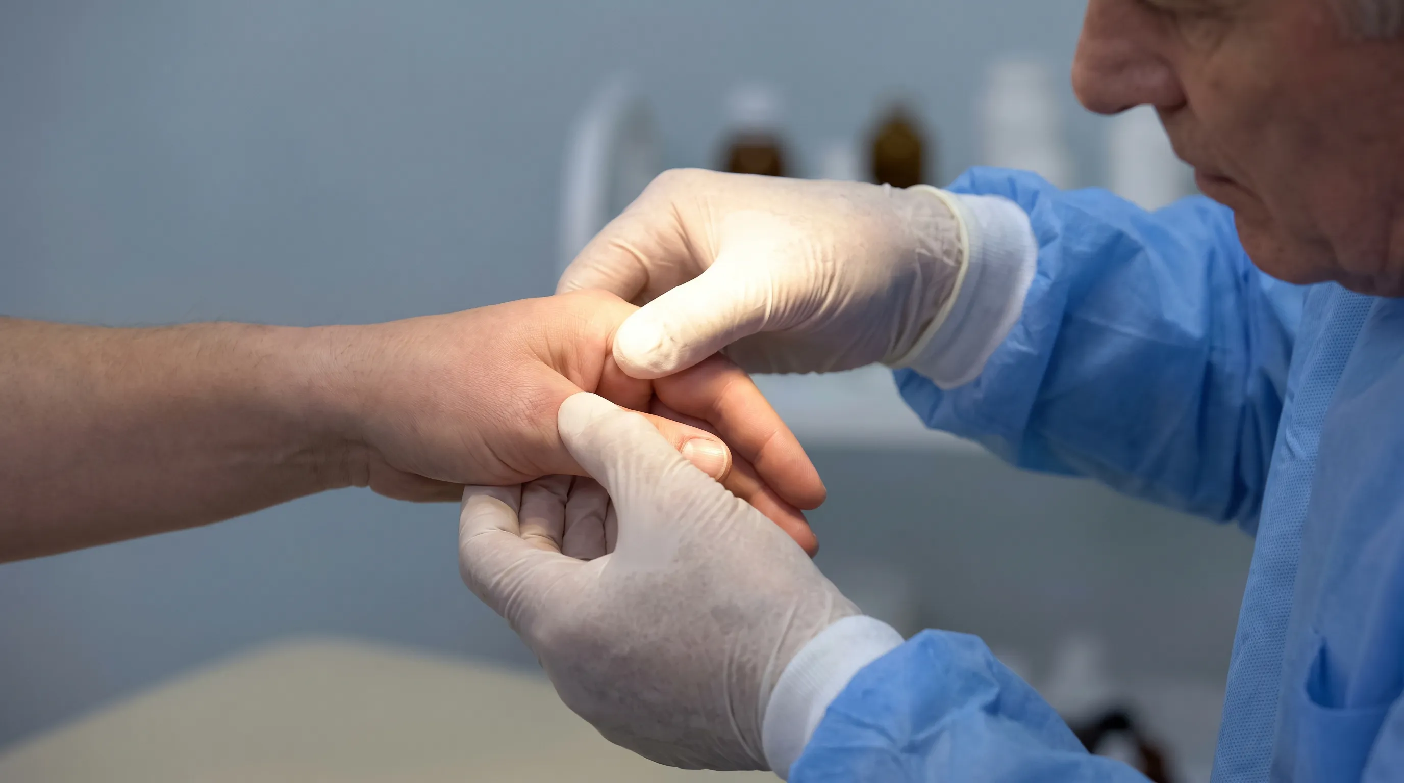
An orthopedic specialist will first assess skin integrity, checking for open wounds that could convert a closed fracture into an open, infection-prone injury. Tendon function is evaluated by asking the patient to flex and extend each joint independently. Neurovascular status is confirmed via capillary refill testing and two-point discrimination. Ligamentous stress testing, such as valgus/varus instability checks at the MCP joint, helps identify associated soft tissue disruption.
Standard Radiographic Imaging
Anteroposterior, lateral, and oblique X-rays are the gold standard initial imaging tools. These three orthogonal views reveal fracture location, displacement magnitude, joint step-off, and rotational deformity. Radiologists specifically measure the CMC joint surface gap; any displacement exceeding two millimeters strongly indicates surgical necessity for intra-articular patterns.
Advanced Imaging Protocols
When standard X-rays prove inconclusive or when planning complex surgical intervention, a computed tomography (CT) scan provides three-dimensional reconstruction of comminuted fragments. CT is invaluable for Rolando and severe Bennett fractures, allowing surgeons to map screw trajectories and plate placement preoperatively. Magnetic resonance imaging (MRI) is reserved for cases where ligamentous rupture, tendon entrapment, or bone contusion is suspected but not visible on CT.
Treatment Options: Conservative vs. Surgical Approaches
The management of a fractured thumb is highly individualized. Orthopedic decision-making balances fracture stability, joint involvement, patient age, activity demands, and bone quality. Below is a comprehensive comparison of the primary treatment pathways.
| Treatment Modality | Indications | Procedure Details | Immobilization Duration | Key Advantages | Potential Limitations |
|---|---|---|---|---|---|
| Closed Reduction & Casting | Non-displaced or minimally displaced fractures, extra-articular patterns | Manual realignment without incision, followed by thumb spica cast application | 4 to 6 weeks | Non-invasive, preserves soft tissue envelope, cost-effective | Requires strict compliance, risk of late displacement, joint stiffness possible |
| Percutaneous Pinning (K-wires) | Moderately displaced fractures, unstable intra-articular fragments | Minimally invasive insertion of stainless steel pins under fluoroscopic guidance | 3 to 5 weeks plus post-pin rehabilitation | Quick fracture stabilization, lower infection risk than open surgery | Pin site infection risk, requires second minor procedure for removal |
| Open Reduction Internal Fixation (ORIF) | Significantly displaced fractures, Bennett/Rolando fractures, joint incongruity | Surgical exposure, direct fragment reduction, fixation with plates, lag screws, or mini-plates | 2 to 4 weeks followed by dynamic therapy | Anatomical restoration, early controlled motion possible, highest union rates | Surgical risks (infection, nerve irritation, hardware prominence), scar formation |
| External Fixation | Severe comminution, open fractures with soft tissue loss, contaminated wounds | Perpendicular pins inserted into proximal and distal segments connected to an external carbon frame | 6 to 8 weeks until callus forms | Allows wound management, adjustable alignment, avoids placing hardware in compromised bone | Bulky apparatus, pin tract care required, potential for joint stiffness |
Non-Surgical Management Protocols
Conservative treatment is the standard of care for stable, well-aligned fractures lacking joint involvement. After a successful closed reduction under local anesthesia, a custom-molded thumb spica cast or rigid orthoplast splint is applied. The wrist is positioned in slight extension to optimize tendon tension, while the thumb remains in functional alignment (the CMC joint in slight abduction, MCP in slight flexion). Follow-up radiographs at one, two, and four weeks ensure fracture alignment is maintained as edema subsides and soft tissues contract.
Surgical Intervention Considerations
Surgery becomes mandatory when conservative methods cannot restore anatomical alignment or maintain joint stability. Intra-articular fractures demand absolute reduction to prevent cartilage degeneration. Modern orthopedic techniques prioritize early mobilization; rigid internal fixation allows surgeons to initiate protected range-of-motion therapy within days rather than weeks, drastically reducing stiffness and adhesion formation. Surgical planning also accounts for bone density; osteoporotic patients may require augmented fixation with bone graft substitutes or specialized locking plates.
Rehabilitation and Recovery Timeline
Rehabilitation is not an afterthought; it is the critical bridge between fracture union and functional restoration. Bone healing follows a predictable biological timeline, but soft tissue stiffness, tendon gliding deficits, and proprioceptive loss can persist long after radiographic union is confirmed.
Phase One: Immobilization and Edema Control (Weeks 0 to 4)
The primary goals during this phase are fracture protection, swelling reduction, and prevention of adjacent joint stiffness. Patients must elevate the injured hand above heart level whenever possible. Gentle active motion of the uninvolved fingers, elbow, and shoulder is strongly encouraged to maintain circulation and prevent complex regional pain syndrome. Compression garments or Coban wrapping may be prescribed once the acute swelling phase subsides. Pain management typically involves NSAIDs or acetaminophen, though prolonged high-dose NSAID use is increasingly debated as it may theoretically interfere with early bone callus formation.
Phase Two: Early Mobilization (Weeks 4 to 6)
Upon radiographic confirmation of bridging callus, the cast or pins are removed. A certified hand therapist guides the patient through gentle passive and active-assisted range of motion exercises. Joint mobilization techniques restore capsular elasticity, while tendon gliding exercises prevent adhesions. Edema control continues, as persistent swelling mechanically blocks full flexion. Heat modalities like warm paraffin wax baths improve tissue extensibility before stretching.
Phase Three: Strengthening and Functional Integration (Weeks 6 to 12)
As pain diminishes and range of motion approaches functional baselines, progressive resistance training begins. Therapy putty, resistance bands, and grip strengtheners are introduced gradually. Patients practice pinch variations (tip, lateral, palmar) and power grip activities. Scar massage techniques desensitize surgical incisions and improve pliability. Ergonomic education helps patients modify daily tasks to avoid repetitive strain during tissue remodeling.
Phase Four: Return to Activity and Long-Term Maintenance (Months 3 to 6+)
Full bone remodeling continues for twelve to eighteen months, but most patients resume light occupational duties and recreational activities by three months. High-impact sports, heavy manual labor, and contact athletics typically require four to six months of conditioning. Ongoing proprioceptive drills, such as blindfolded object manipulation or wobble board stabilization, restore neuromuscular coordination. Patients with intra-articular fractures are advised to maintain lifelong hand strength and joint mobility routines to delay arthritic changes.
Nutritional and Lifestyle Optimization
Bone healing is a metabolically demanding process. Adequate protein intake (1.2 to 1.6 grams per kilogram of body weight) supplies collagen precursors essential for matrix formation. Calcium (1000 to 1200 mg daily) and vitamin D (800 to 2000 IU daily) support mineralization. Vitamin C, zinc, and magnesium act as crucial enzymatic cofactors in osteoblast activity. Crucially, nicotine in any form (cigarettes, vaping, patches) causes profound vasoconstriction, reducing osteoblast perfusion and increasing nonunion risk by two to three times. Complete cessation during recovery is non-negotiable for optimal outcomes.
Prevention Strategies and Long-Term Care
While trauma is often unpredictable, evidence-based preventive measures can significantly reduce the incidence and severity of a fractured thumb, particularly in high-risk populations.
Sports-Specific Protective Measures
Athletes participating in skiing, football, basketball, volleyball, and hockey should utilize sport-appropriate protective equipment. Custom-molded thumb guards, neoprene wrist wraps with integrated splints, and proper tape-job techniques (using athletic tape or pre-wrap to stabilize the MCP and CMC joints) absorb and distribute impact forces. Skiers should invest in modern pole straps that release during forward falls rather than catching on the thumb base.
Bone Health and Metabolic Optimization
Underlying metabolic bone disease drastically increases fracture susceptibility. Individuals over fifty, particularly postmenopausal women, should undergo regular bone density screening. Maintaining optimal vitamin D levels through safe sun exposure or supplementation improves bone mineral density. Weight-bearing exercises and resistance training stimulate osteogenic pathways, creating denser trabecular bone structure that resists traumatic loading.
Ergonomic Adaptations and Fall Prevention
For older adults, home safety modifications prevent low-energy falls that commonly cause upper extremity fractures. Removing tripping hazards, installing grab bars in bathrooms, improving nighttime lighting, and using non-slip footwear drastically reduce incident rates. Workplace ergonomics also matter; repetitive microtrauma weakens bone microarchitecture over time. Incorporating regular hand rests, utilizing shock-absorbing gloves for manual labor, and alternating high-force tasks with low-strain activities preserve long-term thumb resilience.
Frequently Asked Questions
How long does it take for a fractured thumb to heal?
Bone union typically occurs within four to six weeks, but complete functional recovery and full bone remodeling take three to six months. Intra-articular fractures or those requiring surgery often demand longer rehabilitation due to the complexity of joint surface restoration and soft tissue healing.
Can I drive with a thumb spica cast on?
Driving is generally unsafe while your thumb is immobilized. The cast restricts grip strength, reaction time, and steering control, significantly increasing accident risk. Most specialists recommend waiting until cast removal and clearance from your orthopedic provider before resuming driving.
Is surgery always necessary for a broken thumb?
No. Non-displaced, stable fractures without joint involvement are successfully managed with conservative treatment, including closed reduction and casting. Surgery is reserved for displaced fractures, intra-articular involvement, open wounds, or cases where alignment cannot be maintained through splinting alone.
What are the long-term risks of not treating a thumb fracture properly?
Untreated or malunited thumb fractures can cause chronic joint instability, persistent pain, significantly reduced pinch and grip strength, and early-onset post-traumatic osteoarthritis. These complications may eventually require complex reconstructive surgery, joint fusion, or arthroplasty to restore basic hand function.
How can I prevent thumb injuries during sports and daily activities?\Utilize proper protective taping or custom orthotics, maintain adequate calcium and vitamin D intake for bone density, and strengthen intrinsic hand muscles through targeted exercises. Always wear appropriate protective gear in high-impact sports, and modify home or workplace environments to reduce fall and crush injury risks.
Conclusion
A fractured thumb represents a significant orthopedic challenge that demands prompt, specialized medical attention to preserve the critical functionality this digit provides. From understanding the nuanced differences between Bennett, Rolando, and extra-articular patterns to adhering to structured rehabilitation protocols, every phase of recovery requires deliberate, evidence-based action. Whether managed conservatively or surgically, the ultimate goal remains consistent: restoring pain-free motion, robust grip strength, and long-term joint stability. By prioritizing accurate diagnosis, respecting healing timelines, optimizing nutritional support, and committing to professional hand therapy, patients can successfully navigate the complexities of thumb fracture recovery. Remember, early intervention and strict adherence to your orthopedic specialist's guidance are the most reliable predictors of a successful, complication-free outcome. Always seek qualified medical evaluation for any suspected hand trauma to ensure your path to recovery remains on track.
For further clinical reading on fracture management and rehabilitation protocols, consult authoritative resources like AAOS OrthoInfo - Thumb Fractures and the Mayo Clinic - Broken Hand/Thumb patient guidelines.
How we work
Researched and drafted with AI assistance, then reviewed by a human editor against the cited sources.
This article is general health information, not medical advice. Consult a qualified healthcare professional about your own situation.