Pulled Muscle in Forearm: Complete Recovery Guide, Symptoms & Prevention

Dealing with a sudden, sharp pain in your arm while lifting, typing, or swinging can instantly disrupt your daily routine and leave you searching for answers. When you experience a pulled muscle in your forearm, the discomfort often radiates from the inner or outer arm, making even simple tasks like opening a jar or holding a coffee cup feel like an uphill battle. Understanding how these injuries happen, recognizing the early warning signs, and implementing evidence-based recovery strategies are crucial for restoring full function and preventing long-term complications. In this comprehensive guide, we will explore the anatomical complexities of forearm strain, walk you through clinically proven treatment protocols, and provide actionable steps to safely return to your activities. Whether you are an athlete, a dedicated gym-goer, or someone navigating repetitive occupational tasks, knowing exactly how to manage and heal a pulled muscle in forearm will empower you to take control of your recovery journey and regain optimal upper limb performance.
Understanding Forearm Anatomy and Strain Mechanics
The human forearm is a highly intricate anatomical structure composed of twenty distinct muscles, multiple nerve pathways, vascular networks, and complex connective tissue systems. These muscles are primarily organized into two major functional compartments: the anterior flexor group and the posterior extensor group. Understanding this anatomical layout is essential for accurately identifying the location of your injury, predicting recovery timelines, and selecting the most appropriate rehabilitation exercises for a pulled muscle in forearm. The anterior compartment houses muscles responsible for wrist flexion, finger bending, and forearm pronation, while the posterior compartment controls wrist extension, finger straightening, and supination. Both groups originate near the elbow joint and taper into long tendons that cross the wrist and insert into the small bones of the hand.
Free calculatorHigh-Fiber Foods ChartChart of high-fiber foods for digestive healthOpen the calculatorThe Physiology of Muscle Fiber Damage
When a muscle is stretched beyond its physiological capacity, microscopic or macroscopic tearing occurs within the contractile fibers. This process triggers an immediate inflammatory response, which is actually a vital biological mechanism for tissue repair. Blood vessels in the damaged area dilate to deliver immune cells, oxygen, and essential nutrients required for healing. The body initiates a three-phase repair cascade: the inflammatory phase clears cellular debris, the proliferative phase deposits new collagen matrix, and the remodeling phase aligns the fibers along stress lines to restore tensile strength. If this natural healing sequence is disrupted by premature loading, inadequate rest, or improper treatment, scar tissue may form irregularly, leading to reduced mobility and chronic pain. Properly managed rehabilitation ensures that regenerated tissue matches the structural integrity and functional capacity of the original muscle architecture.
Recognizing Symptoms and Severity Grading
Accurately identifying the clinical presentation of your injury is the first step toward effective treatment. While a pulled muscle in forearm shares overlapping symptoms with tendinitis, joint sprains, and nerve compression syndromes, muscle strains exhibit distinct characteristics. Pain is typically localized to a specific muscle belly and worsens with active contraction or direct palpation. You may notice localized swelling, mild bruising, or a feeling of tightness that restricts normal wrist and finger movement. In acute cases, a sudden tearing sensation or popping sound may occur at the moment of injury, followed by rapid onset of stiffness. Differentiating between mild discomfort and significant tissue damage requires understanding clinical grading scales and recognizing red flag symptoms that warrant immediate professional evaluation.
Clinical Classification of Forearm Strains
Medical professionals utilize a standardized three-tier grading system to classify muscle strains based on fiber involvement, functional impairment, and expected healing duration. Grade 1 strains involve less than five percent fiber disruption, resulting in mild pain, minimal swelling, and full strength retention. Individuals with this severity typically experience a pulled muscle in forearm that responds rapidly to conservative care. Grade 2 tears represent moderate damage affecting ten to fifty percent of the muscle cross-section. Patients report sharp pain during movement, noticeable weakness when gripping, and palpable tenderness along the affected region. Grade 3 strains indicate near-complete or total muscle rupture, characterized by severe pain initially, followed by functional weakness, significant bruising, and sometimes a visible defect in the muscle contour. Proper grading guides treatment selection, rehabilitation pacing, and realistic return-to-activity expectations.
Distinguishing Muscle Pain from Other Conditions
Accurate symptom differentiation prevents mismanagement and accelerates recovery. Unlike a muscle strain, lateral epicondylitis primarily causes localized elbow tenderness with pain radiating down the dorsal forearm during resisted wrist extension. Carpal tunnel syndrome presents with numbness, tingling, and burning sensations primarily affecting the thumb, index, and middle fingers, often worsening at night. Forearm compartment syndrome, though rare, involves severe, disproportionate pain, tense swelling, and neurological changes that constitute a medical emergency requiring immediate intervention. If your symptoms persist beyond expected healing windows, progressively worsen, or include neurological deficits, comprehensive medical evaluation becomes necessary. Understanding these clinical distinctions ensures you apply targeted interventions rather than generic pain relief measures that may delay proper healing.
Common Causes and Risk Factors
Forearm muscle injuries rarely occur in isolation; they typically result from cumulative stress, sudden overload, or biomechanical imbalances. Recognizing the underlying etiology helps clinicians design personalized rehabilitation programs and empowers individuals to modify behaviors that predispose them to recurrent damage. Repetitive stress injuries driven by overuse remain the predominant driver of forearm strains in modern populations, particularly among desk workers, musicians, construction professionals, and recreational athletes. The repetitive microtrauma caused by continuous contraction without adequate recovery time gradually fatigues muscle fibers, reduces tissue elasticity, and increases susceptibility to acute tearing. Understanding your personal risk profile allows you to implement preventive strategies before minor discomfort escalates into a debilitating injury.
Overuse and Repetitive Stress Injuries
Occupational activities requiring sustained gripping, typing, or tool manipulation place continuous demand on forearm flexors and extensors. When muscles operate near their fatigue threshold, motor unit recruitment patterns shift, causing secondary muscles to compensate and absorb excessive mechanical stress. Athletes participating in racquet sports, climbing, weightlifting, and throwing disciplines frequently develop forearm strains due to the high-velocity rotational forces and eccentric loading demands placed on the upper limb. Training errors such as rapid progression in volume or intensity, inadequate warm-up routines, and insufficient rest days further compound the risk. Implementing periodized training schedules and listening to early fatigue signals significantly reduces overuse strain incidence.
Trauma and Biomechanical Triggers
Acute injuries often occur during sudden forceful movements, falls, or unexpected heavy lifting when the forearm muscles are unprepared or positioned disadvantageously. Attempting to catch a falling object, performing high-velocity pulling exercises with poor form, or experiencing direct blunt force trauma can instantly exceed tissue tolerance limits. Biomechanical factors such as poor scapular stability, restricted shoulder mobility, or improper wrist alignment during functional movements force the forearm to absorb disproportionate mechanical loads. Weakness in the proximal kinetic chain transfers excessive stress distally, creating a cascade of compensatory patterns that overload smaller stabilizing muscles. Addressing these foundational movement dysfunctions is essential for preventing recurrent injuries and optimizing long-term upper limb health.
Professional Diagnosis and Assessment
While mild discomfort can often be managed safely at home, professional evaluation provides accurate diagnosis, severity grading, and individualized treatment planning. Healthcare providers utilize systematic clinical assessment protocols to evaluate pain location, muscle function, joint mobility, and neurological integrity. A thorough patient history establishes injury mechanism, symptom timeline, previous upper limb issues, and occupational or athletic demands. Physical examination techniques focus on palpation to identify tender points, resisted contraction testing to isolate affected muscles, passive stretching to assess tissue extensibility, and functional movement screens to observe compensatory patterns. This comprehensive approach ensures that treatment targets the specific injured structure rather than generalized arm discomfort.
Physical Examination Techniques
Clinicians employ specialized orthopedic tests to differentiate between muscle, tendon, joint, and nerve involvement. Resisted wrist flexion and extension maneuvers isolate specific muscle groups, reproducing pain at the site of tissue damage when fibers are strained. Passive range-of-motion testing evaluates joint capsule restrictions and muscle flexibility limitations. Grip strength dynamometry provides objective baseline measurements that track functional recovery progression. Neurological screening assesses sensory distribution and reflex integrity to rule out cervical radiculopathy or peripheral nerve entrapment. These standardized assessments generate reliable clinical data that guide treatment selection and establish measurable recovery milestones.
When to Seek Advanced Imaging
Conservative management remains the standard of care for most muscle strains, but certain clinical presentations warrant diagnostic imaging. Magnetic resonance imaging (MRI) provides exceptional soft tissue contrast, clearly delineating fiber disruption severity, hematoma formation, and tendon involvement. Ultrasound imaging offers dynamic, real-time visualization of muscle architecture during contraction, identifying partial tears and scar tissue organization with minimal cost and radiation exposure. X-ray studies rule out associated bony avulsion fractures or stress fractures that may accompany severe muscle contractions. Imaging is typically reserved for high-grade injuries, chronic non-responsive symptoms, surgical candidates, or cases where diagnostic uncertainty impacts treatment decisions. Advanced diagnostic tools ensure precise management planning and reduce the risk of missed pathology.
Evidence-Based Treatment and Rehabilitation
Restoring optimal function following a forearm strain requires a phased, scientifically supported approach that balances tissue protection with progressive loading. Modern rehabilitation protocols emphasize early controlled movement over prolonged immobilization, recognizing that appropriately dosed mechanical stress stimulates cellular regeneration and prevents joint stiffness. Each recovery phase transitions smoothly based on symptom resolution, functional capacity, and objective strength measurements rather than arbitrary timelines. Implementing evidence-based interventions accelerates healing, minimizes scar tissue formation, and reduces re-injury rates. Understanding the rationale behind each treatment component empowers patients to actively participate in their recovery process and make informed decisions about activity modification and therapeutic exercises.
Immediate Acute Phase Management (First 48–72 Hours)
The initial post-injury period focuses on minimizing excessive inflammation, controlling pain, and protecting damaged tissue from further stress. The foundational principles of protection, optimal loading, ice therapy, compression, and elevation guide acute injury management strategies. Avoiding activities that provoke sharp pain prevents additional fiber disruption. Gentle, pain-free movements within comfortable ranges maintain synovial fluid circulation and prevent adhesion formation. Cryotherapy applied intermittently for fifteen to twenty minutes reduces local metabolic demand and limits secondary hypoxic injury. Elastic compression wraps manage edema formation without compromising vascular perfusion. Positioning the arm above heart level utilizes gravity to facilitate lymphatic drainage and reduce tissue swelling. Anti-inflammatory medications may provide symptomatic relief, though emerging research suggests that completely suppressing the inflammatory cascade can delay tissue remodeling when used excessively.
Subacute Recovery and Gentle Mobilization
As acute symptoms subside and tissue integrity improves, the focus shifts to restoring normal mobility, reducing muscle guarding, and initiating low-intensity loading. Stretching exercises should be gentle, sustained, and performed only within pain-free ranges to gradually lengthen shortened muscle fibers without triggering protective spasm responses. Myofascial release techniques, including targeted self-massage with foam rollers or soft tissue tools, improve fascial glide, reduce trigger point sensitivity, and enhance local blood flow. Isometric contractions, where muscles engage against resistance without joint movement, safely stimulate neuromuscular pathways while maintaining low mechanical strain on healing fibers. Progressive active range-of-motion drills restore proprioceptive awareness and coordinate multi-joint movement patterns essential for functional tasks. This transitional phase bridges the gap between tissue protection and active rehabilitation, preparing the musculature for more demanding loading stimuli.
Progressive Strengthening Protocols
Once full pain-free range of motion returns, structured resistance training becomes the primary driver of tissue maturation and functional restoration. Eccentric loading exercises, where muscles lengthen under tension, demonstrate exceptional efficacy in remodeling collagen architecture and increasing tensile strength. Graduated resistance bands, light dumbbells, and specialized grip tools introduce controlled mechanical stress that stimulates sarcomerogenesis and neuromuscular adaptation. Progressive overload principles dictate incremental increases in volume, intensity, and complexity as tissue tolerance improves. Incorporating compound movements that engage the shoulder, elbow, and wrist simultaneously restores kinetic chain integration and prepares the upper limb for real-world demands. Consistency in training frequency, proper exercise selection, and attention to technique remain paramount for achieving complete functional recovery without setbacks.
Prevention Strategies and Long-Term Care
Preventing recurrent forearm injuries requires addressing the underlying biomechanical, ergonomic, and training factors that initially contributed to tissue breakdown. Sustainable prevention strategies integrate seamlessly into daily routines, occupational environments, and fitness programs. Implementing systematic warm-up protocols, optimizing workplace ergonomics, maintaining balanced strength ratios, and adhering to recovery-focused nutrition significantly reduce strain susceptibility. Long-term upper limb health depends on proactive maintenance rather than reactive injury management. Developing habits that support tissue resilience, mobility preservation, and functional strength creates a protective foundation that withstands the physical demands of work, sports, and recreational activities.
Ergonomic Modifications for Work and Hobbies
Workplace ergonomics plays a critical role in reducing cumulative forearm strain, particularly for individuals engaged in prolonged computer use, manual tool operation, or repetitive assembly tasks. Positioning keyboards and mice at neutral wrist alignment minimizes extensor and flexor muscle activation during static postures. Utilizing split keyboard designs, vertical mice, or ergonomic tool handles distributes mechanical stress more evenly across the upper limb. Implementing regular micro-breaks every forty-five minutes allows fatigued muscles to recover and prevents sustained low-level contraction from impairing circulation. Hobbies involving sustained gripping or repetitive wrist motions benefit from equipment modifications, task rotation strategies, and scheduled recovery intervals. These environmental adjustments reduce chronic overload while maintaining productivity and enjoyment.
Prehabilitation and Maintenance Routines
Consistent prehabilitation programs build structural resilience before injuries occur. Incorporating dynamic forearm stretches, grip strength exercises, and proximal stability work into regular fitness routines maintains optimal muscle length-tension relationships and joint mobility. Focusing on balanced development between flexor and extensor compartments prevents strength asymmetries that predispose certain muscles to overwork. Hydration strategies, adequate protein intake, and targeted micronutrient supplementation support ongoing tissue turnover and recovery capacity. Sleep optimization facilitates hormonal regulation essential for muscle repair and nervous system restoration. A proactive approach to upper limb maintenance transforms injury prevention from a reactive obligation into an integral component of overall wellness and performance enhancement.
| Strain Severity | Fiber Disruption | Primary Symptoms | Expected Recovery | Recommended Management |
|---|---|---|---|---|
| Grade 1 | Less than 5% | Mild aching, minimal swelling, full strength | 2-3 weeks | Activity modification, gentle stretching, progressive loading |
| Grade 2 | 10-50% | Sharp pain, noticeable weakness, localized tenderness | 3-6 weeks | Bracing initially, structured physical therapy, isometric progression |
| Grade 3 | 50-100% | Severe initial pain, functional loss, visible defect or bruising | 8-16 weeks | Medical evaluation, possible surgical consult, phased rehab protocol |
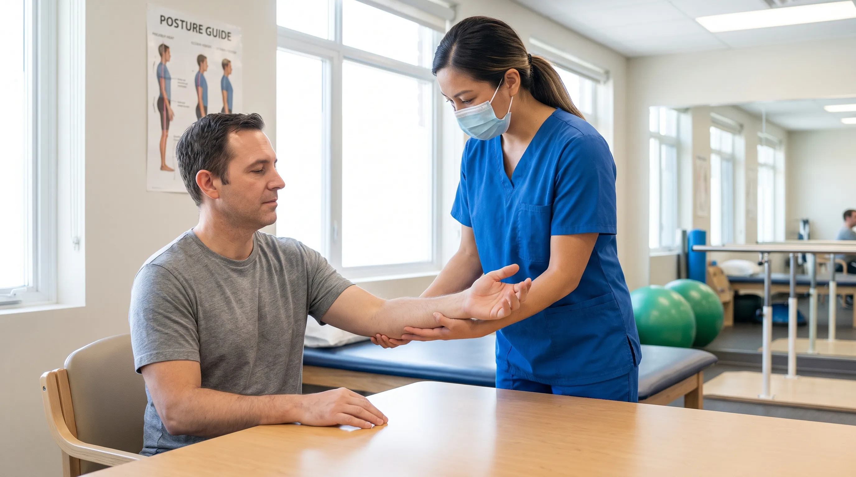
Integrating visual guidance into your recovery process significantly enhances exercise accuracy and reduces the risk of compensatory movements that delay healing. Professional demonstration videos provide clear biomechanical references for proper form, breathing patterns, and movement pacing.
Following structured exercise progressions ensures that each rehabilitation phase builds appropriately upon the previous one, establishing a solid foundation for sustained strength gains. Consistency in technique and gradual intensity increases remain essential for optimal tissue adaptation and long-term upper limb health.

Frequently Asked Questions
How long does it take for a pulled muscle in the forearm to heal completely?
Healing time depends heavily on the severity of the initial tissue damage and your adherence to structured rehabilitation protocols. Mild Grade 1 strains typically resolve within two to three weeks with consistent rest, gentle mobility work, and gradual loading. Moderate Grade 2 tears require three to six weeks of guided physical therapy, incorporating progressive resistance exercises and manual therapy techniques. Severe Grade 3 ruptures involving substantial fiber disruption can take two to four months, occasionally requiring specialized orthopedic intervention. Factors such as age, baseline fitness level, nutritional status, smoking habits, and sleep quality significantly influence cellular repair rates. Following evidence-based recovery guidelines and avoiding premature heavy loading dramatically improves outcomes and minimizes chronic dysfunction risk.
Should I apply heat or ice to a strained forearm muscle?
The application of thermal therapy depends entirely on the injury timeline and tissue healing phase. During the initial acute phase, typically the first forty-eight to seventy-two hours, ice therapy remains the gold standard. Applying cryotherapy for fifteen to twenty minutes at two-hour intervals constricts blood vessels, limits excessive inflammation, and reduces pain signal transmission to the central nervous system. Once acute swelling stabilizes and the injury transitions into the subacute proliferative phase, moist heat application becomes more beneficial. Heat therapy increases local blood flow, enhances tissue extensibility, relaxes hypertonic muscle fibers, and prepares the area for gentle stretching and mobilization exercises. Alternating contrast baths can also stimulate vascular pumping and accelerate metabolic waste clearance during intermediate recovery stages.
When should I see a doctor for forearm pain instead of treating it at home?
Home management remains appropriate for mild discomfort with gradual improvement over several days, but certain clinical presentations require immediate professional evaluation. Seek urgent medical attention if you experience a audible popping sound during injury, rapid and severe swelling, visible muscle deformity, or complete inability to extend or flex your wrist and fingers. Neurological symptoms such as persistent numbness, tingling, burning sensations, or radiating pain extending past the elbow into the hand suggest potential nerve compression or cervical involvement that requires specialized diagnostic testing. Additionally, if pain fails to improve after two weeks of consistent conservative management, worsens despite rest, or severely disrupts sleep and daily functioning, comprehensive medical assessment becomes necessary to rule out fractures, tendon avulsions, or compartment syndromes.
Can I continue light exercise while recovering from a pulled forearm muscle?
Strategic activity modification often accelerates recovery compared to complete immobilization, provided that movements remain within pain-free ranges and respect tissue healing limitations. During the early inflammatory phase, prioritize gentle active range-of-motion exercises, isometric contractions, and proximal joint mobility work that does not stress the injured forearm directly. As swelling decreases and baseline pain subsides, gradually incorporate low-intensity stretching, light resistance band work, and functional movement patterns that promote blood circulation and neuromuscular re-education. Avoid heavy gripping, weight-bearing through the arm, high-velocity pulling motions, or repetitive stress activities until you can perform full-range movements without discomfort or compensatory muscle activation. A licensed physical therapist can design a personalized loading progression that safely challenges healing tissue while maintaining cardiovascular fitness and overall conditioning.
What is the difference between a pulled forearm muscle and tennis elbow?
Understanding the anatomical distinction between these two common conditions ensures appropriate treatment selection and prevents mismanagement. A pulled forearm muscle involves structural damage to the contractile tissue fibers themselves, producing localized aching, swelling, and pain that intensifies during active muscle contraction or direct stretching. The discomfort typically spans a broader area along the muscle belly and improves predictably with rest and conservative care. Tennis elbow, clinically termed lateral epicondylitis, represents a degenerative tendinopathy affecting the common extensor tendon attachment site at the outer elbow bone. This condition generates sharp, localized pain directly over the lateral epicondyle that worsens specifically during resisted wrist extension or forceful gripping, without significant muscle belly swelling. Treatment protocols differ substantially, with muscle strains responding well to progressive loading and mobility restoration, while tendinopathy requires eccentric strengthening, load management, and targeted tissue regeneration strategies.
Conclusion
Recovering from a pulled muscle in forearm requires patience, strategic planning, and consistent adherence to evidence-based rehabilitation principles. While the initial discomfort can feel overwhelming, understanding the biological healing process empowers you to implement targeted interventions that support tissue regeneration rather than hinder it. Prioritizing proper acute management, transitioning smoothly through structured mobility and strengthening phases, and addressing underlying biomechanical or ergonomic risk factors creates a comprehensive recovery pathway that minimizes complications and prevents recurrence. Remember that true functional restoration extends beyond mere pain elimination; it encompasses restored strength, improved flexibility, enhanced proprioception, and the confidence to return to daily activities without restriction. By integrating progressive loading protocols, optimizing workplace and training environments, maintaining balanced nutrition, and consulting healthcare professionals when symptoms persist, you can successfully navigate the healing journey and emerge with a stronger, more resilient upper limb. Your forearm muscles are remarkably adaptable; treating them with respect, providing adequate recovery time, and following scientifically supported rehabilitation strategies will ensure you regain full functionality and prevent future injuries.
How we work
Researched and drafted with AI assistance, then reviewed by a human editor against the cited sources.
This article is general health information, not medical advice. Consult a qualified healthcare professional about your own situation.