Understanding and Relieving Flat Feet Soreness: A Complete Evidence-Based Guide

Key points
- Flat feet soreness typically stems from posterior tibial tendon dysfunction, ligament laxity, or mechanical overload rather than the arch shape alone.
- Conservative management, including orthotics, supportive footwear, calf stretching, and intrinsic foot strengthening, resolves symptoms in the majority of patients.
- Rigid flatfoot, sudden arch collapse, or unilateral symptoms require prompt orthopedic evaluation to rule out tendon rupture or neurological pathology.
- Consistent lifestyle modifications, weight management, and surface awareness are critical for preventing symptom recurrence and preserving long-term foot function.
- Surgical intervention remains a highly effective last-resort option for severe structural deformities, offering reliable pain relief and restored alignment when conservative methods are exhausted.
Experiencing persistent discomfort in the arches, ankles, or lower legs can significantly impact your daily mobility, but understanding the underlying mechanics of flat feet soreness is the first step toward lasting relief. The human foot is a marvel of biomechanical engineering, designed to absorb shock, distribute weight evenly, and propel us forward with every step. When the medial longitudinal arch collapses, however, this intricate system becomes overloaded. While many individuals live their entire lives with flat feet and never notice a difference, a substantial subset experiences recurring inflammation, muscular fatigue, and joint strain. This comprehensive guide explores the clinical realities of arch collapse, breaks down the most effective evidence-based interventions, and provides actionable strategies to restore comfort and function. Whether your discomfort stems from a congenital trait, gradual tendon degeneration, or acute overuse, understanding the root causes of flat feet soreness empowers you to make informed decisions about treatment, footwear, and long-term foot health.
Understanding the Anatomy and Biomechanics of Arch Collapse
To properly address flat feet soreness, it is essential to first understand how a healthy foot functions under load. The foot contains three primary arches: the medial longitudinal arch (most prominent), the lateral longitudinal arch, and the transverse arch. These structures act as natural shock absorbers, converting rigid levers for propulsion into flexible mechanisms for ground adaptation. When you stand or walk, weight is transferred from the heel through the midfoot and into the forefoot. In an optimally arched foot, the arches compress slightly upon impact, storing elastic energy that is then released during push-off. This kinetic chain relies heavily on the integrity of tendons, ligaments, and the precise alignment of twenty-six bones per foot.
Free calculatorBMI CalculatorCalculate Body Mass IndexOpen the calculatorThe Role of the Arches in Shock Absorption
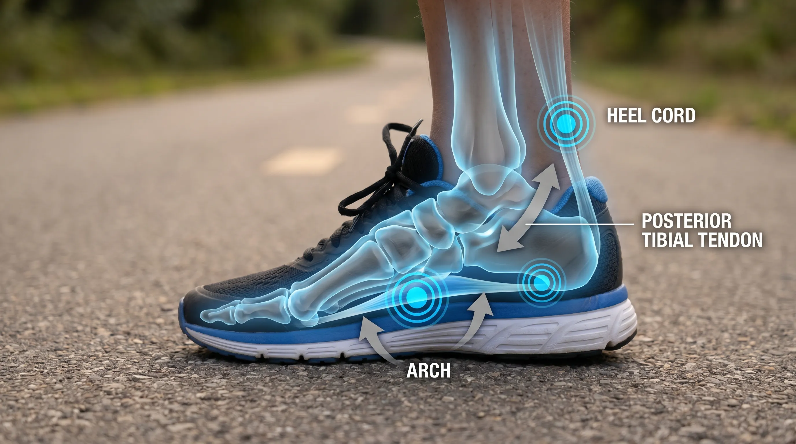
The medial arch is primarily supported by the plantar fascia, the spring ligament (plantar calcaneonavicular ligament), and the posterior tibial tendon (PTT). When these structures weaken or stretch beyond their physiological limits, the arch loses its ability to maintain its curved contour during weight-bearing activities. As the arch flattens, the calcaneus (heel bone) tilts outward into a valgus position, and the forefoot abducts. This misalignment alters the entire kinetic chain, forcing the knees, hips, and lower back to compensate. The resulting mechanical inefficiency is a primary driver of flat feet soreness, as tissues that were never designed to bear excessive tensile or compressive loads are forced to adapt. Over time, microtrauma accumulates, leading to localized inflammation, tendon degeneration, and chronic discomfort.
Flexible vs. Rigid Flatfoot: What’s the Difference?
Clinicians categorize arch collapse into distinct phenotypes, each requiring different management approaches. Flexible flatfoot is the most common presentation, particularly in children. In this variation, the arch is clearly visible when the foot is non-weight-bearing (such as when sitting or standing on tiptoes) but disappears when the individual stands flat. This condition is typically benign and painless in younger populations, though it can progress into symptomatic flat feet soreness if muscular imbalances or excessive pronation are left unaddressed. Conversely, rigid flatfoot lacks an arch even when the foot is off the ground. This presentation often indicates a more serious structural abnormality, such as tarsal coalition (where two or more foot bones are fused), arthritic changes, or severe posterior tibial tendon rupture. Rigid deformities frequently cause significant pain, restricted ankle dorsiflexion, and require comprehensive orthopedic evaluation to rule out underlying congenital or degenerative pathology.
Adult-Acquired Flatfoot and Posterior Tibial Tendon Dysfunction
The progressive nature of arch collapse in adulthood is frequently termed Progressive Collapsing Foot Deformity (PCFD), formerly known as adult-acquired flatfoot. According to the American Academy of Orthopaedic Surgeons, PCFD is a complex, multi-stage condition that begins with posterior tibial tendon dysfunction (PTTD). The PTT originates behind the medial malleolus (inner ankle bone) and fans out across the arch, acting as the primary dynamic stabilizer that prevents arch collapse and controls foot alignment during the gait cycle. When the PTT becomes inflamed, frayed, or ruptured due to repetitive microtrauma, systemic inflammation, or acute injury, the arch sags. This initiates a biomechanical vicious cycle: as the PTT weakens, the spring ligament stretches, transferring excessive load to surrounding joints and secondary tendons. These structures, unable to handle the new mechanical demands, also begin to deteriorate, placing even greater stress on the already compromised PTT. Understanding this degenerative cascade is critical for interrupting the progression of flat feet soreness before it leads to irreversible joint deformities.
Recognizing the Symptoms and Clinical Warning Signs
Symptoms of arch-related discomfort rarely present in isolation. They typically evolve gradually, beginning as mild fatigue and progressing into localized pain, swelling, and functional limitations. The Mayo Clinic and MedlinePlus (NIH) consistently highlight several hallmark indicators that clinicians use to differentiate benign arch variation from pathological flat feet soreness.
Primary Areas of Discomfort
The most common site of pain is directly along the medial longitudinal arch and the inside of the foot and ankle. Patients frequently report a deep, aching sensation that intensifies with prolonged standing, walking on uneven terrain, or participating in high-impact sports. As the condition advances, pain often radiates anteriorly into the medial midfoot or posteriorly along the course of the posterior tibial tendon. Swelling along the inner ankle is another classic sign, particularly noticeable at the end of the day or after extended periods of physical activity. In some cases, the pain migrates laterally to the outside of the ankle or even into the lateral midfoot, caused by compression of the sinus tarsi joint due to the forefoot abduction that accompanies severe arch collapse.
Gait Alterations and Secondary Pain Patterns
Biomechanical compensation rarely stays localized. As flat feet soreness persists, individuals unconsciously alter their walking patterns to minimize discomfort. Common adaptations include excessive inward rolling (overpronation), shortened stride length, or toeing-out to increase base of support. These compensatory mechanisms shift stress upward into the kinetic chain. The lower leg may develop medial shin pain (often mistaken for tibial stress syndrome), the knees can experience valgus stress leading to patellofemoral tracking issues, and hip internal rotation may increase, contributing to lower back strain. Additionally, shoes tend to wear unevenly, with accelerated breakdown along the medial edge of the sole and the inner heel counter. This rapid shoe deterioration serves as a visible external indicator of the internal biomechanical breakdown causing flat feet soreness.
Pediatric vs. Adult Symptom Presentation
Children and adults experience arch-related symptoms differently. Most pediatric flat feet are flexible and asymptomatic, resolving naturally as foot muscles strengthen and fat pads redistribute. The National Health Service (NHS) emphasizes that intervention in asymptomatic children is unnecessary and may even delay natural developmental milestones. However, children who complain of foot pain, fatigue during play, frequent tripping, or reluctance to participate in physical activities warrant clinical assessment. In adults, symptoms are more directly tied to degenerative changes, occupational demands, or systemic health factors. Adult flat feet soreness rarely resolves spontaneously and typically requires proactive management to prevent progressive tendon failure and joint degeneration.
Primary Causes and Risk Factors Driving Discomfort
Arch collapse rarely occurs without contributing factors. While some individuals are born with naturally lower arches due to genetic inheritance, most cases of symptomatic flat feet soreness result from a combination of mechanical stress, tissue degeneration, and systemic health conditions. Recognizing these risk factors is essential for targeted prevention and effective treatment planning.
Genetic Predisposition and Congenital Factors
Family history plays a substantial role in foot morphology. Genetic conditions that affect connective tissue elasticity and joint stability significantly increase susceptibility to arch collapse. Disorders such as Ehlers-Danlos Syndrome and Marfan Syndrome compromise collagen synthesis, leading to hypermobility and ligamentous laxity that cannot adequately support the medial arch. Similarly, conditions like cerebral palsy or spina bifida can cause muscle imbalance or neurological deficits that alter weight distribution and tendon function, predisposing individuals to rigid or flexible flat feet. Congenital bone malformations, where the tarsal bones do not develop properly in utero, can also create rigid structural flatfoot that requires early orthopedic intervention.
The Impact of Weight and Systemic Conditions
Excess body weight dramatically increases the mechanical load on foot structures with every step. Obesity is one of the most significant modifiable risk factors for flat feet soreness, as the plantar fascia, PTT, and midfoot ligaments must absorb substantially higher ground reaction forces. Over time, this chronic overload leads to tissue elongation and inflammatory microtears. Systemic diseases further compound this risk. Rheumatoid arthritis triggers autoimmune-mediated joint inflammation that destroys supportive cartilage and ligaments. Diabetes mellitus contributes to peripheral neuropathy and impaired microvascular healing, making tendons more prone to silent degeneration. Aging naturally reduces tendon vascularity and collagen elasticity, explaining why adult-acquired flatfoot is most prevalent in individuals over fifty. Hormonal changes during pregnancy can also temporarily relax ligaments via relaxin secretion, occasionally triggering transient arch flattening and discomfort.
Trauma, Overuse, and the Progressive Degenerative Cycle
Acute injuries, such as ankle sprains, fractures, or direct tendon trauma, can disrupt the delicate balance of midfoot stability. However, gradual overuse injuries are far more common culprits in flat feet soreness. Repetitive high-impact activities (running, jumping, prolonged standing on hard surfaces) subject the posterior tibial tendon to millions of micro-stress cycles. Without adequate recovery, the tendon undergoes tendinosis (degeneration without significant inflammation), losing its structural integrity. The AAOS OrthoInfo framework describes this as a biomechanical downward spiral: initial tendon weakening allows subtle arch lowering, which alters the lever arm and increases the force required from the PTT during push-off. The tendon, already compromised, must work harder under worse mechanical conditions, accelerating tissue failure. Environmental factors, such as consistently walking on uneven ground or wearing completely flat, unsupported footwear (like flip-flops), exacerbate this cycle by removing external stabilization and forcing intrinsic foot muscles to overcompensate.
Evidence-Based Strategies to Manage Flat Feet Soreness
Conservative, non-surgical interventions form the cornerstone of treatment for most individuals experiencing arch-related discomfort. Clinical guidelines from the Mayo Clinic and NHS strongly recommend a stepped approach that prioritizes activity modification, biomechanical support, and targeted rehabilitation before considering invasive procedures. The goal is to reduce inflammation, restore optimal load distribution, and strengthen the musculotendinous unit to prevent further degeneration.
Optimizing Footwear and Orthotic Support
Selecting appropriate footwear is the most immediate and effective way to mitigate flat feet soreness. Shoes should feature a firm heel counter, adequate midfoot torsional rigidity (meaning the shoe shouldn't twist easily when held at both ends), and a structured medial post to control overpronation. Avoid completely flat shoes, sandals with minimal support, or overly cushioned running shoes that lack structural guidance. Arch-support orthotics serve as a bridge between shoe and foot, providing customized load redistribution. Over-the-counter arch supports work well for mild symptoms, offering general medial cushioning and heel stabilization. Custom prescription orthotics, fabricated from 3D scans or plaster molds, address specific biomechanical deficiencies, accommodate asymmetrical foot shapes, and incorporate precise posting angles to realign the subtalar joint. The table below compares these interventions to help guide decision-making:
| Intervention Type | Best For | Pros | Cons |
|---|---|---|---|
| Over-the-Counter Insoles | Mild soreness, temporary relief, active lifestyles | Affordable, immediately available, adaptable to multiple shoes | Less precise, materials degrade quickly, limited structural correction |
| Custom Prescription Orthotics | Moderate to severe PCFD, asymmetrical gait, chronic tendon issues | Precise anatomical fit, durable materials, targeted biomechanical correction | Higher cost, requires professional fitting, initial adaptation period |
| Supportive Motion-Control Shoes | Daily wear, occupational standing, high-impact sports | Integrated stability, reduces reliance on separate inserts, cost-effective over time | Heavier design, limited aesthetic options, may not suit narrow feet |
Targeted Stretching and Strengthening Protocols
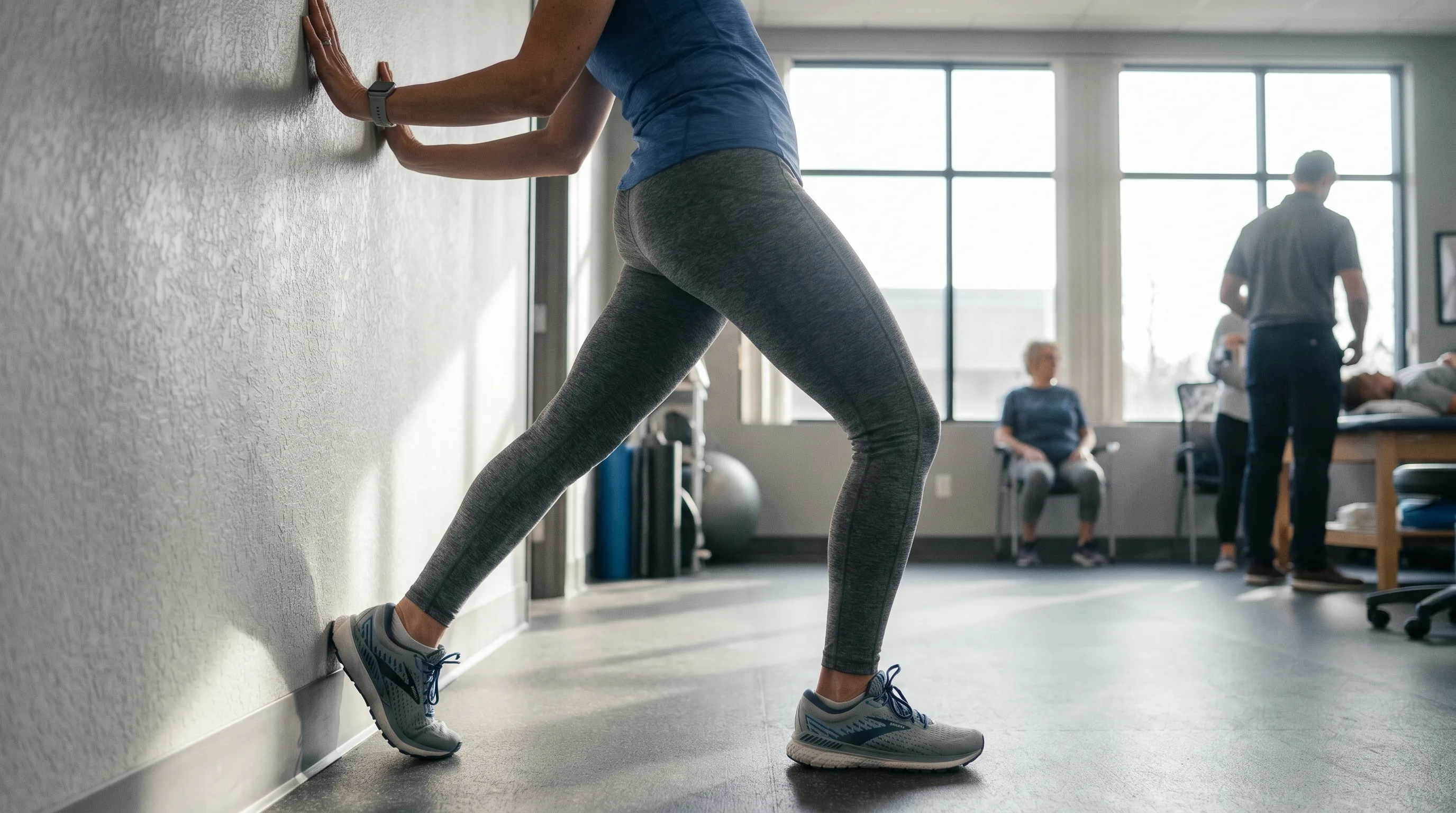
Restoring tissue elasticity and muscular endurance is critical for long-term management of flat feet soreness. The calf muscles (gastrocnemius and soleus) often become chronically shortened in individuals with flat arches, restricting ankle dorsiflexion and increasing strain on the posterior chain during gait. The American Academy of Orthopaedic Surgeons recommends a standardized heel cord stretch: lean forward against a wall with one leg positioned in front of the other. Keep the back leg straight, press the heel firmly into the floor, and bend the front knee. Maintain this position for 15 to 30 seconds while keeping both heels flat. Point the toes of the rear foot slightly toward the heel of the front foot to maximize stretch along the deep calf fascia. Repeat this exercise three times per leg, daily. Additionally, intrinsic foot strengthening exercises such as toe curls, towel scrunches, and short-foot exercises (actively raising the arch without curling the toes) help rebuild the muscular corset that supports the medial arch. Consistency with these protocols typically yields measurable improvements in arch height, push-off strength, and overall pain reduction within two months.
Physical Therapy and Modalities for Pain Relief
When home exercises and footwear modifications provide insufficient relief, structured physical therapy becomes necessary. Licensed therapists employ manual techniques to mobilize restricted tarsal joints, perform instrument-assisted soft tissue mobilization (IASTM) to break down tendon adhesions, and apply therapeutic taping to offload the PTT during the healing phase. Modalities such as ultrasound therapy, cryotherapy, and low-level laser therapy may be utilized to reduce localized inflammation and promote tissue regeneration. Gait retraining is a crucial component, as therapists analyze walking patterns using pressure plates or video analysis to identify and correct biomechanical faults. By addressing muscular imbalances, improving proprioception, and teaching proper load management, physical therapy addresses the root mechanical drivers of flat feet soreness rather than merely masking symptoms with medication.
Advanced Medical Interventions and Surgical Options
Despite comprehensive conservative care, a subset of patients will progress to stages of PTTD or rigid flatfoot that require surgical intervention. Surgery is not a first-line treatment but rather a carefully considered option when structural deformities prevent functional rehabilitation, or when tendon rupture has occurred. The MedlinePlus (NIH) and AAOS guidelines emphasize that surgical success heavily depends on accurate staging of the disease and selecting the appropriate procedural approach.
When Conservative Management Fails
Indications for surgical consultation include persistent, debilitating pain that severely limits activities of daily living despite six months of dedicated physical therapy and orthotic use; complete posterior tibial tendon rupture; rigid deformities unresponsive to bracing; or secondary joint arthritis in the subtalar or talonavicular joints. Imaging studies such as weight-bearing X-rays, MRI, or ultrasound are essential to assess tendon integrity, joint alignment, and ligamentous damage. In the early stages (Stage I and II), where the tendon is inflamed or moderately elongated but still functional, procedures focus on tendon debridement, synovectomy (removal of inflamed tissue), or tendon transfer (typically the flexor digitorum longus) to restore dynamic arch support. These joint-sparing techniques preserve natural mobility while reconstructing the stabilizing mechanism.
Reconstructive Procedures and Recovery Expectations
Advanced deformities (Stage III and IV) often require more extensive reconstructive surgery. Osteotomies, where bones are carefully cut and realigned, are commonly combined with ligamentous reconstructions to restore proper arch height and hindfoot alignment. In cases of severe joint degeneration, arthrodesis (joint fusion) may be performed to eliminate painful arthritic motion by permanently fusing the talonavicular or subtalar joints. While the prospect of foot surgery can be daunting, modern techniques utilize internal fixation devices, bone grafting, and accelerated rehabilitation protocols that significantly improve recovery timelines. Post-operative care typically involves six to eight weeks of non-weight-bearing immobilization in a cast or boot, followed by gradual physical therapy focused on scar mobilization, range-of-motion restoration, and progressive weight-bearing. Full functional recovery can take six to twelve months, but the majority of patients report dramatic reductions in flat feet soreness and restored ability to walk comfortably on level ground.
Lifestyle Modifications and Long-Term Foot Health
Sustainable management of arch-related conditions extends far beyond clinical treatments. Daily habits, environmental factors, and proactive foot care routines play a pivotal role in preventing symptom recurrence and maintaining structural integrity. Integrating these strategies into your lifestyle ensures that progress made through therapy and orthotics is preserved long-term.
Activity Modification and Surface Awareness
The surfaces you walk on significantly impact the stress placed on compromised arches. Concrete, asphalt, and hard tile offer zero shock absorption, transmitting ground reaction forces directly into the midfoot. Whenever possible, incorporate walking routes with softer surfaces like grass, dirt trails, or rubberized tracks. If your occupation requires prolonged standing on hard floors, invest in anti-fatigue mats that encourage micro-muscle engagement and distribute pressure more evenly. Activity modification also means balancing high-impact exercises with low-impact alternatives. Swapping daily running sessions for swimming, cycling, or elliptical training allows tendons to recover while maintaining cardiovascular fitness. Avoid sudden spikes in training volume, as rapid increases in mileage or intensity are classic triggers for tendon overload and exacerbated flat feet soreness.
Maintenance Routines for Sustainable Relief
Foot maintenance should be treated as a non-negotiable aspect of overall wellness. Implement a nightly routine that includes gentle calf and plantar fascia rolling using a frozen water bottle or specialized massage ball to reduce tissue tension. Rotate between two pairs of supportive shoes to allow midsole foams to fully decompress, preventing premature material fatigue. Monitor shoe wear patterns monthly; excessive medial breakdown indicates that your current footwear is no longer providing adequate pronation control. Maintain a healthy body weight through balanced nutrition and regular exercise, as even a modest reduction in body mass significantly decreases the mechanical load on foot structures. Finally, schedule periodic check-ins with a podiatrist or physical therapist to assess orthotic integrity, monitor tendon health, and adjust rehabilitation protocols as needed. Consistent attention to these details transforms passive symptom management into active, lifelong foot preservation.
Frequently Asked Questions
Can flat feet soreness be permanently fixed without surgery?
In many cases, flat feet soreness can be effectively managed and significantly reduced through conservative treatments like custom orthotics, targeted physical therapy, and supportive footwear. While surgery is reserved for severe structural deformities or tendon tears that fail to respond to non-invasive care, most individuals achieve lasting relief without operative intervention.
Why does flat feet soreness worsen when walking barefoot?
Walking barefoot removes external arch support, forcing the posterior tibial tendon and plantar ligaments to bear the full load of body weight. This lack of cushioning and structural reinforcement increases strain on the arch, leading to accelerated fatigue, microtrauma, and heightened soreness after prolonged activity.
Are custom orthotics better than store-bought insoles for flat feet?
Custom orthotics are generally more effective because they are molded to your exact foot anatomy, gait mechanics, and pressure distribution patterns. Over-the-counter inserts provide generalized support and may work well for mild symptoms, but rigid or highly pronated feet often require prescription-grade materials and precise biomechanical alignment for optimal soreness reduction.
How long does it take for physical therapy to relieve flat feet soreness?
Most patients experience noticeable improvement within 6 to 12 weeks of consistent physical therapy. Strengthening the intrinsic foot muscles, stretching tight heel cords, and correcting gait mechanics takes time, and adherence to a home exercise program significantly accelerates the timeline for lasting symptom relief.
Should children with flat feet wear orthotics to prevent soreness?
No, routine orthotic use in asymptomatic children is generally not recommended. According to pediatric guidelines, most flat feet in young children are flexible and painless, naturally resolving as the foot arches develop between ages 3 and 10. Orthotics are only indicated if the child experiences persistent pain, frequent tripping, or restricted ankle mobility.
Conclusion
Navigating flat feet soreness requires patience, consistency, and a clear understanding of how your foot mechanics interact with daily demands. While arch collapse is common, it does not have to dictate a life of chronic pain or limited mobility. By integrating supportive footwear, targeted rehabilitation, and proactive lifestyle adjustments, you can effectively interrupt the degenerative cycle, restore functional stability, and reclaim comfortable movement. Remember that early intervention significantly improves long-term outcomes; ignoring progressive symptoms only allows structural deterioration to advance. Consult a healthcare professional if your pain limits daily activities, appears suddenly, or affects only one foot. With evidence-based care and dedicated self-management, the journey from arch-related discomfort to resilient foot health is entirely achievable.
Key Takeaways
- Flat feet soreness typically stems from posterior tibial tendon dysfunction, ligament laxity, or mechanical overload rather than the arch shape alone.
- Conservative management, including orthotics, supportive footwear, calf stretching, and intrinsic foot strengthening, resolves symptoms in the majority of patients.
- Rigid flatfoot, sudden arch collapse, or unilateral symptoms require prompt orthopedic evaluation to rule out tendon rupture or neurological pathology.
- Consistent lifestyle modifications, weight management, and surface awareness are critical for preventing symptom recurrence and preserving long-term foot function.
- Surgical intervention remains a highly effective last-resort option for severe structural deformities, offering reliable pain relief and restored alignment when conservative methods are exhausted.
How we work
Researched and drafted with AI assistance, then reviewed by a human editor against the cited sources.
This article is general health information, not medical advice. Consult a qualified healthcare professional about your own situation.