How Do U Know If Toe Is Broken: Symptoms, Diagnosis & Recovery Guide

Understanding exactly how do u know if toe is broken can save you from unnecessary pain, chronic mobility issues, and long-term joint complications. The human foot contains twenty-six bones, with fourteen located exclusively in the toes. Explore detailed foot anatomy and structural vulnerabilities to better understand why these digits are prone to injury. These small but structurally vital phalanges endure immense daily pressure during walking, running, and balancing. When trauma strikes, whether from dropping a heavy object, stubbing your foot against furniture, or experiencing high-impact sports injuries, the resulting damage often mimics other conditions. This overlap in symptom presentation is why proper evaluation matters so much. A fractured toe is not merely an inconvenience; it disrupts your kinetic chain, alters your gait, and forces compensatory movements that can strain your knees, hips, and lower back. If left untreated or improperly managed, even minor fractures can develop into malunions, chronic arthritis, or recurrent pain that limits athletic performance and daily activities. By understanding the clinical markers, differentiating between soft tissue injuries and bony damage, and following evidence-based management protocols, you can navigate recovery safely and efficiently. Whether you are an athlete seeking to return to competition or a professional needing to maintain mobility, recognizing the precise indicators of a fracture is the critical first step toward optimal healing.
Understanding Toe Anatomy and Why Fractures Occur
To truly grasp the nature of toe injuries, it is essential to examine the anatomical architecture that makes these digits both resilient and vulnerable. Each toe consists of three phalangeal bones: the proximal, middle, and distal phalanges. The exception is the great toe, or hallux, which contains only two phalanges but bears approximately forty percent of the weight during normal gait cycles. These bones articulate at the interphalangeal joints and connect proximally to the metatarsals via the metatarsophalangeal joints. The structural integrity of the toes relies heavily on a complex network of tendons, ligaments, and the intrinsic plantar and dorsal muscles of the foot. When trauma exceeds the physiological load capacity of this system, bone failure occurs.
Free calculatorFoot Reflexology ChartFoot reflexology points and corresponding organsOpen the calculatorThe Structural Role of Phalanges
The phalanges serve multiple biomechanical functions beyond mere weight distribution. They act as levers during the push-off phase of walking, provide sensory feedback through dense nerve endings, and assist in maintaining balance on uneven surfaces. The cortical bone of the toes is thinner than in larger weight-bearing bones, making them particularly susceptible to direct impact trauma. Additionally, the rich vascular supply surrounding the phalanges means that fractures often present with rapid and pronounced swelling and bruising. The plantar plate ligament, a thick fibrocartilaginous structure beneath the metatarsophalangeal joint, works synergistically with the phalanges to stabilize toe movement. When this stabilizer is compromised alongside a fracture, joint instability becomes a significant risk factor that complicates recovery timelines.
Common Mechanisms of Injury
Trauma to the toes typically follows three primary pathways. Direct impact injuries account for the majority of fractures, occurring when heavy objects fall onto the foot or when the toe strikes a rigid surface at high velocity. Axial loading injuries happen when the toe is forcefully driven backward into the metatarsal, commonly seen during sports where feet are abruptly halted while running forward. Repetitive stress fractures, though less common in the toes compared to the metatarsals, can develop in athletes engaged in high-volume jumping or running activities without adequate recovery periods. Environmental factors also play a role; cold temperatures can stiffen tissues, reducing shock absorption and increasing fracture risk during winter sports. Footwear choices further influence injury likelihood, as shoes with narrow toe boxes or insufficient structural support fail to protect the digits during sudden impacts.
Recognizing the Key Symptoms of a Broken Toe
Identifying a fracture requires careful observation of clinical signs and an understanding of how your body responds to skeletal trauma. Review the established clinical signs of toe fractures to distinguish serious injuries from minor impacts. Pain is the universal signal, but its characteristics, timing, and associated features determine whether you are dealing with a simple contusion or a genuine fracture. The acute inflammatory response following bone injury follows a predictable physiological cascade, releasing cytokines, histamines, and prostaglandins that trigger localized swelling, increased blood flow, and nerve sensitization. Recognizing these patterns early allows for timely intervention and prevents secondary complications.
Pain Characteristics and Severity
Fracture pain is typically sharp, localized, and immediately follows the traumatic event. Unlike muscle strains that may develop gradually or dull aches that fluctuate with weather changes, bone pain is often described as deep, throbbing, and exacerbated by any direct pressure or movement. The intensity usually peaks within the first twenty-four hours as edema expands within the confined tissue compartments of the digit. Pain that radiates along the entire foot or intensifies when the toe is passively flexed or extended strongly suggests structural compromise. Nighttime pain is another hallmark; as inflammation increases while you lie still, pressure builds within the periosteum, the highly innervated membrane covering the bone, causing persistent discomfort that disrupts sleep. If you find yourself constantly asking how do u know if toe is broken, pay close attention to whether the pain persists beyond forty-eight hours of basic rest and elevation, as this indicates ongoing structural stress rather than superficial tissue trauma.
Visible Signs: Swelling, Bruising, and Deformity
External manifestations provide immediate visual clues. Swelling typically envelops the entire toe within hours, sometimes extending to adjacent digits and the forefoot. The inflammatory process causes fluid accumulation in the interstitial spaces, creating a taut, shiny appearance over the skin. Ecchymosis, or bruising, follows as damaged blood vessels leak hemoglobin breakdown products into surrounding tissues. Bruising may migrate distally over several days due to gravity, appearing on the sole or between toes. Deformity is the most definitive sign of a displaced fracture. This may present as an unnatural angle, overlapping alignment, rotation, or shortening of the digit. Subungual hematomas, characterized by dark purple or black blood pooling beneath the toenail, frequently accompany phalangeal fractures and indicate significant trauma to the nail bed and underlying bone. While these visual markers are highly suggestive, their absence does not completely rule out a hairline or non-displaced fracture, which can present with minimal external changes despite underlying structural damage.
Functional Limitations and Mobility Issues
Beyond pain and appearance, functional impairment serves as a critical diagnostic indicator. A healthy toe contributes to balance and propulsion, allowing smooth weight transfer during gait cycles. When fractured, the digit loses its mechanical efficiency. You may notice difficulty pushing off the ground, an altered walking pattern, or an inability to wear standard footwear due to pressure sensitivity. Attempting to stand on tiptoes or perform single-leg balance tests becomes markedly challenging or painful. Compensatory gait alterations, such as limping, favoring the lateral foot edge, or walking on the heel, place abnormal stress on other musculoskeletal structures. These adaptations, while protective in the short term, can lead to secondary injuries in the ankle, knee, or hip if maintained for extended periods. Monitoring functional decline helps clinicians gauge fracture severity and determines whether immobilization or surgical stabilization is required to restore normal biomechanics.
Differentiating a Broken Toe from a Sprain or Stubbed Toe
Many patients struggle with the question of how do u know if toe is broken versus experiencing a soft tissue injury or minor impact trauma. The clinical presentation of these conditions overlaps significantly, making self-diagnosis challenging. Sprains involve ligamentous stretching or tearing, contusions damage capillaries and subcutaneous tissue, and fractures disrupt the osseous matrix. While initial management often aligns across these injuries, accurate differentiation prevents inappropriate treatment timelines and ensures optimal healing conditions for the specific damaged tissue.
Overlapping Symptoms Explained
Both sprains and fractures cause immediate pain, rapid swelling, and restricted range of motion. The inflammatory response does not discriminate between tissue types; any structural damage triggers identical biochemical pathways. Bruising appears in both scenarios as blood escapes damaged vessels into surrounding tissues. This physiological similarity explains why minor fractures are frequently dismissed as severe bruises. However, ligamentous injuries typically present with joint laxity, instability, and pain localized to the connective tissue rather than deep within the bone. Stubbed toes cause acute, sharp pain that usually diminishes significantly within a few hours. Persistent discomfort, worsening swelling, or inability to bear weight beyond the first day strongly suggests skeletal involvement. The key differentiator lies in symptom trajectory: soft tissue injuries generally follow a rapid improvement curve with basic care, while fractures maintain or escalate symptoms until proper immobilization and time allow osteogenesis to commence.
The Ottawa Toe Rules: When Imaging Is Necessary
Clinical decision rules help standardize evaluation and reduce unnecessary radiographic exposure. The Ottawa Foot Rules, widely adopted in emergency medicine, recommend X-ray imaging when specific criteria are met. For toe injuries, imaging is indicated if there is bone tenderness at the base of the fifth metatarsal, the navicular bone, or any of the phalanges. Additionally, inability to bear weight immediately after injury and during clinical examination warrants radiographic evaluation. These criteria achieve high sensitivity for detecting fractures, minimizing missed diagnoses while preventing over-imaging. Understand when diagnostic imaging is medically necessary to confirm structural damage. When you wonder how do u know if toe is broken, applying these clinical markers provides an evidence-based framework for determining whether professional imaging is necessary. Physical examination techniques, including gentle palpation along the bone shafts and assessment of passive and active joint mobility, complement these rules to create a comprehensive diagnostic picture.
| Feature | Broken Toe | Sprained Toe | Stubbed Toe |
|---|---|---|---|
| Pain Duration | Persists beyond 48 hours, often worsening at night | Gradually improves over 3-7 days | Subsides significantly within 12-24 hours |
| Swelling Pattern | Rapid, extensive, often extends to forefoot | Localized to joint area, moderate severity | Mild, localized to impact site |
| Deformity | Possible angulation, rotation, or misalignment | None; joint remains structurally aligned | None |
| Weight Bearing | Severely limited, sharp pain on pressure | Tolerable with discomfort, unstable feeling | Normal after brief rest |
| Bruising Timeline | Appears within 24 hours, migrates over days | May develop, usually confined to ligament area | Minimal or absent |
| Bone Tenderness | Direct, sharp pain when pressing along bone shaft | Diffuse joint discomfort, ligament-specific | Focal impact point pain |
Medical Diagnosis: What to Expect at the Doctor’s Office
Professional evaluation transforms suspicion into certainty. When you seek clinical assessment, healthcare providers follow systematic protocols to confirm fractures, classify injury severity, and rule out associated complications. Understanding the diagnostic process reduces anxiety and prepares you for what to expect during your appointment. Accurate classification directly influences treatment pathways, recovery timelines, and long-term functional outcomes.
Physical Examination Techniques
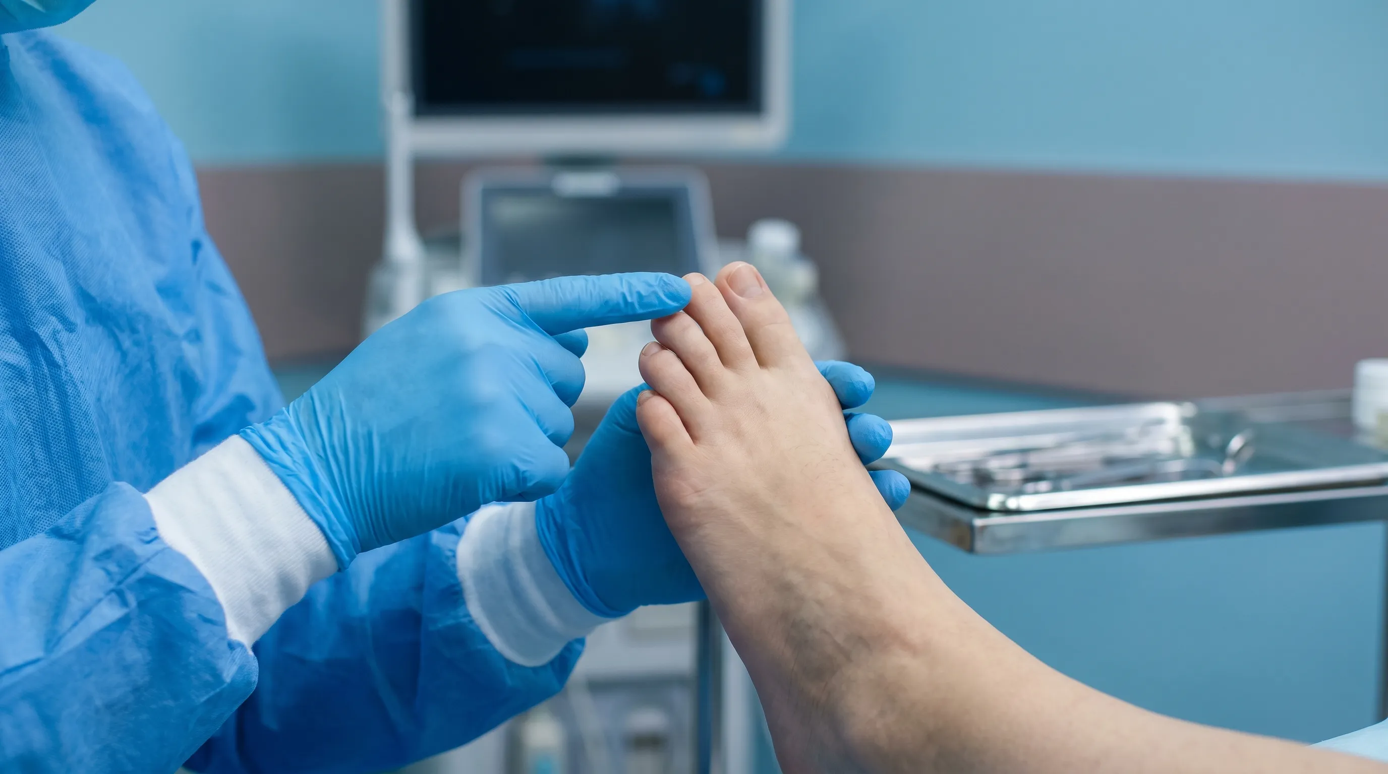
The initial assessment begins with a thorough history and observation. Clinicians document the injury mechanism, symptom onset, prior foot conditions, and any relevant medical history affecting bone health, such as osteoporosis or diabetes. Visual inspection evaluates swelling magnitude, skin integrity, nail condition, and toe alignment. Palpation proceeds systematically from proximal to distal segments, identifying precise points of maximal tenderness, crepitus, or step-off deformities. Range of motion testing assesses joint stiffness and determines whether movement restrictions stem from pain, swelling, or mechanical blockage. Neurovascular examination checks capillary refill time, sensation distribution, and pulse quality to ensure adequate blood flow and nerve function. This comprehensive physical evaluation establishes a clinical baseline and determines whether conservative management or advanced imaging is appropriate.
Imaging Modalities: X-rays, CT, and MRI
Standard radiography remains the gold standard for initial fracture detection. Multiple views, including anteroposterior, lateral, and oblique projections, visualize bone continuity, alignment, and joint space integrity. Hairline fractures may not appear on initial X-rays due to microscopic crack size and overlapping bone structures. In such cases, clinicians may recommend follow-up imaging in ten to fourteen days, allowing bone resorption at the fracture site to make the injury visible on repeat radiographs. Computed tomography scans provide three-dimensional visualization, essential for complex intra-articular fractures, comminuted injuries, or preoperative planning. Magnetic resonance imaging excels at detecting occult fractures, bone bruises, and associated soft tissue damage such as ligament tears or tendon injuries. Your provider selects the appropriate modality based on clinical suspicion, injury mechanism, and potential surgical requirements, ensuring comprehensive diagnostic accuracy.
Immediate First Aid and Home Management Strategies
Once you understand how do u know if toe is broken, implementing immediate protective measures becomes critical. Follow CDC-recommended first aid protocols to manage acute musculoskeletal trauma effectively. The first seventy-two hours represent the acute inflammatory phase, during which proper intervention significantly influences swelling magnitude, pain levels, and subsequent healing efficiency. Evidence-based self-care protocols prioritize tissue protection, edema control, and early mobilization within pain-free parameters.
The R.I.C.E. Protocol Explained
Rest, Ice, Compression, and Elevation form the cornerstone of acute injury management. Rest involves minimizing weight-bearing activities, avoiding sports, and reducing unnecessary foot movement to prevent further tissue damage. Ice application constricts blood vessels, reducing hemorrhage and inflammatory mediator release. Apply cold packs wrapped in thin cloth for fifteen to twenty minutes every two to three hours during the first forty-eight hours. Compression utilizes elastic bandages to limit interstitial fluid accumulation; ensure wrapping is snug but not constrictive to avoid vascular compromise. Elevation requires positioning the foot above heart level, leveraging gravity to facilitate venous and lymphatic drainage. Recent sports medicine research has expanded this framework to the PEACE & LOVE protocol, emphasizing Protection, Elevation, Avoidance of anti-inflammatories in early phases, Compression, and Education, followed by Load management, Optimism, Vascularization, and Exercise. Both approaches prioritize controlled inflammation management while preventing secondary tissue breakdown.
Buddy Taping: Step-by-Step Guide
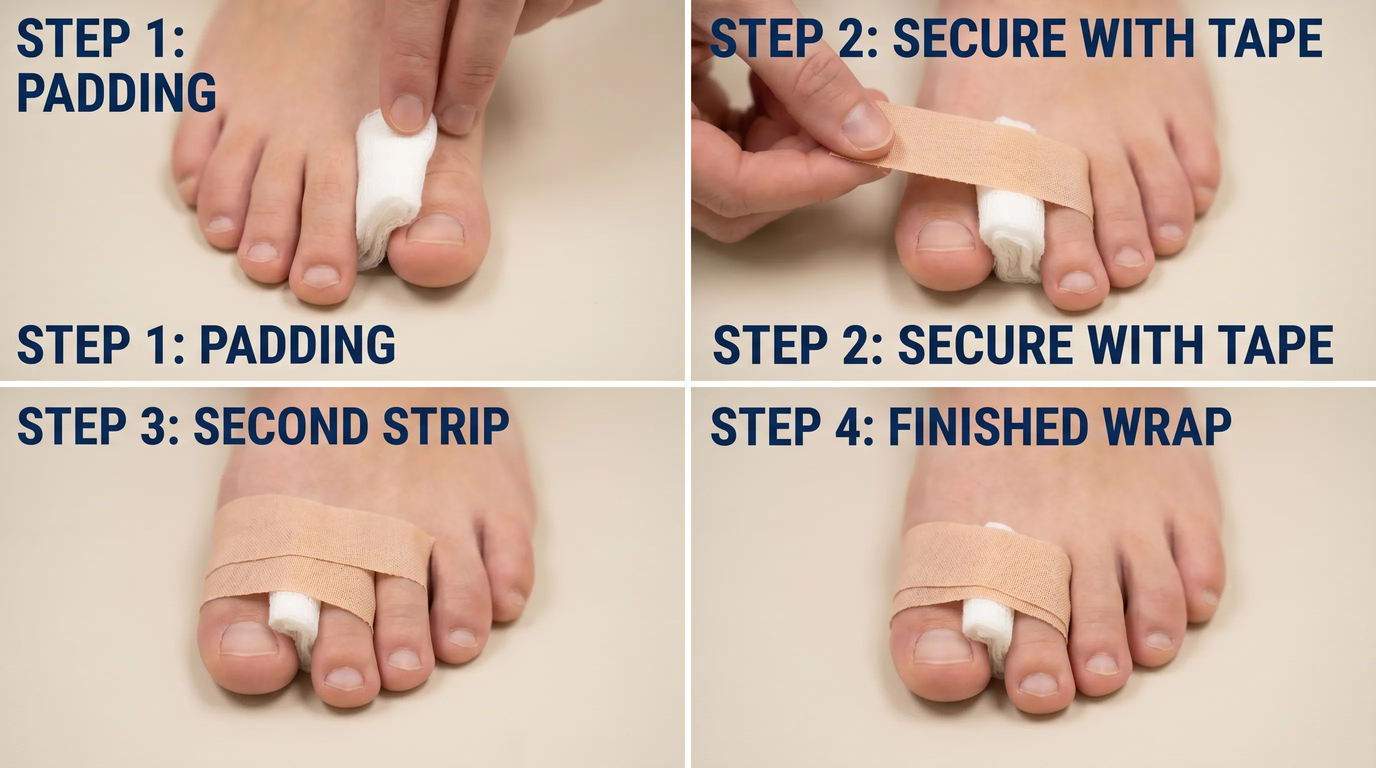
Buddy taping stabilizes the fractured toe by securing it to an adjacent healthy digit, utilizing the intact toe as a natural splint. Begin by cleansing and thoroughly drying both digits. Place a small cotton ball or gauze pad between the toes to prevent moisture accumulation and skin maceration. Align the injured toe carefully with its neighbor, ensuring natural anatomical positioning without excessive pulling or rotation. Wrap medical tape around both toes, avoiding the joint spaces to preserve circulation. Apply two to three secure wraps, maintaining gentle compression without restricting blood flow. Check distal perfusion by pressing the nail bed; color should return within two seconds. Replace the tape every two to three days or if it becomes loose, wet, or irritating. This technique provides mechanical stability, reduces movement-induced pain, and allows controlled weight-bearing during daily activities. Proper application is essential, as overly tight taping can cause ischemia, while loose taping fails to provide adequate immobilization.
Footwear Modifications for Comfort
Standard footwear often exacerbates toe fractures by compressing inflamed tissues and restricting necessary swelling accommodation. Transition to wide-toe-box shoes with stiff soles to limit toe flexion during walking. Postoperative shoes or rigid walking sandals provide structured support, distributing pressure across the midfoot and heel rather than the forefoot. Avoid soft, flexible sneakers or minimalist shoes that allow excessive toe movement. Sock selection matters equally; choose seamless, moisture-wicking fabrics that reduce friction and prevent blister formation. Elevating the toe slightly within the shoe using soft padding can prevent direct pressure on the nail bed and dorsal surface. These modifications create a protective environment that minimizes mechanical stress while allowing natural healing processes to proceed without interference.
Medical Treatments and When Surgery Is Required
While many toe fractures heal successfully with conservative management, certain injury patterns require professional intervention. Learn when medical intervention is necessary for toe injuries to prevent long-term complications. Understanding when and how medical treatments are applied ensures you seek appropriate care rather than attempting prolonged self-management for injuries that exceed home treatment capacity.
Realignment (Reduction) Procedures
Displaced fractures with angular deformity or joint incongruity often necessitate closed reduction. Under local anesthesia, the healthcare provider manually manipulates the toe to restore anatomical alignment. Gentle traction counteracts muscle spasms, followed by precise positioning of bone fragments. Post-reduction, buddy taping or specialized splints maintain alignment during initial healing. Follow-up radiographs confirm successful reduction and rule out secondary displacement. This procedure is typically performed in outpatient settings and requires minimal downtime beyond standard immobilization protocols. Proper realignment prevents long-term joint dysfunction, abnormal wear patterns, and chronic pain associated with malunited fractures.
Surgical Intervention for Complex Fractures
Open fractures, severely comminuted injuries, intra-articular fractures with step-off deformities exceeding two millimeters, or fractures involving the hallux that compromise weight-bearing mechanics often require surgical fixation. Operative techniques utilize K-wires, mini-screws, or low-profile plates to stabilize bone fragments. Surgery restores joint congruity, allows earlier rehabilitation, and reduces malunion risk. Postoperative care involves strict immobilization, wound monitoring, and progressive weight-bearing restrictions based on radiographic healing progression. Surgical candidates typically require physical therapy referral to address stiffness, muscle atrophy, and gait retraining. While invasive, these interventions prevent permanent mobility limitations when conservative management proves insufficient.
Pain Management and Medication Guidelines
Effective pain control facilitates rest, improves sleep quality, and enables early mobilization within tolerance limits. Nonsteroidal anti-inflammatory drugs provide dual analgesic and anti-inflammatory effects but may theoretically interfere with early bone healing if used excessively during the first two weeks. Acetaminophen offers reliable analgesia without impacting osteogenesis. Topical analgesics and local cold therapy provide adjunctive relief without systemic side effects. For severe pain, short-term prescription medications may be utilized under medical supervision. Medication selection should consider individual contraindications, gastrointestinal tolerance, and renal function. Gradual transition from pharmacological management to activity-based pain modulation supports long-term recovery while minimizing dependency risks.
Recovery Timeline and Long-Term Healing Process
Bone healing follows a highly coordinated biological sequence. Study the physiological stages of bone repair according to the National Institutes of Health. Understanding each phase allows you to align expectations with physiological reality, preventing premature return to high-impact activities that jeopardize structural integrity. Patience and adherence to progressive loading protocols determine whether recovery completes successfully or results in chronic complications.
Phase 1: Acute Healing (Weeks 1-2)
The inflammatory phase dominates the first fourteen days. Hematoma formation at the fracture site initiates cellular recruitment. Macrophages clear debris, fibroblasts produce collagen scaffolding, and osteoprogenitor cells migrate to the injury zone. Pain and swelling gradually subside as new vascular networks establish circulation to the regenerating tissue. Immobilization remains critical during this window, protecting the fragile fibrin clot from mechanical disruption. Gentle ankle pumps and non-weight-bearing toe wiggles, if pain-free, prevent excessive stiffness and promote lymphatic drainage. Nutritional support emphasizing protein, vitamin C, zinc, and calcium accelerates cellular proliferation. Patients often report significant symptom reduction by the end of this phase, though structural stability remains minimal.
Phase 2: Bone Remodeling (Weeks 3-6)
Cartilaginous callus transforms into woven bone through endochondral ossification. The fracture site gains mechanical strength, allowing gradual increase in weight-bearing activities. Swelling continues to resolve, and bruising dissipates completely. Clinical examination reveals reduced tenderness and improved passive range of motion. Protective footwear and buddy taping may be discontinued under medical guidance. Physical therapy introduces gentle stretching, proprioceptive exercises, and progressive resistance training. Radiographic evaluation typically shows bridging callus formation, confirming structural continuity. Return to low-impact activities becomes feasible, though high-velocity movements remain contraindicated until cortical bone matures fully.
Phase 3: Functional Rehabilitation (Weeks 7-12)
Woven bone undergoes continuous remodeling, replacing disorganized trabecular architecture with strong, lamellar bone aligned along mechanical stress lines. Full weight-bearing tolerance returns, and gait mechanics normalize. Rehabilitation focuses on restoring full range of motion, strengthening intrinsic foot muscles, and improving balance coordination. Toe curls, marble pickups, and resisted band exercises rebuild neuromuscular control. Gradual reintroduction to sport-specific drills ensures safe transition to competitive activities. Most fractures achieve clinical union by eight weeks, though complete radiographic remodeling may continue for several months. Patients often question how do u know if toe is broken has fully healed during this phase; the answer lies in pain-free full range of motion, normalized gait patterns, and radiographic confirmation of cortical continuity.
Potential Complications and When to Seek Emergency Care
While most toe fractures resolve uneventfully, certain warning signs indicate complications requiring immediate medical intervention. Identify red flags that warrant urgent orthopedic care to prevent irreversible damage. Recognizing these red flags prevents irreversible damage and ensures timely treatment adjustments.
Malunion and Nonunion Risks
Improper alignment during healing leads to malunion, resulting in toe deformity, abnormal joint mechanics, and secondary osteoarthritis. Symptoms include persistent pain, visible angulation, difficulty wearing shoes, and altered gait patterns. Nonunion occurs when bone fragments fail to bridge, often due to excessive movement, poor vascular supply, or underlying metabolic conditions like diabetes or smoking-related vasoconstriction. Chronic pain, mobility restriction, and persistent tenderness beyond twelve weeks suggest nonunion. Both conditions may require corrective osteotomy or surgical stabilization to restore function and alleviate discomfort.
Infection and Open Fracture Warnings
Skin integrity breach near the fracture site transforms a closed injury into an open fracture, dramatically increasing infection risk. Signs include purulent drainage, escalating redness, warmth, fever, and foul odor. Open fractures require urgent irrigation, debridement, and intravenous antibiotics to prevent osteomyelitis, a severe bone infection difficult to eradicate and potentially requiring multiple surgical interventions. Even minor nail bed lacerations with underlying fractures warrant professional wound management to ensure proper closure and prevent chronic paronychia.
Secondary Foot Problems (Bunions, Arthritis)
Altered toe biomechanics following fracture can disrupt load distribution across the metatarsal heads, increasing pressure on adjacent joints. Over time, this compensatory stress may contribute to hallux valgus development, transfer metatarsalgia, or accelerated degenerative joint disease. Proper footwear selection, orthotic support, and early rehabilitation mitigate these secondary risks. Regular monitoring ensures timely intervention if chronic pain or structural changes emerge, preserving long-term foot health and mobility.
Frequently Asked Questions
Can a broken toe heal without medical treatment?
Many minor, non-displaced toe fractures can heal naturally with proper rest, elevation, and protective footwear over 4 to 6 weeks. However, medical evaluation is strongly recommended to rule out joint involvement, alignment issues, or open fractures that require professional intervention. Self-management without diagnostic confirmation risks improper healing and chronic complications.
How long should I keep my toe buddy-taped?
Buddy taping is typically maintained for 2 to 4 weeks, depending on the severity of the fracture and your healing progress. It should be repositioned weekly to maintain skin health, and discontinued if skin irritation or circulation issues develop. Your healthcare provider will determine the appropriate duration based on clinical reassessment and radiographic healing.
Is it normal for a broken toe to feel numb?
Mild numbness or tingling can occur initially due to swelling compressing peripheral nerves. Persistent, worsening, or complete loss of sensation warrants immediate medical attention, as it may indicate nerve damage or severe vascular compromise. Monitoring capillary refill and skin temperature helps distinguish between benign edema-related symptoms and serious neurovascular injury.
When should I see a doctor for toe pain?
Seek immediate medical care if you experience visible deformity, an open wound near the fracture site, severe unrelenting pain, inability to bear any weight, coldness or discoloration of the toe, or if symptoms fail to improve after 3 to 5 days of home care. Early professional evaluation prevents complications and establishes an accurate treatment pathway.
Can I walk on a broken toe?
Walking is generally possible but discouraged in the acute phase. If walking is necessary, wear a stiff-soled or postoperative shoe to limit toe flexion. Prolonged weight-bearing can delay healing, worsen displacement, and increase pain and swelling. Gradual reintroduction to weight-bearing activities should follow clinical clearance and pain-free tolerance.
Conclusion
Navigating a toe fracture requires a balance of immediate protective action, accurate symptom recognition, and adherence to evidence-based recovery protocols. When you understand how do u know if toe is broken, you empower yourself to make informed decisions about home care, medical evaluation, and rehabilitation pacing. Explore global injury prevention guidelines to proactively protect your foot health. While minor fractures often heal well with conservative management, dismissing persistent symptoms risks long-term mobility limitations and chronic pain. Prioritize proper immobilization, follow structured progression timelines, and consult healthcare professionals when warning signs emerge. Foot health is foundational to overall musculoskeletal wellness; protecting these vital digits ensures you maintain active, pain-free movement throughout every stage of life. Commit to patience, respect your body's healing timeline, and implement preventive strategies to safeguard against future injuries.
How we work
Researched and drafted with AI assistance, then reviewed by a human editor against the cited sources.
This article is general health information, not medical advice. Consult a qualified healthcare professional about your own situation.