Dark Spots in Bottom of Feet: Causes, Diagnosis, and Treatment Guide

Noticing unexpected changes on your skin can be unsettling, and discovering dark spots in bottom of feet often triggers immediate concern. The soles of our feet bear our weight, endure constant friction, and remain largely concealed, which makes sudden discoloration difficult to monitor and easy to dismiss. However, plantar skin changes are rarely random. They typically reflect an underlying physiological process, whether it is a harmless response to mechanical stress, a localized inflammatory reaction, or a more serious dermatological condition requiring prompt medical intervention. Understanding the root causes, recognizing warning signs, and implementing evidence-based management strategies can protect both your skin health and your overall mobility. In this comprehensive guide, we will explore the science behind plantar pigmentation, differentiate between benign and potentially serious conditions, review clinical evaluation protocols, and provide actionable daily care routines. By the end, you will have a clear roadmap for assessing dark spots in bottom of feet confidently and knowing exactly when to consult a healthcare professional.
Understanding Skin Discoloration on the Soles
The plantar surface possesses unique anatomical and histological characteristics that directly influence how pigmentation develops, resolves, or progresses. Unlike most other body regions, the soles are protected by an exceptionally thick stratum corneum, the outermost layer of the epidermis. This dense keratinized barrier is designed to absorb impact, reduce shear forces, and shield underlying vascular and nervous structures. Because melanocytes—the pigment-producing cells responsible for skin coloration—are distributed throughout the basal layer of the epidermis, any alteration in their activity or any disruption to the overlying skin matrix can result in visible darkening.
Free calculatorA1C CalculatorA1C to average blood glucoseOpen the calculatorAnatomy of the Plantar Skin
The sole contains specialized glabrous skin, which lacks hair follicles and sebaceous glands but features a high concentration of eccrine sweat glands and dense sensory nerve endings. This structural composition means that plantar skin responds differently to environmental triggers compared to sun-exposed or thin-skinned areas. When repetitive pressure or microtrauma occurs, keratinocytes produce additional layers of protective cells. Over time, this thickened skin can trap melanin or oxidize hemoglobin from minor capillary ruptures, creating a visually darker region. Furthermore, the absence of hair follicles eliminates a common route for topical product absorption, which explains why superficial discolorations on the soles often resolve more slowly than identical marks on the arms or legs. Understanding this anatomical baseline is crucial when evaluating why dark spots in bottom of feet appear and persist longer than anticipated.
How Melanin Production Works
Melanin synthesis is a tightly regulated biochemical cascade controlled by the enzyme tyrosinase. When skin cells detect ultraviolet radiation, inflammatory mediators, or hormonal fluctuations, they signal melanocytes to increase melanosome production and transfer these pigment granules to neighboring keratinocytes. On the plantar surface, melanin accumulation is typically stimulated by mechanical friction rather than sun exposure. However, certain systemic factors, including genetic predisposition, endocrine imbalances, or medication side effects, can amplify baseline melanin production across all skin layers. Recognizing that dark spots in bottom of feet often stem from localized melanin clustering rather than widespread pigmentary disorders helps clinicians and patients narrow down potential diagnoses more efficiently.
Common Benign Causes of Dark Spots in Bottom of Feet
The vast majority of plantar pigmentation falls into non-threatening categories. Friction-induced changes, post-inflammatory responses, and localized infections are responsible for most clinical presentations. While these conditions generally resolve with conservative care, proper identification prevents unnecessary anxiety and guides appropriate self-management.
Friction and Pressure-Induced Hyperpigmentation
Mechanical stress remains the most frequent catalyst for localized skin darkening. Daily activities like prolonged standing, high-impact exercise, or wearing ill-fitting footwear create repetitive shear forces across specific pressure points, typically the heels, metatarsal heads, and lateral arch. The epidermis responds by increasing keratin production and trapping melanin within the thickened stratum corneum, resulting in a flat, painless, hyperpigmented patch. These areas often feel slightly rough but do not exhibit bleeding, scaling beyond normal callus formation, or rapid morphological changes. Evidence-based podiatric guidelines recommend addressing biomechanical imbalances through custom orthotics and proper footwear selection, cushioned insoles, and rotational shoe wear to redistribute plantar pressure. When mechanical triggers are minimized, the skin gradually sheds the hyperkeratotic layer over four to eight weeks, allowing natural pigmentation to return. Monitoring dark spots in bottom of feet during this adaptation period is essential to confirm they fade as expected rather than persisting or expanding.
Post-Inflammatory Hyperpigmentation from Injuries
Any disruption to the plantar epidermis—including minor cuts, blistering, insect bites, or thermal exposure—triggers an inflammatory cascade that frequently leaves residual pigmentation. As macrophages clear damaged tissue and keratinocytes repair the barrier, excess melanin is deposited along the wound margins or within the newly formed epithelial layer. Post-inflammatory hyperpigmentation (PIH) appears as flat, brown to gray-brown patches that precisely match the original injury site. Unlike malignant lesions, PIH borders remain geometrically consistent with the initial trauma, and the color gradually lightens over three to twelve months depending on individual healing capacity. Topical treatments containing niacinamide, azelaic acid, or gentle retinoids may accelerate pigment clearance by promoting epidermal turnover, though patients must avoid harsh peeling agents that could compromise the weight-bearing surface. Tracking the evolution of dark spots in bottom of feet following an injury helps differentiate normal PIH from abnormal cellular proliferation.
Plantar Warts and Viral Infections
Human papillomavirus (HPV) infections frequently manifest on weight-bearing surfaces, creating thickened, hyperpigmented nodules that mimic calluses. Plantar warts typically feature a rough, cauliflower-like texture, interrupted skin lines, and pinpoint black dots representing thrombosed capillaries. These lesions result from viral invasion into the basal epidermal layer, prompting abnormal keratinization and localized immune responses that darken the area. Unlike melanocytic growths, warts demonstrate tenderness when squeezed laterally rather than compressed directly. Conservative treatments include salicylic acid applications, cryotherapy, and immunomodulating therapies, while refractory cases may require minor surgical removal. Patients should never attempt to shave or aggressively scrape suspected warts, as this can spread the virus and increase the risk of secondary bacterial infection. Understanding the viral origin of certain dark spots in bottom of feet prevents misdiagnosis and ensures appropriate antiviral or keratolytic interventions.
When Dark Spots Signal a Medical Concern
While most discolorations are harmless, certain patterns require immediate clinical attention. Plantar skin changes can reflect systemic diseases, vascular compromise, or malignant transformations. Recognizing red flags enables timely intervention and significantly improves long-term outcomes.
Acral Lentiginous Melanoma
Acral lentiginous melanoma (ALM) is a rare but aggressive subtype of cutaneous melanoma that predominantly affects palms, soles, and nail beds. Unlike UV-driven melanomas, ALM develops independently of sun exposure and occurs across all skin tones, though it represents a higher percentage of melanoma cases in individuals with darker complexions. Early signs include a slowly enlarging, asymmetric patch with irregular, scalloped borders, variegated pigmentation ranging from tan to black, and occasional bleeding or ulceration. Because the thick plantar stratum corneum can mask early cellular atypia, lesions are frequently diagnosed at later stages. Dermatoscopic evaluation reveals characteristic patterns such as parallel ridge pigmentation, irregular network disruption, and blotchy melanin distribution. Clinical guidelines emphasize that any new or evolving dark spot on the sole warrants professional biopsy. Early surgical excision with appropriate margins remains the gold standard for localized disease, while advanced cases require systemic therapies including immunotherapy and targeted kinase inhibitors.
Peripheral Vascular Disease and Poor Circulation
Chronic arterial insufficiency fundamentally alters plantar tissue viability. Reduced oxygen delivery and impaired waste clearance lead to dependent rubor, hemosiderin deposition, and eventual tissue necrosis. Hemosiderin, an iron-storage complex released during capillary breakdown, leaves a distinct reddish-brown discoloration that typically affects the lower legs but can extend to the soles when edema and venous congestion are severe. Patients often report cold extremities, diminished pedal pulses, and cramping during ambulation. Vascular studies such as ankle-brachial index (ABI) testing and duplex ultrasonography confirm perfusion deficits. Managing circulatory dark spots requires a multidisciplinary approach involving smoking cessation, supervised exercise programs, antiplatelet therapy, and, in advanced cases, endovascular revascularization. Ignoring vascular-related pigmentation can lead to non-healing ulcers and limb-threatening ischemia.
Diabetic Dermopathy and Nerve Damage
Diabetes mellitus induces profound microvascular and neuropathic changes that frequently manifest on the plantar surface. Diabetic dermopathy presents as round, light-brown to dark-brown atrophic patches, often bilateral, resulting from basement membrane thickening and localized collagen alterations. Peripheral neuropathy eliminates protective pain sensation, allowing repetitive trauma to accumulate unnoticed. Consequently, calluses develop beneath which microhemorrhages occur, creating darkened, hemorrhagic centers that mimic melanoma but actually represent localized tissue breakdown. The CDC strongly recommends daily comprehensive foot inspections for all diabetic patients. Preventive strategies include glycemic optimization, neuropathic pain management, offloading pressure with diabetic-certified footwear, and professional debridement to prevent ulceration beneath hyperpigmented calluses.
How to Examine and Monitor Changes Safely
Effective self-assessment requires systematic methodology, proper lighting, and consistent documentation. Home monitoring bridges the gap between routine care and clinical evaluation, enabling early detection of concerning transformations.
The ABCDE Rule for Skin Lesions
The ABCDE framework provides a standardized approach to evaluating pigmented abnormalities. Asymmetry refers to uneven distribution of color and shape across imaginary central axes. Border irregularity manifests as notched, scalloped, or blurred margins rather than smooth, well-defined edges. Color variation includes multiple shades of brown, black, blue, red, or white within a single lesion. Diameter traditionally flags lesions larger than six millimeters, though ALM frequently presents smaller initially. Evolution encompasses any change in size, shape, color, elevation, or symptoms such as itching, tenderness, or bleeding over weeks to months. Patients should photograph suspicious areas monthly using consistent distance and lighting, creating a visual timeline that assists clinicians in assessing growth velocity. Applying this structured evaluation to dark spots in bottom of feet dramatically improves early recognition rates.
Dermoscopy and Professional Evaluation
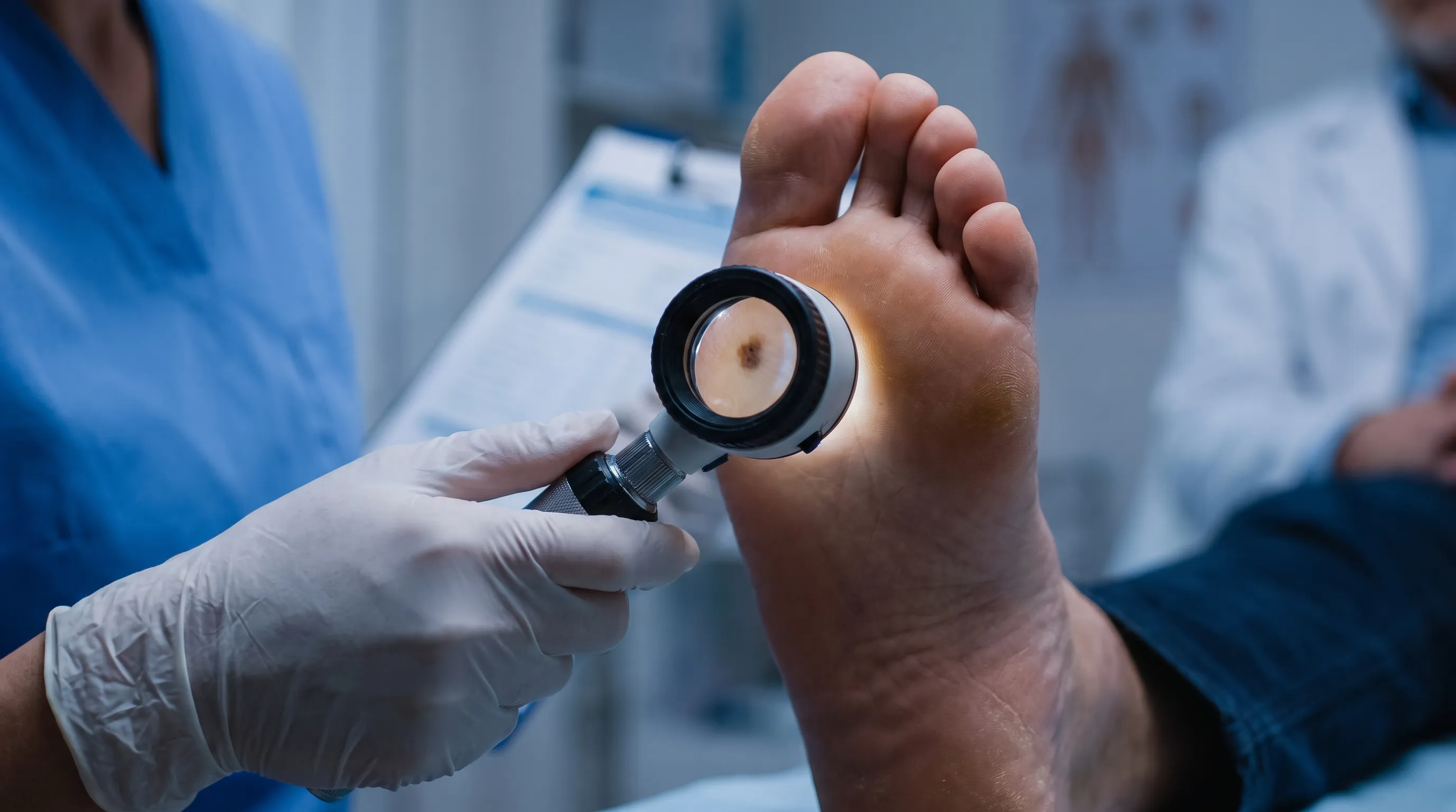
While the naked eye provides limited resolution, dermoscopy employs polarized or non-polarized light and specialized magnification to visualize subsurface pigment networks and vascular structures invisible during standard examination. Dermatologists utilize dermoscopy to differentiate melanocytic proliferation from hemorrhage, callus, or viral wart patterns. If structural atypia suggests malignancy, a full-thickness skin biopsy confirms histopathological diagnosis. Biopsy types include punch, shave, and excisional techniques, selected based on lesion depth and suspected pathology. Patients should request referrals to board-certified dermatologists or podiatric dermatologists when encountering persistent, painful, or rapidly changing pigmentation. Professional evaluation ensures accurate diagnosis, appropriate staging if necessary, and coordinated treatment planning.
Evidence-Based Treatment and Management Strategies
Once etiology is established, targeted interventions restore skin integrity and prevent recurrence. Treatment selection depends entirely on underlying pathology, lesion characteristics, and patient comorbidities.
Topical Depigmenting Agents
For benign hyperpigmentation, prescription and over-the-counter topicals modulate melanocyte activity and accelerate epidermal renewal. Hydroquinone inhibits tyrosinase, effectively reducing melanin synthesis, though prolonged use requires medical supervision to prevent exogenous ochronosis. Alternative agents include kojic acid, licorice root extract, tranexamic acid, and vitamin C derivatives, which interfere with pigment transfer or promote antioxidant-mediated skin brightening. Retinoids normalize keratinocyte differentiation and facilitate shedding of melanin-loaded cells. Because plantar skin absorbs products slowly, formulation penetration enhancers and consistent application regimens improve outcomes. Patients must combine topical therapy with pressure redistribution and moisturization to prevent reactivation of friction-induced pathways.
Laser Therapy and Cryotherapy
When topical treatments prove insufficient, energy-based devices and controlled tissue destruction offer faster resolution. Q-switched Nd:YAG lasers target melanin granules with photomechanical energy, fragmenting pigment clusters without damaging surrounding tissue. This modality suits post-inflammatory and friction-related hyperpigmentation but requires multiple sessions and strict sun avoidance post-procedure. Cryotherapy utilizes liquid nitrogen to induce cellular necrosis in viral warts, actinic keratoses, or localized dysplastic growths. Proper freeze-thaw cycles minimize scarring while ensuring adequate lesion clearance. Both modalities demand specialized training to prevent thermal injury or depigmentation, particularly on weight-bearing surfaces where tissue regeneration is slower.
Surgical Excision for Suspicious Lesions
Biopsy-confirmed melanoma or rapidly proliferating atypical lesions require prompt surgical intervention. Wide local excision with margin assessment ensures complete removal while preserving functional plantar anatomy. Mohs micrographic surgery may be indicated for complex or recurrent growths where tissue conservation is critical. Postoperative rehabilitation includes strict offloading protocols, antimicrobial wound care, and gradual return to weight-bearing activities. Multidisciplinary teams coordinate surgical management with oncologic staging, adjuvant therapy, and long-term surveillance. Addressing malignant dark spots in bottom of feet through timely surgery significantly improves disease-free survival and quality of life.
Daily Foot Care and Prevention Guidelines
Preventive maintenance remains the most cost-effective strategy for avoiding recurrent plantar discoloration. Integrating structured routines into daily life protects skin integrity and reduces mechanical, vascular, and infectious risks.
Proper Footwear and Pressure Redistribution
Shoe selection directly impacts plantar stress distribution. Optimal footwear features a roomy toe box, adequate arch support, and shock-absorbing midsoles. Replace athletic shoes every 300 to 500 miles as cushioning degrades, increasing ground reaction forces transmitted to the skin. Custom orthotics correct biomechanical abnormalities like pes planus or hallux valgus, redirecting pressure away from hyperkeratotic zones. Alternating shoe pairs daily allows materials to decompress and dry, reducing friction and moisture accumulation. Patients should avoid thin-soled footwear or going barefoot outdoors, which significantly elevates risk of microtrauma and subsequent pigmentation.
Sun Protection for Exposed Areas
Although the soles rarely encounter direct ultraviolet radiation, beach outings, poolside activities, and sandal wear expose plantar surfaces to UVB and UVA rays. Chronic, intermittent sun exposure contributes to cumulative photodamage and melanocyte stimulation. Applying broad-spectrum sunscreen with SPF 30 or higher to the soles during extended outdoor activities, wearing water shoes, and avoiding peak UV hours between 10 a.m. and 4 p.m. substantially reduces photoaging and pigmentary risks. Consistent photoprotection preserves skin resilience and minimizes unpredictable melanin activation.
Moisturization and Callus Management
Hydration prevents excessive keratin accumulation and maintains epidermal flexibility. Apply urea-based creams (10 to 40 percent concentration) daily to the soles, focusing on heels and metatarsal pads. Urea dissolves corneodesmosomes, softening calluses while drawing moisture into the stratum corneum. Avoid sharp blades or aggressive scraping, which trigger inflammatory rebounds and post-inflammatory hyperpigmentation. Instead, gently file softened keratin once weekly after bathing, followed by heavy occlusive moisturizers. Maintain balanced humidity levels, manage systemic edema through sodium moderation and leg elevation, and schedule routine podiatric visits for professional callus debridement. These combined practices prevent the cycle of friction, thickening, and darkening that frequently produces dark spots in bottom of feet.
Frequently Asked Questions
Are dark spots in bottom of feet always a sign of skin cancer?
No, the overwhelming majority of plantar pigmentation stems from benign mechanical stress, post-inflammatory healing, or localized infections. However, because acral lentiginous melanoma can occur on non-sun-exposed surfaces, any lesion demonstrating asymmetry, irregular borders, color variation, diameter expansion, or evolution over time requires professional evaluation. Routine self-examination combined with clinical dermoscopy provides definitive differentiation between harmless and concerning growths.
Can diabetes cause dark patches on the soles?
Yes, diabetes frequently alters plantar skin appearance through multiple pathways. Microvascular disease promotes hemosiderin deposition and impaired wound healing, while peripheral neuropathy eliminates protective pain feedback, leading to unrecognized repetitive trauma. Diabetic dermopathy and hemorrhagic calluses manifest as darkened patches that require comprehensive glycemic control, specialized offloading footwear, and regular podiatric debridement to prevent ulceration. Patients with diabetes should conduct daily foot inspections and report any new pigmentation promptly to their healthcare provider.
How do dermatologists diagnose suspicious foot spots?
Dermatologists employ a stepwise diagnostic protocol beginning with thorough history taking and visual inspection. Dermoscopy enhances magnification to evaluate subsurface pigment networks, vascular patterns, and structural disruption. If clinical suspicion persists, a biopsy provides histopathological confirmation, identifying cellular atypia, invasion depth, and mitotic activity. Vascular assessments may accompany dermatological evaluation if circulation deficits are suspected. This multimodal approach ensures accurate diagnosis and guides appropriate treatment selection.
What home care routines help prevent discoloration on the soles?
Preventive care centers on pressure management, barrier protection, and consistent hydration. Wear properly fitted shoes with adequate cushioning, rotate footwear daily, and utilize custom orthotics if biomechanical imbalances exist. Apply urea-based moisturizers daily to prevent excessive keratin buildup, and gently file softened calluses weekly. Protect feet from extreme temperatures, avoid barefoot walking on rough surfaces, and apply broad-spectrum sunscreen during prolonged sun exposure. These evidence-based practices significantly reduce the likelihood of developing friction-related or inflammatory pigmentation.
When should I see a doctor about a dark spot on my foot?
Schedule an appointment if a spot exhibits rapid growth, irregular borders, multiple color variations, surface ulceration, bleeding, or persistent tenderness. Additionally, seek evaluation if pigmentation fails to fade after six to eight weeks of conservative care, interferes with walking, or occurs alongside systemic symptoms like cold extremities, diminished pulses, or unexplained swelling. Early professional assessment enables timely intervention, prevents unnecessary complications, and provides peace of mind through accurate diagnosis.
Conclusion
Discovering dark spots in bottom of feet triggers understandable concern, but systematic evaluation typically reveals manageable, benign causes. Friction, post-inflammatory healing, and localized infections account for the majority of cases, while proper footwear, daily moisturization, and consistent self-monitoring effectively prevent recurrence. However, plantar skin also serves as a window into systemic health, reflecting vascular compromise, diabetic neuropathy, or, rarely, aggressive malignancies like acral lentiginous melanoma. Recognizing warning signs, applying the ABCDE assessment framework, and seeking timely dermatological evaluation transform anxiety into actionable care. By integrating biomechanical support, targeted topical treatments, and evidence-based surgical or laser interventions when necessary, patients preserve both skin integrity and functional mobility. Ultimately, proactive foot maintenance, combined with professional guidance, ensures that dark spots in bottom of feet remain a manageable aspect of overall wellness rather than a source of prolonged uncertainty. Prioritize routine inspections, protect plantar surfaces from excessive stress, and never hesitate to consult a healthcare provider when changes persist or progress—your feet carry you through life, and they deserve vigilant, science-backed care.

How we work
Researched and drafted with AI assistance, then reviewed by a human editor against the cited sources.
This article is general health information, not medical advice. Consult a qualified healthcare professional about your own situation.