Understanding a Bump on Back of Heel: Causes, Treatments, and Expert Relief Strategies

Noticing a painful or tender lump developing near your Achilles tendon can quickly derail your daily routine, turning simple walks or workouts into uncomfortable challenges. This specific anatomical concern, often described by patients as a bump on back of heel, affects millions of individuals worldwide and spans across age groups, from active teenagers to older adults, as documented by the National Institute of Arthritis and Musculoskeletal and Skin Diseases (NIAMS). The discomfort typically begins subtly, perhaps as mild irritation after wearing dress shoes or following a long run, but without proper intervention, it can progress into chronic inflammation, persistent swelling, and significant mobility limitations. Understanding why this condition develops is the first step toward effective management. The posterior heel is a biomechanically complex junction where the calcaneus bone, Achilles tendon, and retrocalcaneal bursa intersect. Repetitive stress, improper footwear, and structural foot mechanics can disrupt this delicate balance, leading to tissue irritation and bony remodeling. Fortunately, modern podiatric medicine and orthopedic science offer a wide spectrum of evidence-based interventions. From targeted stretching protocols and gait-correcting footwear to advanced physical therapy and minimally invasive surgical techniques, most individuals can achieve lasting relief without extensive downtime. In this comprehensive guide, we will explore the underlying causes, diagnostic pathways, and clinically proven treatment strategies to help you regain pain-free mobility and confidently return to an active lifestyle.
What Is a Bump on Back of Heel?
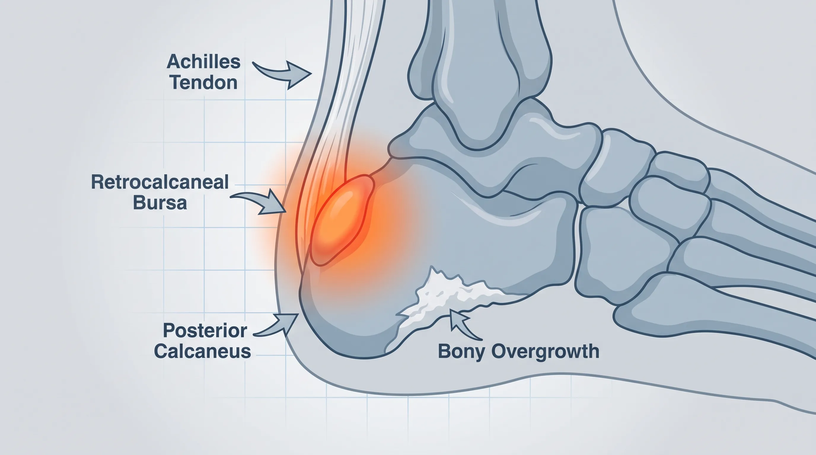
The posterior heel region is a high-stress anatomical crossroads. When a palpable lump develops in this area, it typically signals a localized response to mechanical friction, chronic inflammation, or structural bone changes. The most widely recognized clinical term associated with this presentation is Haglund’s deformity, a condition thoroughly outlined by the Mayo Clinic, but the condition encompasses a broader spectrum of pathologies. To effectively manage the symptoms, it is crucial to understand the precise anatomical structures involved and the physiological mechanisms that drive tissue enlargement.
Free calculatorFoot Reflexology ChartFoot reflexology points and corresponding organsOpen the calculatorUnderstanding Haglund’s Deformity
Haglund’s deformity is an acquired bony enlargement on the posterior-superior aspect of the calcaneus (heel bone). It frequently occurs when a prominent heel bone repeatedly rubs against stiff footwear or when abnormal foot biomechanics increase tension along the Achilles tendon insertion point. Over time, this chronic mechanical irritation stimulates osteoblast activity, prompting the body to deposit additional bone tissue in an attempt to stabilize the stressed area. This process creates a hard, noticeable bump on back of heel that is often accompanied by localized tenderness and swelling. The condition is sometimes referred to colloquially as "pump bump" because it historically correlates with rigid-backed women’s heels, though it affects runners, hikers, and individuals in occupations requiring prolonged standing just as frequently. The enlarged bone protrudes into the space normally occupied by the retrocalcaneal bursa and the Achilles tendon, creating a painful impingement that worsens with ankle dorsiflexion.
Other Common Causes of Heel Protuberances
While Haglund’s deformity is the primary structural culprit, a bump on back of heel can also stem from several soft-tissue and inflammatory conditions. Retrocalcaneal bursitis occurs when the fluid-filled sac situated between the Achilles tendon and heel bone becomes inflamed due to repetitive friction or trauma, as noted by the Cleveland Clinic. Unlike a bony spur, a bursitis-related swelling feels spongier and often presents with pronounced redness and warmth. Achilles tendinopathy, particularly insertional tendinopathy, involves degenerative changes within the tendon fibers as they attach to the calcaneus. Chronic tendon overload can lead to calcification within the tendon itself, forming a hardened nodule that mimics a bone growth. Additionally, localized ganglion cysts, lipomas, rheumatoid nodules, and even stress fractures can manifest as posterior heel swellings. In adolescents, Sever’s disease (calcaneal apophysitis) involves inflammation of the growth plate, which may sometimes present with mild swelling and tenderness at the heel’s posterior edge. Accurate differentiation among these conditions is essential, as treatment protocols vary significantly depending on the underlying pathology.
When to Seek Professional Medical Evaluation
Self-diagnosis is rarely sufficient for posterior heel abnormalities. You should consult a podiatrist or orthopedic specialist if the swelling persists beyond two to three weeks of conservative home care, if pain intensifies during routine activities, or if you notice visible deformity progression. Red flags requiring immediate clinical attention include sudden sharp pain accompanied by a "pop" sound (suggestive of an Achilles tendon rupture), severe redness and warmth extending up the lower calf (indicative of infection or deep vein thrombosis), numbness or tingling radiating into the foot (possible nerve entrapment), and an inability to bear weight or stand on your tiptoes. Early diagnostic intervention prevents chronic tendon degeneration, reduces the likelihood of compensatory gait abnormalities, and preserves long-term joint function.
Recognizing the Signs and Diagnostic Process
A thorough clinical evaluation combines detailed patient history, targeted physical examination techniques, and advanced imaging modalities. Because multiple conditions can produce similar symptoms, healthcare providers rely on systematic assessment protocols to differentiate between bone, tendon, and bursal involvement.
Clinical Symptoms and Physical Examination
Patients with a bump on back of heel typically report localized pain that worsens with shoe wear, especially when the heel counter presses directly against the prominence. Other common symptoms include visible swelling at the tendon’s insertion site, stiffness after periods of rest (particularly upon first steps in the morning), tenderness to direct palpation, and reduced ankle range of motion during dorsiflexion. During the physical exam, clinicians often perform the Fowler-Philip angle assessment to evaluate the posterior calcaneal contour. A normal angle ranges between 50 and 65 degrees; values exceeding this threshold indicate a high, prominent calcaneal tuberosity. Providers may also compress the retrocalcaneal bursa from both sides of the Achilles tendon to reproduce pain, a maneuver that strongly suggests bursitis. Neurological screening of the sural nerve and assessment of Achilles tendon integrity through the Thompson squeeze test are routinely conducted to rule out nerve compression or partial tearing.
Imaging Modalities and Differential Diagnosis
Radiographic evaluation remains the cornerstone of definitive diagnosis. Lateral weight-bearing X-rays reveal the exact size and position of the bony prominence, highlight calcaneal spurs, and allow precise angle measurements. Ultrasound imaging offers dynamic, real-time visualization of tendon integrity, bursa fluid accumulation, and surrounding soft tissue vascularity. It is particularly valuable for guiding therapeutic injections and assessing tendon thickness. When soft-tissue detail is paramount, magnetic resonance imaging (MRI) provides exceptional contrast resolution, identifying early tendinopathy, bone marrow edema, and partial tendon tears that X-rays may miss. Advanced diagnostic pathways help clinicians distinguish between Haglund’s deformity, chronic bursitis, tendinosis, and rare neoplastic conditions, ensuring that treatment aligns with the precise underlying pathology, consistent with guidelines from the American College of Radiology and NIH clinical resources.
Comprehensive Treatment Pathways
Management of a bump on back of heel follows a graduated approach, prioritizing conservative, non-invasive therapies before considering procedural interventions. Evidence consistently demonstrates that over 80 percent of patients achieve meaningful symptom resolution through structured rehabilitation and mechanical offloading, according to clinical data from the Cleveland Clinic.
First-Line Conservative Management
The foundation of conservative care revolves around reducing mechanical stress, controlling inflammation, and restoring tissue elasticity. Initial protocols emphasize the RICE principle (Rest, Ice, Compression, Elevation) during acute flare-ups. Nonsteroidal anti-inflammatory drugs (NSAIDs) like ibuprofen or naproxen provide short-term pain relief and help modulate localized inflammation. Heel lifts are strategically placed inside footwear to slightly elevate the calcaneus, decreasing tensile strain on the Achilles tendon and reducing friction against the posterior prominence. A physical therapist typically introduces a progressive loading program that includes eccentric calf raises, which have been extensively studied for their ability to stimulate collagen realignment and tendon remodeling. Manual therapy techniques such as instrument-assisted soft tissue mobilization (IASTM) and deep tissue massage improve fascial pliability and break down adhesions. Gait retraining addresses underlying biomechanical faults like excessive pronation, tight calf musculature, or inadequate hip mobility that contribute to abnormal heel loading patterns.
Advanced Medical Interventions
When conservative measures plateau, clinicians may introduce advanced therapeutic modalities. Extracorporeal shockwave therapy (ESWT) delivers focused acoustic pulses to the affected region, stimulating neovascularization, accelerating tissue healing, and disrupting calcified deposits. Platelet-rich plasma (PRP) injections utilize concentrated autologous growth factors to promote regenerative healing within degenerated tendon tissue, offering a safer alternative to corticosteroids, which carry a known risk of tendon weakening and rupture. Therapeutic ultrasound and laser therapy provide targeted thermal and photobiomodulation effects to reduce inflammation and accelerate cellular repair. Corticosteroid injections are occasionally administered with extreme caution directly into the bursa, avoiding the tendon substance, but their use is increasingly restricted due to long-term tissue vulnerability. Comprehensive rehabilitation programs integrate these modalities with supervised exercise progressions to optimize functional recovery, as supported by recent orthopedic literature and the Mayo Clinic.
Surgical Options and Post-Operative Rehabilitation
Surgical intervention is reserved for patients who experience persistent pain, significant functional impairment, and radiographic confirmation of a severe structural deformity after at least six to twelve months of rigorous conservative care. The most common procedure is a posterior calcaneal exostectomy, where the surgeon carefully shaves down the prominent bone. If the retrocalcaneal bursa is severely degenerated, a bursectomy is performed concurrently. In cases where the Achilles tendon has become severely degenerative or calcified within its substance, an extensive debridement and possible tendon reattachment (often utilizing suture anchors) may be necessary. Minimally invasive endoscopic techniques have gained popularity, offering smaller incisions, reduced postoperative pain, and faster tissue healing compared to open procedures. Post-operative rehabilitation follows a strict phased protocol: immobilization in a cast or walker boot for the first two to four weeks, followed by progressive range-of-motion exercises, gradual weight-bearing, and eventual strengthening. Full return to high-impact activities typically requires four to nine months, depending on the extent of surgical intervention and adherence to rehabilitation guidelines.
Footwear Science and Biomechanical Modifications
Footwear selection plays a pivotal role in both preventing and managing a bump on back of heel. The structural design of shoes directly influences posterior heel friction, Achilles tendon tension, and overall foot biomechanics.
How Shoe Design Impacts Posterior Heel Stress
The heel counter, the rigid cup structure at the rear of a shoe, provides stability but can become a source of chronic irritation if it is too stiff, improperly shaped, or too narrow. When a shoe’s heel counter compresses against a prominent calcaneus, it creates sustained pressure that exacerbates bursal inflammation and accelerates bony remodeling. Patients are advised to avoid shoes with rigid, non-compliant heel backs, particularly during the acute inflammatory phase. Instead, footwear with soft, padded, or flexible heel collars significantly reduces shear forces. Open-back shoes, such as clogs, slide sandals, and certain slip-on athletic models, eliminate direct posterior contact entirely. Additionally, the internal pitch of a shoe matters; a slight heel-to-toe drop (typically 8–12 mm) reduces the stretch demand on the Achilles tendon during walking, whereas completely flat shoes or those with extreme heel elevation can worsen symptoms. Proper length and width sizing are equally critical, as cramped footwear forces the foot to slide forward, increasing friction at the heel counter during the gait cycle.
The Role of Custom Orthotics and Heel Lifts
Custom-fabricated orthotic devices address underlying biomechanical imbalances that contribute to excessive posterior heel stress. For individuals with high arches (pes cavus), custom orthotics redistribute pressure away from the calcaneus and stabilize midfoot alignment, preventing abnormal heel strike patterns. Those with overpronation benefit from medial posting that controls excessive inward rolling, which can alter Achilles pull vectors. Heel lifts, typically ranging from 1/8 to 1/4 inch, provide immediate mechanical relief by elevating the calcaneus within the shoe. This slight elevation decreases the angle of ankle dorsiflexion required during stance phase, thereby reducing Achilles tension and minimizing friction against the posterior prominence. Silicone heel cushions or donut-shaped pads can be strategically placed around the bump to create a protective void, allowing the affected area to rest without bearing direct weight. Combining orthotic support with appropriate heel elevation creates a synergistic offloading effect that significantly accelerates symptom resolution.

Preventative Strategies and Long-Term Foot Care
Proactive foot care and lifestyle modifications are essential for preventing recurrence and maintaining long-term Achilles health. Chronic heel issues rarely develop overnight; they are typically the cumulative result of repetitive microtrauma, poor mechanical habits, and inadequate recovery.
Daily Stretching and Achilles Conditioning
A consistent flexibility and strengthening routine forms the cornerstone of preventative care. The calf-Achilles complex adapts to mechanical stress, but tight gastrocnemius and soleus muscles dramatically increase insertional tension. Daily stretching protocols should include both knee-straight and knee-bent calf stretches, held for 30 seconds per repetition and repeated three to five times. Eccentric loading exercises, where the muscle lengthens under tension, are particularly effective. Performing slow, controlled calf drops off a stair edge (three seconds down, neutral position up) for three sets of fifteen repetitions twice daily stimulates collagen synthesis and improves tendon resilience. Strengthening the intrinsic foot muscles and proximal hip stabilizers further optimizes lower-extremity kinetics, ensuring that the heel absorbs impact efficiently rather than bearing compensatory stress. Maintaining ankle dorsiflexion range of motion prevents the compensatory gait mechanics that drive chronic heel irritation.
Activity Modification for Active Lifestyles
Runners, athletes, and highly active individuals must strategically periodize their training loads to avoid Achilles overload. Sudden increases in mileage, intensity, or hill work frequently precipitate posterior heel flare-ups. The widely accepted 10 percent rule suggests increasing weekly training volume gradually, allowing connective tissues time to adapt. Cross-training activities like swimming, cycling, and elliptical training maintain cardiovascular fitness while eliminating impact stress on the heel, aligning with CDC physical activity recommendations. Surface selection matters as well; consistently running on hard concrete amplifies ground reaction forces, whereas softer trails or tracks provide better shock absorption. Proper warm-up routines that include dynamic mobility drills and gradual progression into high-impact activities prepare the Achilles for strenuous demands. Adequate recovery, including sleep optimization and nutritional support rich in collagen-building amino acids, ensures tissue repair keeps pace with mechanical stress. Listening to early warning signs like morning stiffness or post-exercise soreness allows for timely intervention before a bump on back of heel develops into a chronic condition.
Home Care Protocols and Symptom Management
Effective self-management bridges the gap between clinical visits and accelerates recovery. Implementing structured at-home protocols reduces inflammation, protects vulnerable tissues, and empowers patients to take an active role in their healing process.
Effective At-Home Pain Relief Techniques
Cryotherapy remains one of the most accessible and effective anti-inflammatory strategies. Applying an ice pack wrapped in a thin towel to the posterior heel for fifteen to twenty minutes, three to four times daily, constricts blood vessels, reduces metabolic activity, and limits edema formation. Ice massage, where a frozen water cup is gently rubbed over the affected area in circular motions for seven to ten minutes, provides targeted cooling combined with mild myofascial release. Contrast hydrotherapy, alternating between warm and cold water immersion, promotes vasodilation and vasoconstriction cycles that flush out metabolic waste and enhance circulation. Night splints or dorsiflexion braces worn during sleep maintain the ankle at a neutral angle, preventing the Achilles tendon from tightening overnight and reducing the sharp pain associated with first morning steps. Over-the-counter topical analgesics containing menthol, arnica, or diclofenac provide localized pain relief without systemic side effects. Consistency in applying these techniques, rather than sporadic use during peak pain, yields the most meaningful symptom reduction, as recommended by the Cleveland Clinic.
Proper Application of Protective Padding
Strategic padding shields the posterior heel from direct shoe friction and distributes pressure more evenly. Silicone heel cups conform to the natural contour of the foot, cushioning the calcaneus while providing gentle compression that stabilizes the bursa. Donut-shaped felt or foam pads feature a central cutout that positions the opening directly over the prominence, ensuring that the surrounding healthy tissue bears the load while the bump rests in a pressure-free zone. It is crucial to secure padding properly to prevent shifting, which can create new friction points and worsen irritation. Patients should avoid adhesive pads that leave residue or stretch the delicate skin around the heel. Moisture-wicking, seamless socks reduce shear forces and prevent blister formation over the irritated area. Regularly replacing worn padding ensures optimal cushioning and support. When combined with appropriate footwear and targeted exercises, protective padding serves as a simple yet highly effective component of a comprehensive self-care regimen.
Comparison of Conservative vs. Advanced Treatment Approaches
| Feature | Conservative Management | Advanced Medical/Surgical Intervention |
|---|---|---|
| Primary Focus | Reducing inflammation, mechanical offloading, tissue remodeling | Correcting structural deformity, removing damaged tissue |
| Typical Timeline | 3–6 months for significant improvement | 4–9 months for full functional recovery |
| Invasiveness | Non-invasive, self-directed or supervised therapy | Invasive, requires clinical procedure or operation |
| Risks & Side Effects | Minimal; occasional muscle soreness or temporary discomfort | Surgical complications, infection, tendon weakness, scar tissue |
| Success Rate | 70–85% achieve functional relief with compliance | 85–95% structural correction success for refractory cases |
| Cost Considerations | Low; covers therapy, OTC meds, footwear modifications | Higher; imaging, procedural fees, post-op care, rehab |
| Best Candidates | Early-stage symptoms, mild-moderate bump on back of heel | Severe deformity, chronic pain unresponsive to conservative care |
Frequently Asked Questions
Is a bump on back of heel always Haglund’s deformity?
No, it is not. While Haglund’s deformity represents a prominent calcaneal bone growth, the swelling could also be caused by retrocalcaneal bursitis, insertional Achilles tendinopathy, localized cysts, lipomas, or rheumatoid nodules. Accurate diagnosis requires clinical examination and imaging to determine whether the lump is bony, cystic, or tendinous in origin.
Can a bump on back of heel go away without surgery?
Absolutely. The vast majority of cases respond favorably to non-surgical interventions. Through strategic footwear modification, targeted eccentric exercises, custom heel lifts, physical therapy, and activity pacing, patients can significantly reduce inflammation, improve tissue elasticity, and achieve lasting pain relief. Surgery is typically considered only after six to twelve months of dedicated conservative care fail to resolve symptoms.
How long does it take to recover from heel bump surgery?
Recovery duration depends on the surgical scope. A straightforward exostectomy (bony shaving) usually allows protected weight-bearing within two weeks and a return to standard footwear within four to six weeks. If the Achilles tendon requires debridement or reattachment, the rehabilitation timeline extends significantly. Patients typically require immobilization in a boot for several weeks, followed by gradual loading and strengthening. Full return to running or high-impact sports generally takes six to nine months.
Are cortisone injections safe for treating a bump on back of heel?
Cortisone injections carry notable risks when administered near the Achilles tendon due to the potential for tendon weakening, collagen degeneration, and increased rupture susceptibility. While they may offer temporary pain relief, most orthopedic specialists reserve them for severe bursal inflammation and avoid injecting directly into the tendon. Safer alternatives like platelet-rich plasma (PRP) or extracorporeal shockwave therapy (ESWT) provide therapeutic benefits without compromising tendon integrity.
What type of shoes are best if I have a bump on back of heel?
Ideal footwear features a soft, flexible, or open-back design that eliminates direct pressure on the posterior calcaneus. Look for shoes with padded heel counters, adequate width to accommodate swelling, and a moderate heel-to-toe drop (8–12 mm) to reduce Achilles tension. Avoid rigid dress shoes, high heels, or narrow athletic models with stiff heel cups. Incorporating silicone heel cushions or custom orthotics further enhances comfort and prevents friction-related flare-ups.
Conclusion
Developing a bump on back of heel can feel frustrating, but it is highly manageable with the right approach and consistent care. Whether the underlying cause is a structural bone prominence, chronic bursal inflammation, or insertional tendon degeneration, evidence-based treatments provide clear pathways to pain reduction and restored mobility. Prioritizing conservative strategies like proper footwear selection, targeted stretching, and mechanical offloading addresses the root biomechanical factors driving the condition. When symptoms persist, advanced therapies and surgical interventions offer reliable solutions backed by decades of orthopedic research. By staying proactive, adhering to rehabilitation protocols, and making informed lifestyle adjustments, you can protect your Achilles complex, prevent future flare-ups, and confidently maintain an active, healthy routine in line with global WHO physical activity guidelines. Always consult a qualified podiatrist or orthopedic specialist for personalized evaluation, especially if pain worsens or mobility becomes restricted. Early intervention remains the most powerful tool for preserving long-term foot health and ensuring that every step remains pain-free and supported.
How we work
Researched and drafted with AI assistance, then reviewed by a human editor against the cited sources.
This article is general health information, not medical advice. Consult a qualified healthcare professional about your own situation.