Why Does My Tongue Look White? Causes, Diagnosis, and Proven Treatments
Waking up to a strange coating on your tongue can be unsettling, prompting the immediate question of why does my tongue look white. While a sudden change in your oral appearance often triggers anxiety about serious health issues, the reality is usually far more manageable. The tongue is a dynamic organ composed of muscle tissue, mucous membranes, and thousands of tiny projections that play a crucial role in taste, speech, and digestion. When the surface becomes discolored, it is typically a reflection of localized oral changes, hygiene habits, hydration levels, or occasionally, an underlying medical condition. Understanding the root causes requires a closer look at how oral health intersects with your overall wellness. In this comprehensive guide, we will explore the physiological reasons behind tongue discoloration, differentiate between harmless buildup and signs that warrant medical attention, and provide actionable, evidence-based strategies to restore your oral health. Whether you are experiencing mild morning coating or persistent white patches, knowing what your body is communicating will empower you to make informed decisions and seek appropriate care when necessary.
The Science Behind Tongue Surface Changes
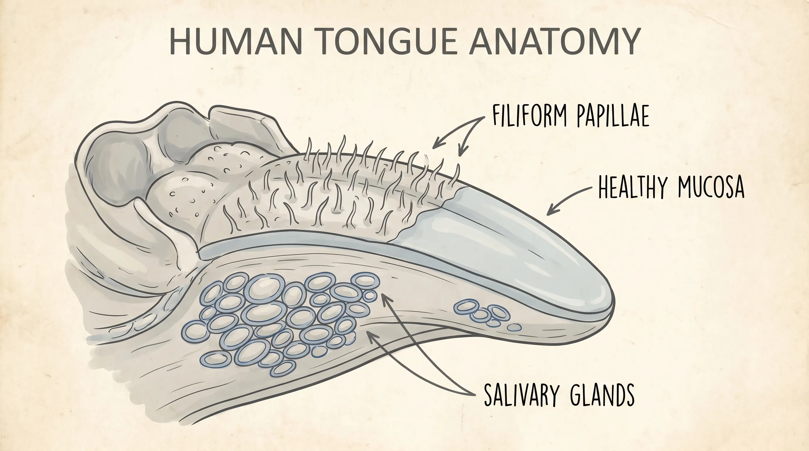
To truly understand why does my tongue look white, it is essential to examine the intricate anatomy of the organ itself and the microscopic processes that govern its appearance. The dorsal surface of the tongue is covered with lingual papillae, which give it a slightly rough, textured appearance. These papillae vary in type and function, with filiform papillae being the most numerous and responsible for the tongue’s characteristic texture. Unlike fungiform or circumvallate papillae that house taste buds, filiform papillae are heavily keratinized, meaning they contain a tough structural protein also found in hair and nails. When these structures become elongated or fail to shed properly due to mechanical friction, dietary habits, or cellular changes, they trap food particles, dead epithelial cells, and oral bacteria. This trapped debris oxidizes and forms a visible biofilm that reflects light as a white or off-white layer.
Free calculatorWater Intake CalculatorCalculate daily water needsOpen the calculatorSaliva plays a critical role in maintaining a healthy oral microbiome and washing away cellular debris. The average adult produces approximately one to one and a half liters of saliva daily, which contains antimicrobial enzymes like lysozyme and lactoferrin that keep pathogenic microbes in check. When salivary flow decreases, the natural cleansing mechanism is impaired, allowing keratin and bacterial colonies to accumulate rapidly on the papillae. The oral cavity hosts a complex ecosystem of microorganisms, primarily bacteria and fungi, that normally exist in a delicate symbiotic balance. Disruptions to this equilibrium—whether from diet changes, medication use, or immune system fluctuations—can trigger an overgrowth of specific microbes that produce visible coatings. Research published by the National Institutes of Health highlights that transient tongue coating affects nearly twenty percent of the adult population at some point, often resolving with improved hydration and mechanical cleaning. By recognizing the biological processes that govern tongue appearance, you can better distinguish between normal physiological variations and clinical signs that require targeted intervention.
Common and Harmless Triggers
Many individuals discover a coated tongue after periods of disrupted routine, travel, or minor lifestyle shifts. These benign triggers account for the vast majority of clinical presentations and typically resolve without prescription medication or invasive procedures. Recognizing them early can prevent unnecessary worry and guide you toward simple, effective self-care strategies that restore comfort and appearance.
Poor Oral Hygiene Routine
The most straightforward explanation for a coated tongue is inadequate daily cleaning. While many people diligently brush their teeth twice daily, they frequently overlook the tongue’s surface entirely or clean it too aggressively. Bacteria, dead mucosal cells, and microscopic food remnants settle deep between the elongated papillae, creating a thick biofilm that reflects ambient light as a white layer. Without consistent mechanical removal, this layer can thicken, especially after consuming sugary, dairy-based, or starchy foods that provide rapid fuel for oral bacteria. Establishing a comprehensive routine that includes gentle tongue brushing or specialized scraping is often the fastest way to restore a clean, pink appearance. Clinical guidelines from the Mayo Clinic recommend incorporating tongue cleaning into your twice-daily oral hygiene practice to prevent plaque migration, reduce volatile sulfur compounds that cause halitosis, and maintain optimal taste receptor sensitivity.
Dehydration and Reduced Saliva Flow
Water is fundamental to every physiological process, including oral mucosal health. When you become dehydrated, your salivary glands produce less fluid, leading to xerostomia, commonly known as dry mouth. According to the Cleveland Clinic, saliva acts as a natural detergent, breaking down food particles, neutralizing acids, and flushing away dead keratinized cells before they accumulate into a visible layer. Without adequate moisture, the tongue’s surface dries out, causing papillae to clump together and appear pale, chalky, or rough. Factors contributing to dehydration include inadequate daily fluid intake, excessive caffeine or alcohol consumption, vigorous exercise without electrolyte replenishment, and chronic mouth breathing during sleep. Mouth breathing, often linked to seasonal allergies, deviated septum, or undiagnosed sleep apnea, significantly accelerates mucosal drying. Replenishing fluids with water or herbal infusions and addressing respiratory habits can dramatically improve tongue appearance within forty-eight hours.
Tobacco Use and Alcohol Consumption
Smoking cigarettes, vaping, and chewing tobacco are well-documented contributors to oral tissue changes, including persistent tongue discoloration. The chemical irritants in tobacco products, including nicotine, tar, and various aldehydes, trigger a localized inflammatory response that accelerates the growth and hyperkeratinization of lingual papillae. Additionally, tobacco residue itself leaves a visible film that physically alters the tongue’s natural color and creates a favorable environment for pathogenic bacteria. Similarly, excessive alcohol consumption acts as a systemic dehydrant, stripping moisture from the oral mucosa while simultaneously disrupting the local microbiome through pH fluctuations. Both substances impair the body’s natural cellular turnover processes, meaning recovery takes longer even after discontinuation. Quitting tobacco and moderating alcohol intake not only improves tongue appearance but also significantly reduces the long-term risk of periodontal disease, tooth decay, and oral squamous cell carcinoma, as emphasized in World Health Organization guidelines on systemic and oral health integration.
Infectious and Inflammatory Conditions
When lifestyle factors and hygiene habits are optimized but the discoloration persists or worsens, infectious or inflammatory processes may be driving the symptomatology. These conditions often present with additional clinical markers that distinguish them from simple debris accumulation. Recognizing these patterns is crucial for determining when professional medical treatment is necessary to prevent progression.
Oral Candidiasis (Thrush)
Oral thrush is a localized fungal infection caused by an opportunistic overgrowth of Candida species, primarily Candida albicans. While this yeast naturally resides in the oral cavity, gastrointestinal tract, and skin of healthy individuals, immune system fluctuations, broad-spectrum antibiotic use, inhaled corticosteroids, or uncontrolled diabetes mellitus can disrupt the competitive microbial balance, allowing fungal colonies to proliferate unchecked. The classic clinical presentation of oral thrush includes thick, creamy white patches that resemble cottage cheese or milk curds on the tongue, inner buccal mucosa, and soft palate. Unlike harmless debris, these patches cannot be easily scraped away and often reveal erythematous, bleeding tissue beneath when disturbed. Patients frequently report a persistent cottony sensation in the mouth, metallic or altered taste perception, and mild burning during meals. High-risk populations include neonates, older adults, denture wearers who do not remove appliances nightly, and individuals on immunosuppressive regimens. The Centers for Disease Control and Prevention emphasizes that recurrent or severe thrush warrants comprehensive medical evaluation to rule out systemic immune deficiencies or underlying metabolic dysregulation. Topical antifungal medications, such as nystatin suspension or clotrimazole troches, are highly effective when prescribed and completed as directed.
Leukoplakia and Lichen Planus
Leukoplakia is characterized by hyperkeratotic white patches that form on the tongue, gingival margins, or buccal mucosa in direct response to chronic irritation. Unlike thrush, leukoplakia lesions are firmly adherent and resist scraping. While the majority of leukoplakia cases are reactive and benign, the condition carries a documented potential for dysplastic transformation into oral carcinoma, making histopathological biopsy essential for definitive diagnosis. The etiology is strongly associated with tobacco exposure, frequent alcohol consumption, and mechanical irritation from fractured dental restorations or maloccluded teeth. Oral lichen planus, on the other hand, is a T-cell-mediated chronic autoimmune disorder that targets the basal layer of oral squamous epithelium. It often presents as bilateral, lace-like white streaks known as Wickham striae, frequently accompanied by reticular, erosive, or atrophic mucosal changes. Symptoms may wax and wane, often exacerbating with psychological stress, sleep deprivation, or consumption of acidic and spicy foods. Management typically involves high-potency topical corticosteroids, calcineurin inhibitor rinses, avoidance of identified triggers, and biannual monitoring by an oral medicine specialist. Both conditions underscore the non-negotiable importance of professional evaluation when a white coating appears persistent, irregular in borders, or symptomatic.
Systemic Health Links and Nutritional Factors
The oral cavity is frequently referred to by clinicians as a diagnostic window to systemic health, and tongue discoloration can sometimes reflect broader physiological imbalances or micronutrient deficiencies. Understanding these interconnections ensures that localized symptoms are not dismissed when they signal deeper metabolic or immune-related needs.
Digestive Disorders and Acid Reflux
Gastrointestinal health and oral symptomatology are intricately intertwined through shared neurological pathways and microbiome cross-talk. Chronic acid reflux or gastroesophageal reflux disease (GERD) can cause gastric acid to travel upward into the pharynx and oral cavity during recumbent periods, altering local pH balance and disrupting normal commensal bacterial populations. This acidic microenvironment promotes the overgrowth of acid-tolerant microorganisms and triggers low-grade mucosal inflammation, resulting in a pale, thickened, or white-coated tongue. Additionally, conditions like Helicobacter pylori infection, small intestinal bacterial overgrowth (SIBO), or functional dyspepsia have been clinically associated with measurable changes in salivary and oral microbial composition. Patients experiencing frequent heartburn, regurgitation, epigastric discomfort, or bloating alongside persistent tongue changes should consult a gastroenterologist. Dietary modifications, such as reducing trigger foods like caffeine, chocolate, and fatty meals, eating smaller portions, maintaining a two to three-hour fasting window before bedtime, alongside targeted medical management of reflux, often lead to noticeable improvements in both digestive comfort and mucosal appearance, as detailed in NIH digestive health resources.
Vitamin Deficiencies and Hematological Imbalances
Nutritional status profoundly impacts epithelial integrity and immune surveillance within the oral cavity. Deficiencies in dietary iron, vitamin B12, folate, or zinc can compromise cellular turnover, reduce hemoglobin oxygen delivery to peripheral tissues, and weaken local immune defenses. Iron deficiency anemia, for example, may cause tongue pallor due to reduced oxygenated hemoglobin concentration in mucosal capillaries, giving the clinical appearance of a pale or whitish tongue. Vitamin B12 deficiency often initially presents with a smooth, glossy, beefy-red tongue (glossitis) due to papillary atrophy, but chronic untreated cases can lead to secondary bacterial colonization and white coating as the compromised tissue becomes increasingly susceptible to infection. Individuals following strict vegan or vegetarian diets without appropriate fortified supplementation, those with autoimmune gastritis, or patients with malabsorption disorders like celiac disease or inflammatory bowel disease are at elevated risk. A comprehensive metabolic and hematological blood panel can identify these micronutrient gaps rapidly, and targeted clinical supplementation typically restores mucosal vitality and normal papillary architecture within three to six weeks, according to clinical guidance from the Mayo Clinic.

Diagnostic Approaches and Clinical Evaluation
While many presentations of tongue discoloration resolve with improved mechanical hygiene and hydration, persistent, painful, or rapidly progressive cases require structured medical assessment. Understanding the standard diagnostic workflow helps patients prepare for appointments, provide accurate histories, and ensures that underlying pathology is identified efficiently.
Clinical Examination and Comprehensive Medical History
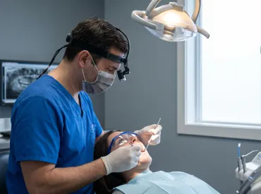
The foundational step in any professional evaluation involves a meticulous visual and tactile inspection of the entire oral cavity. Dentists or primary care physicians will assess the coating’s thickness, topography, distribution, and adherence to the underlying epithelium. They will carefully document whether patches are easily removable, if the exposed tissue appears inflamed, atrophic, or ulcerated, and whether similar lesions extend to the hard palate, tonsillar pillars, or floor of the mouth. A detailed medical history follows, covering current and recent medications (particularly antibiotics, antihistamines, diuretics, and anticholinergics), dietary patterns, tobacco and alcohol consumption history, recent systemic illnesses, and pre-existing conditions like diabetes mellitus, autoimmune disorders, or immunosuppressive states. This comprehensive clinical synthesis frequently reveals the primary etiological factor without necessitating expensive or invasive testing. Patients are strongly encouraged to document symptom onset, duration, fluctuations, and any accompanying systemic signs like dysphagia, odynophagia, weight changes, or cervical lymphadenopathy to assist clinicians in constructing an accurate differential diagnosis.
Laboratory Testing and Specialized Multidisciplinary Referrals
When initial clinical assessment suggests active infection, systemic metabolic imbalance, or potential cellular dysplasia, targeted diagnostic investigations are warranted. For suspected candidiasis, a simple clinical swab of the lesion is examined under potassium hydroxide microscopy or cultured on selective fungal media to identify the exact Candida strain and determine antifungal susceptibility. Comprehensive blood work evaluating complete blood count with differential, serum ferritin, vitamin B12, folate levels, and fasting glucose or HbA1c helps definitively rule out nutritional deficits, anemia, and metabolic dysregulation. If leukoplakia or erosive lichen planus is strongly suspected based on clinical appearance and risk profile, an incisional biopsy under local anesthesia may be performed to examine tissue histology and grade for dysplasia. In complex, refractory, or systemically linked cases, referral to an otolaryngologist (ENT specialist), board-certified oral pathologist, or clinical gastroenterologist ensures multidisciplinary management. Early and precise diagnosis remains the critical determinant for implementing the most effective therapeutic pathway and preventing irreversible mucosal damage.
| Condition/Cause | Key Visual Features | Associated Symptoms | Primary Management Approach |
|---|---|---|---|
| Poor Oral Hygiene | Thin, easily removable coating | Mild halitosis, no pain or sensitivity | Daily tongue scraping, improved brushing technique |
| Dehydration | Dry, chalky or pale appearance | Thirst, sticky saliva, rough texture | Increased water intake, saliva stimulants, address mouth breathing |
| Oral Thrush | Thick, cottage-cheese-like patches | Burning, altered taste, cotton mouth sensation | Prescription antifungals, review antibiotic/steroid use, manage diabetes |
| Leukoplakia | Firm, adherent white plaques | Usually asymptomatic, rough or thickened surface | Eliminate tobacco/irritants, serial monitoring, biopsy if dysplastic |
| Lichen Planus | Lacy white streaks (Wickham striae) | Sensitivity to spicy/acidic foods, occasional erosive ulcers | Topical corticosteroids, stress reduction, regular oral pathology follow-up |
| Nutritional Deficiency | Pale, thin coating or atrophic papillae | Fatigue, glossodynia, brittle hair or nails | Targeted micronutrient supplementation, dietary optimization, hematological testing |
Evidence-Based Management and Self-Care Protocols
Addressing why does my tongue look white effectively requires a synergistic combination of professional clinical guidance and consistent, evidence-based home care. The following therapeutic strategies are supported by contemporary dental and medical literature and can be safely adapted to individual health profiles.
Optimizing Daily Oral Hygiene Practices
Mechanical removal of accumulated keratin and biofilm is the absolute cornerstone of managing benign tongue coating. Using a dedicated, ergonomically designed tongue scraper or a soft-bristled manual toothbrush, gently clean the dorsal surface from posterior to anterior in three to five controlled strokes twice daily. It is imperative to avoid excessive downward pressure, which can cause epithelial microtrauma, capillary rupture, and trigger compensatory hyperkeratinization that paradoxically worsens the white appearance over time. Always sanitize scraping tools with warm soapy water or isopropyl alcohol after each use and replace them completely every four to six weeks to prevent bacterial reintroduction. Pair mechanical cleaning with an alcohol-free, pH-neutral antimicrobial mouthwash to reduce microbial load without inducing mucosal desiccation. Many patients report significant improvement by switching to a mild, sodium lauryl sulfate-free (SLS-free) toothpaste, as this surfactant can strip protective mucins and trigger irritation in sensitive individuals. Consistency remains paramount; most individuals observe visible clinical improvement within three to five days of establishing a disciplined, low-irritation routine.
Hydration, Dietary Optimization, and Targeted Probiotic Support
Maintaining optimal salivary secretion is non-negotiable for long-term tongue mucosal health. Aim to consume a minimum of two to three liters of filtered water daily, dynamically adjusting for ambient temperature, physical exertion, body mass, and medication side effects. Integrating water-dense botanical foods like cucumbers, celery, zucchini, and citrus fruits provides supplementary hydration alongside bioavailable electrolytes. Actively reducing dietary sugar, refined grains, and ultra-processed foods starves acidogenic oral bacteria and opportunistic fungi, while regularly incorporating raw fibrous vegetables naturally stimulates salivary flow and provides gentle mechanical debridement of the papillae. Emerging peer-reviewed research highlights the therapeutic potential of oral-specific probiotics containing well-characterized bacterial strains such as Streptococcus salivarius K12 and M18, which competitively inhibit pathogenic colonization, modulate local immune responses, and produce beneficial bacteriocins. These targeted supplements, typically formulated as slow-dissolving lozenges or sublingual melts, can be particularly advantageous for individuals experiencing recurrent oral mycosis or medication-induced xerostomia, as supported by Cleveland Clinic recommendations on oral microbiome management.
Pharmacological Interventions and Clinical Therapies
When conservative home care measures prove insufficient, targeted medical treatments directly address the underlying pathophysiology. For laboratory-confirmed oral candidiasis, topical antifungals like nystatin oral suspension or miconazole dental paste are typically prescribed, with standard treatment durations spanning seven to fourteen days to prevent relapse. Leukoplakia management heavily emphasizes complete cessation of causative irritants, with surgical excision, laser ablation, or cryotherapy reserved strictly for histologically confirmed dysplastic or malignant lesions. Oral lichen planus frequently requires prescription-strength topical corticosteroids (e.g., clobetasol or fluocinonide), off-label calcineurin inhibitor mouth rinses, or short courses of systemic immunomodulators during severe erosive flares. Documented nutritional deficiencies are corrected through medically supervised supplementation protocols tailored to baseline serum levels. Patients taking essential medications with strong anticholinergic properties may benefit from FDA-approved secretagogues, artificial saliva substitutes, or coordinated dose adjustments after thorough consultation with their prescribing physician. A collaborative, patient-centered approach between dental hygienists, oral pathologists, and primary care providers ensures comprehensive, safe, and individualized therapeutic outcomes.
Long-Term Prevention and Systemic Wellness Integration
Preventing the recurrent development of tongue discoloration requires a proactive, holistic approach that seamlessly integrates oral hygiene into your broader physiological wellness lifestyle. By systematically addressing modifiable systemic risk factors and maintaining unwavering self-care consistency, you can sustain a resilient, healthy oral environment indefinitely.
Routine Dental Surveillance and Professional Prophylaxis
Regular dental evaluations every six months allow licensed professionals to continuously monitor mucosal integrity, identify microscopic early signs of abnormal cellular changes, and perform ultrasonic cleanings that disrupt subgingival deposits inaccessible to home instruments. During these preventive visits, clinicians systematically screen for early-stage premalignant and malignant conditions, objectively assess unstimulated and stimulated salivary flow rates, and deliver customized hygiene coaching tailored to your unique anatomical and lifestyle profile. Patients managing chronic dry mouth, full or partial dentures, or systemic immunosuppression typically require quarterly maintenance intervals. Maintaining a personal symptom log and transparently sharing it with your interdisciplinary healthcare team dramatically facilitates early intervention and minimizes diagnostic delays. Preventive stomatology remains the most clinically effective, cost-efficient, and reliable strategy for preserving optimal oral and systemic health across the lifespan.
Lifestyle Modifications, Sleep Hygiene, and Chronic Stress Management
Chronic psychological and physiological stress persistently elevates circulating cortisol and catecholamine levels, which can directly suppress localized mucosal immune responses, alter salivary protein composition, and shift the oral microbiome toward a dysbiotic state. Evidence-based practices such as daily mindfulness meditation, structured cognitive behavioral therapy for insomnia, maintaining a consistent seven to nine-hour sleep cycle, and engaging in regular moderate-intensity cardiovascular exercise significantly support systemic immune resilience and promote healthy epithelial regeneration. Consistently avoiding known mucosal irritants—including all tobacco products, excessive ethanol consumption, highly thermal foods, and harsh chemical whitening rinses—further protects delicate squamous tissues from chronic inflammation. If you clinically suspect undiagnosed sleep apnea or chronic nasal obstruction is contributing to compensatory mouth breathing and subsequent tongue coating, a formal polysomnography sleep study or comprehensive ENT consultation can deliver targeted solutions such as mandibular advancement appliances, CPAP therapy, or nasal valve reconstruction. Integrating these foundational lifestyle adjustments creates an unshakable biological framework for enduring oral vitality.
Frequently Asked Questions
Can stress cause a white coating on my tongue?
Yes, chronic psychological stress can indirectly contribute to tongue discoloration through neuroendocrine pathways. Elevated cortisol levels may temporarily suppress local immune surveillance in the oral cavity, disrupt the competitive balance of commensal bacteria, and reduce parasympathetically-mediated salivary flow. Stress frequently leads to unconscious mouth breathing, nocturnal bruxism, and poor dietary choices, all of which create a stagnant microenvironment where dead keratinocytes and microbial colonies accumulate more readily. Managing stress through structured relaxation techniques, consistent sleep scheduling, and regular physical activity can rapidly restore oral homeostasis and accelerate natural coating resolution.
Should I scrape my tongue every day, and can aggressive scraping cause tissue damage?
Daily tongue cleaning is strongly recommended by dental professionals, provided it is performed using correct technique and appropriate pressure. Utilizing a clinically tested tongue scraper or soft-bristled brush, gently glide from the posterior third toward the tip in two to four controlled strokes. Excessive downward force or using rigid, non-medical tools can cause epithelial microtears, capillary bleeding, and reactive hyperkeratinization, which may paradoxically worsen the appearance over subsequent days. Always thoroughly clean your device after use with warm water and mild soap, and replace it regularly to maintain hygienic standards. If you encounter persistent pain, erythema, or bleeding despite gentle technique, pause use and consult your dentist.
How do I clinically differentiate between thrush and simple bacterial debris?
The definitive clinical distinction lies in adherence characteristics and underlying mucosal response. Harmless food and cellular debris typically wipes or scrapes away effortlessly, revealing uniformly pink, healthy epithelium beneath. Oral candidiasis manifests as densely adherent, creamy plaques that firmly bind to the surface; mechanical removal frequently exposes raw, erythematous, or mildly hemorrhagic tissue. Thrush is additionally characterized by sensory alterations including burning dysesthesia, metallic dysgeusia, and a pervasive cotton-mouth feeling. If candidiasis is suspected, refrain from prolonged reliance on alkaline home rinses alone and seek prompt professional evaluation for accurate fungal identification and targeted prescription therapy.
Which specific dietary patterns exacerbate persistent tongue coating?\Certain nutritional habits directly promote dysbiosis, mucosal drying, or low-grade epithelial inflammation. Diets high in refined sucrose and processed carbohydrates rapidly ferment into acids that feed opportunistic pathogens. Excessive caffeine and ethanol consumption act as profound diuretics, significantly diminishing protective salivary volume, while highly acidic, extremely hot, or heavily spiced foods can chemically irritate the tongue dorsum, delaying natural desquamation. Conversely, actively incorporating prebiotic fibers, raw vegetables, and clinically validated oral probiotics supports robust microbial diversity and natural papillary cleansing. Maintaining optimal systemic hydration with plain water remains the single most effective nutritional intervention for sustaining a physiologically clean tongue surface.
At what point should I transition from home care to professional medical evaluation?
While the majority of presentations are transient and highly responsive to improved hygiene, specific clinical red flags mandate timely medical assessment. Schedule an appointment if the coating remains unchanged after fourteen days of diligent home care, if you develop progressive pain, odynophagia, or dysphagia, if plaques resist gentle scraping, or if you note concurrent unexplained weight loss, cervical lymphadenopathy, or spontaneous bleeding. Individuals managing diabetes, undergoing oncological therapies, or utilizing long-term immunosuppressive agents should seek evaluation immediately, as these physiological states dramatically lower mucosal defense thresholds. Proactive diagnostic consultation ensures precise etiology identification and prevents avoidable complications.
Conclusion
Discovering why does my tongue look white is often the crucial first step toward reclaiming your confidence, comfort, and overall oral health. In the overwhelming majority of clinical presentations, a white coating simply reflects temporary disruptions in daily hygiene routines, hydration status, or environmental habits that can be rapidly corrected with consistent, scientifically grounded self-care practices. By thoroughly understanding the functional role of filiform papillae, the protective mechanisms of salivary flow, and the delicate balance of the oral microbiome, you can confidently implement targeted interventions such as gentle mechanical scraping, strategic fluid intake, and microbiome-supportive dietary adjustments that restore your tongue’s natural pink vitality. When coating persists beyond two weeks, alters in texture, or accompanies systemic warning signs, professional medical evaluation becomes absolutely essential to accurately rule out opportunistic infections, autoimmune mucosal conditions, or undiagnosed nutritional deficits. Prioritizing routine dental surveillance, proactively managing chronic stress, and maintaining an integrated approach to both stomatological and systemic wellness guarantees enduring mucosal health. Your mouth serves as a highly responsive indicator of overall physiological balance, and attentively monitoring these localized signals empowers you to make timely, health-optimizing decisions. With accurate knowledge, disciplined daily care, and professional partnership when needed, you can effectively resolve discoloration and sustain a vibrant, healthy oral environment for decades to come.
How we work
Researched and drafted with AI assistance, then reviewed by a human editor against the cited sources.
This article is general health information, not medical advice. Consult a qualified healthcare professional about your own situation.