Hard Lump on Gum: Causes, Diagnosis, and Evidence-Based Treatment Guide
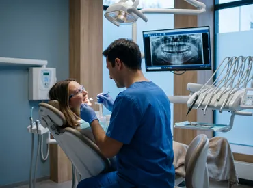
Noticing a hard lump on your gum can be unsettling, but understanding its potential causes and knowing when to seek professional evaluation is the first step toward peace of mind and optimal oral health. Your mouth is a complex ecosystem of soft tissues, bone, nerves, and blood vessels, and any persistent abnormality warrants careful attention. While many hard growths are entirely benign and easily managed, others can indicate underlying infections, dental trauma, or more serious pathological conditions that require timely intervention. By recognizing the distinct characteristics of different oral lesions, you can make informed decisions about your dental care and avoid unnecessary complications. This comprehensive guide explores the anatomy behind gum abnormalities, outlines the most common causes, provides actionable self-care strategies, and details evidence-based treatment pathways recommended by leading dental and medical organizations. Whether you are experiencing mild discomfort or simply observing an unexpected growth, this resource will equip you with the knowledge needed to navigate your next steps confidently.
Understanding Oral Anatomy and Gum Health
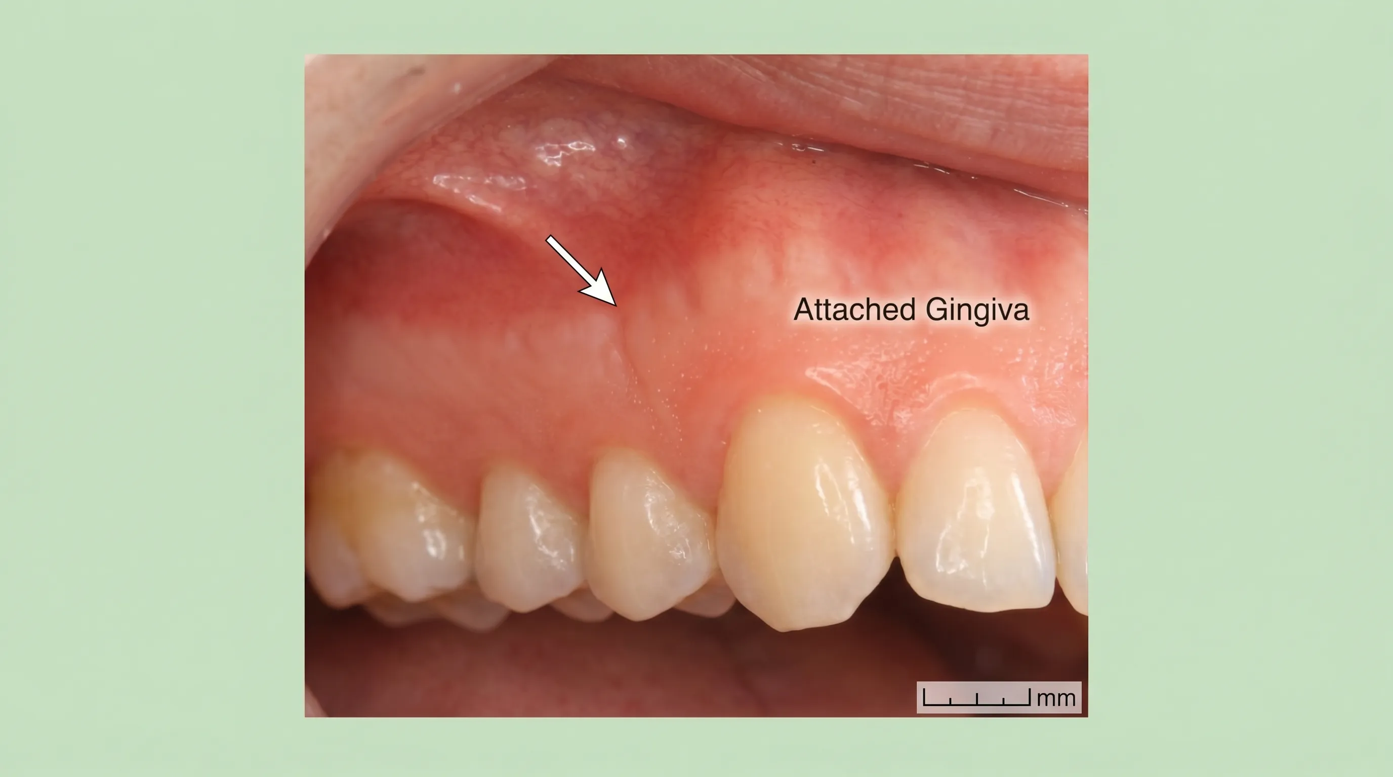
To fully comprehend why a hard lump on gum tissue develops, it is essential to first understand the underlying anatomy. The gingiva, commonly known as the gums, is composed of specialized oral mucosa that firmly attaches to the jawbone and surrounds the teeth. This tissue acts as a protective barrier, preventing pathogens from entering the underlying periodontal structures and alveolar bone. Beneath the superficial epithelial layer lies a dense connective tissue matrix rich in collagen fibers, blood vessels, and nerve endings. The bone structure immediately underneath, known as the alveolar process, houses the tooth roots and maintains dental alignment.
Free calculatorHeadache Location ChartIdentify headache types by locationOpen the calculatorWhen you notice a hard lump on gum tissue, you are typically observing changes within one or more of these anatomical layers. Hard consistency usually indicates mineralized tissue such as bone, calcified cystic material, dense fibrous scarring, or a localized inflammatory response that has organized into firm tissue. Soft, fluctuant lumps, by contrast, typically suggest fluid-filled cysts or acute abscesses. Understanding this distinction is crucial because it directly influences diagnostic pathways and treatment strategies. The American Dental Association emphasizes that consistent monitoring of oral tissues is a cornerstone of preventive dentistry. Any deviation from the normal smooth, pink, stippled appearance of healthy gingiva should be documented and professionally evaluated, especially if the lesion changes in size, color, or texture over time.
Primary Causes of a Hard Lump on Gum
A hard lump on the gum rarely develops without a specific trigger. The oral cavity is highly susceptible to mechanical trauma, bacterial accumulation, and physiological changes that can stimulate localized tissue proliferation or calcification. Below are the most clinically recognized etiologies that dental professionals investigate when evaluating persistent gingival masses.
Dental Abscesses and Periodontal Infections
One of the most frequent reasons for a firm, tender mass along the gumline is a localized dental infection. Abscesses typically originate from untreated dental caries, cracked teeth, or advanced periodontal disease that allows bacteria to penetrate deep into the periodontal ligament and surrounding bone. The body's immune response attempts to wall off the infection, creating a pocket of purulent material surrounded by inflamed tissue. In chronic cases, the overlying gum tissue may become fibrotic and feel surprisingly hard rather than soft, especially when the infection has been present for an extended period. Patients often report throbbing pain, sensitivity to temperature, and occasionally a bad taste in the mouth due to intermittent drainage. According to the Mayo Clinic, prompt drainage and antimicrobial therapy are essential to prevent the infection from spreading to adjacent facial spaces or entering the bloodstream. Mayo Clinic - Dental Abscess
Benign Bone Growths (Oral Tori)
Not every hard lump on the gum represents a disease process. Oral tori are benign, exophytic bone growths that develop slowly along the mandible (torus mandibularis) or the roof of the mouth (torus palatinus). These growths are covered by thin, sometimes ulcerated mucosa but feel rock-hard to the touch because they are composed entirely of cortical bone. Tori are highly prevalent in certain populations and often have a genetic predisposition. They do not transform into cancer, cause pain, or require removal unless they interfere with prosthetic appliances like dentures, cause recurrent ulceration due to food trauma, or hinder effective oral hygiene. Radiographic imaging clearly distinguishes tori from other lesions because they appear as well-defined, dense osseous projections arising directly from the alveolar ridge. The National Institute of Dental and Craniofacial Research confirms that monitoring is the standard of care for asymptomatic tori, as they remain stable or grow very gradually over decades.
Dental Cysts and Granulomas
Periapical granulomas and dentigerous cysts frequently manifest as firm swellings along the gumline. These lesions develop as a reaction to non-vital teeth or impacted tooth structures. A periapical granuloma forms when chronic inflammation at the root tip stimulates the proliferation of granulation tissue and fibroblasts. Over time, this can become firm or even calcify. Dentigerous cysts surround the crowns of unerupted teeth and may expand slowly, causing noticeable bony hardening of the overlying gingiva. While many cysts are initially asymptomatic, they can resorb surrounding bone, displace adjacent teeth, and eventually become infected if left untreated. Surgical enucleation or marsupialization is often required to remove the cyst lining and prevent recurrence. Comprehensive clinical guidelines recommend cone-beam computed tomography (CBCT) to accurately assess cyst size, bone involvement, and proximity to vital nerves before intervention. Cleveland Clinic - Dental Cysts
Epulis and Irritation Fibromas
Chronic mechanical irritation is a leading cause of reactive gingival overgrowths. Fibromas are the most common benign soft-tissue tumors of the oral cavity, typically triggered by repeated trauma from dental appliances, aggressive brushing, or occlusal friction. These lesions consist of dense collagen bundles that give them a firm, rubbery consistency. While they rarely cause pain, a hard lump on gum tissue that has been present for months and shows no signs of shrinking is highly likely to be a fibroma. Excisional biopsy is both diagnostic and curative, with recurrence rates remaining exceptionally low when the inciting irritant is removed. The World Health Organization classifies these reactive lesions as non-neoplastic, emphasizing their completely benign nature.
Impacted Wisdom Teeth and Pericoronitis
Third molars frequently erupt at abnormal angles, becoming trapped beneath the gum tissue. This impaction can create a hard, raised flap of tissue known as an operculum, which may feel firm due to underlying inflammatory changes and fibrosis. Food debris and bacteria easily accumulate beneath this tissue, leading to pericoronitis, a localized infection that causes swelling, trismus, and discomfort. If the impacted tooth presses against the alveolar ridge, it can create a noticeable hard prominence along the posterior gumline. Oral surgeons evaluate panoramic radiographs to determine the angulation, root development, and proximity to the inferior alveolar nerve before recommending extraction or conservative monitoring.
Oral Cancer and Malignant Growths
Although statistically less common than benign conditions, malignancies must always be considered when evaluating a persistent hard lump on gum tissue. Squamous cell carcinoma accounts for over ninety percent of oral cancers and can initially present as a firm, indurated mass that may be accompanied by ulceration, erythema, or leukoplakia. Risk factors include tobacco use, heavy alcohol consumption, human papillomavirus (HPV) infection, chronic sun exposure to the lips, and prolonged immune suppression. The American Cancer Society stresses that any lesion that does not resolve within two to three weeks warrants a biopsy. Early detection dramatically improves prognosis, with localized oral cancers boasting five-year survival rates exceeding eighty percent when treated promptly. American Cancer Society - Oral Cancer Detection
How to Distinguish Between Harmless and Serious Lumps
Differentiating between benign anatomical variations and potentially pathological lesions requires a systematic approach to self-assessment followed by professional verification. While self-examination cannot replace clinical diagnostics, it can help you gauge urgency and prepare for your dental consultation.
Symptom Checklist and Self-Assessment
Begin by observing the lesion's physical characteristics without applying excessive pressure. Note its exact location, size in millimeters, color, and surface texture. Benign tori are typically midline, symmetrically bilateral, and unchanged over years. Reactive fibromas align directly with areas of friction, such as near clasp retainers or sharp tooth edges. Painless, hard, stable masses are generally less concerning than those that are rapidly expanding, ulcerated, fixed to deeper tissues, or accompanied by unexplained bleeding. Monitor for systemic indicators like fever, fatigue, or cervical lymphadenopathy, which suggest an active infectious process or more widespread pathology. Keep a written or photographic log of any changes, as progression over time is a critical diagnostic metric for dental professionals.
Professional Diagnostic Procedures
When you schedule an appointment, your dentist or oral surgeon will perform a thorough intraoral examination, palpating the lesion to assess mobility, consistency, and tenderness. Transillumination may be used to differentiate fluid-filled from solid masses. Intraoral periapical radiographs or panoramic X-rays are standard for evaluating underlying bone changes, root integrity, and the presence of radiolucent cystic spaces or radiopaque calcifications. Advanced cases often require CBCT scanning for three-dimensional assessment of lesion borders and anatomical relationships. If malignancy or atypical cellular activity is suspected, an incisional or excisional biopsy will be performed, sending a tissue sample to an oral pathologist for histological grading. This multi-tiered diagnostic protocol ensures accurate classification and appropriate treatment planning.
When to Seek Immediate Dental or Medical Care
Timing plays a crucial role in preventing complications and ensuring optimal outcomes. Recognizing the threshold between routine monitoring and urgent care can save you from unnecessary pain, bone loss, or systemic infection.
Emergency Red Flags
You should seek emergency dental care or visit an urgent care facility if the hard lump on gum tissue is accompanied by severe swelling that impairs breathing or swallowing, a high fever exceeding 101°F, rapid facial asymmetry, or pus drainage into the mouth or neck. Spreading infections from the oral cavity can quickly compromise the airway or lead to cavernous sinus thrombosis, a life-threatening condition requiring intravenous antibiotics and hospitalization. Additionally, sudden numbness in the lower lip, chin, or tongue indicates potential nerve involvement that requires immediate radiographic evaluation and specialist referral.
What to Expect During a Dental Visit
During your consultation, the clinician will review your medical and dental history, current medications, and symptom timeline. They will document vital signs, perform a comprehensive oral examination, and order necessary imaging. You may be asked to provide a saliva sample if a viral or fungal etiology is suspected. Treatment discussions will include risk-benefit analysis, estimated recovery timelines, and financial considerations. Many dental offices now offer telehealth preliminary assessments, allowing you to share photographs and describe symptoms before scheduling an in-person visit. Always bring a complete list of supplements and prescriptions, as certain medications like bisphosphonates or immunosuppressants can significantly influence treatment protocols and healing trajectories.
Clinical Treatment Pathways
Treatment for a hard lump on gum tissue is entirely dependent on the definitive diagnosis. Modern dentistry offers a range of conservative to surgical interventions, all designed to eliminate pathology while preserving healthy tissue and function.
Pharmacological Management
When bacterial infection is the primary driver, antimicrobial therapy forms the cornerstone of initial management. Broad-spectrum antibiotics such as amoxicillin, clindamycin, or azithromycin are prescribed based on clinical severity, allergy history, and local resistance patterns. Topical antiseptic mouthwashes containing chlorhexidine gluconate help reduce surface bacterial load and promote gingival healing. For pain and inflammation management, nonsteroidal anti-inflammatory drugs like ibuprofen are commonly recommended, as they address both discomfort and underlying inflammatory mediators. Pharmacological treatment is rarely a standalone cure for structural lesions but serves as a vital bridge to definitive intervention.
Minimally Invasive and Surgical Interventions
Structural abnormalities typically require physical removal. Dental abscesses are treated through incision and drainage, followed by addressing the source via root canal therapy or extraction. Benign fibromas and cysts are excised using precise surgical techniques under local anesthesia, with the wound often closed using resorbable sutures that eliminate the need for removal appointments. Oral tori that require reduction are contoured using piezoelectric or traditional rotary instruments, minimizing thermal damage to surrounding tissues. Recovery protocols emphasize gentle oral hygiene, saline rinses, and dietary modifications to protect the surgical site during initial mucosal reattachment.
Biopsy and Histopathological Analysis
When clinical appearance or growth patterns raise concern, tissue sampling becomes mandatory. The biopsy procedure involves administering local anesthetic, excising a representative tissue fragment, and achieving hemostasis with cautery or sutures. The specimen is preserved in formalin, processed, and examined under high-magnification microscopy. Pathologists evaluate cellular architecture, nuclear atypia, and tissue invasion patterns to provide a definitive diagnosis. Histological results typically return within seven to fourteen days, at which point your clinician will discuss the findings and any subsequent treatment requirements. Early biopsy remains the gold standard for ruling out dysplasia and ensuring patient safety.
Evidence-Based Home Care and Symptom Relief
While professional treatment is essential for structural lesions, implementing safe home care practices can significantly reduce discomfort, prevent secondary infections, and support overall healing. These strategies are supported by current clinical guidelines and patient education resources.
Oral Hygiene Optimization
Maintaining meticulous oral hygiene is non-negotiable when managing any gingival abnormality. Brush twice daily using a soft-bristled toothbrush with gentle, circular motions to avoid traumatizing sensitive tissue. Replace electric or manual brushes every three months. Floss daily using waxed tape or interdental brushes tailored to your spacing. Incorporate an alcohol-free, therapeutic mouthwash containing cetylpyridinium chloride or essential oils to reduce plaque biofilm without causing mucosal dryness. Avoid aggressive scraping over any lump, as mechanical irritation can trigger inflammation, ulceration, or secondary bacterial colonization. The American Dental Association's evidence-based hygiene guidelines emphasize consistency over intensity when managing reactive tissue changes.
Dietary Modifications for Gum Health
Nutrition directly impacts immune function, tissue repair, and microbial balance in the oral cavity. Increase intake of vitamin C-rich foods like citrus, bell peppers, and broccoli, as vitamin C is essential for collagen synthesis and capillary integrity. Consume adequate protein to support cellular regeneration and wound closure. Incorporate omega-3 fatty acids from fatty fish, flaxseeds, and walnuts to modulate inflammatory pathways. Simultaneously, eliminate refined sugars, acidic beverages, and highly processed foods that fuel cariogenic bacteria and compromise pH balance. Chew primarily on the opposite side of the mouth to prevent direct trauma to the affected area. Staying well-hydrated promotes salivary flow, which naturally buffers acids and contains antimicrobial enzymes like lysozyme and lactoferrin.
Natural and Over-the-Counter Soothing Methods
Several safe, accessible remedies can provide temporary relief while awaiting professional evaluation. Warm saline rinses prepared by dissolving half a teaspoon of non-iodized salt in eight ounces of lukewarm water help draw out excess fluid, reduce swelling, and create a hypertonic environment unfavorable to pathogenic bacteria. Swish gently for thirty seconds, three to four times daily. Over-the-counter oral gels containing benzocaine or lidocaine can temporarily numb discomfort, though they should be used sparingly to avoid masking worsening symptoms. Cold compresses applied externally to the cheek for fifteen-minute intervals can reduce localized inflammation and provide analgesic effects. Avoid placing aspirin directly against the gum tissue, as this causes chemical burns and delays healing.
Prevention Strategies and Long-Term Oral Wellness
Preventing future oral abnormalities requires a proactive, multifaceted approach that integrates routine clinical care, lifestyle optimization, and vigilant self-monitoring. The following protocols are designed to maintain gingival resilience and minimize risk factors associated with common dental pathologies.
Routine Dental Care Protocols
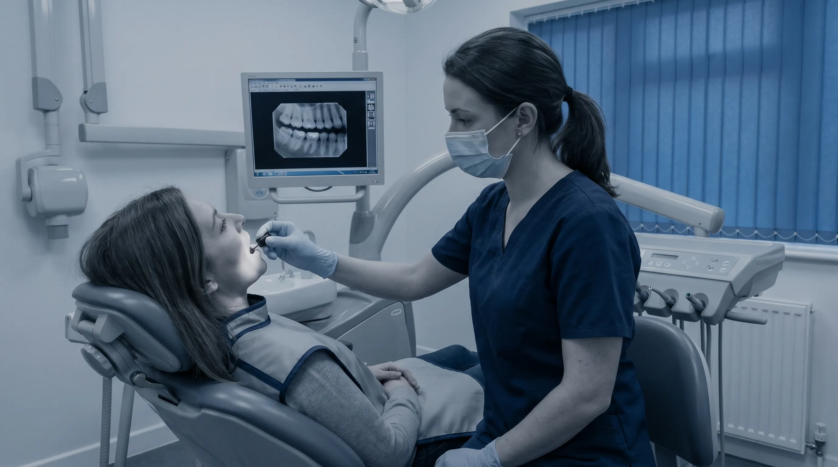
Schedule professional cleanings and comprehensive examinations at least twice annually. Prophylactic appointments allow hygienists to remove calculus deposits that home care cannot eliminate, significantly reducing periodontal disease risk. Your dentist will measure periodontal pocket depths, evaluate occlusal wear patterns, and screen for early signs of mucosal changes. Individuals with a history of recurrent infections, bruxism, or systemic conditions like diabetes may benefit from quarterly maintenance visits. Early detection of microscopic lesions or bone changes allows for minimally invasive interventions that preserve natural dentition and prevent extensive surgical requirements.
Systemic Health Connections
Oral health is deeply intertwined with systemic wellness. Poor glycemic control in diabetic patients dramatically increases susceptibility to periodontal infections and impairs tissue regeneration. Cardiovascular disease shares bidirectional inflammatory pathways with chronic gingivitis, meaning untreated gum disease may exacerbate systemic vascular inflammation. Hormonal fluctuations during pregnancy, menopause, or puberty can increase gingival vascularity, making tissues more reactive to plaque accumulation. Managing underlying medical conditions, maintaining healthy BMI ranges, avoiding tobacco in all forms, and limiting alcohol consumption collectively strengthen your body's ability to resist oral pathogens and heal efficiently from trauma or surgical procedures.
Comparative Analysis of Common Gum Abnormalities
| Feature | Dental Abscess | Oral Torus | Irritation Fibroma | Periapical Cyst |
|---|---|---|---|---|
| Consistency | Firm to fluctuant | Rock-hard | Firm, rubbery | Firm, bony if calcified |
| Onset | Days to weeks | Months to years | Weeks to months | Months to years |
| Pain Level | Moderate to severe | Painless | Usually painless | Painless unless infected |
| Response to Antibiotics | Significant improvement | No effect | No effect | Minimal (requires drainage/surgery) |
| Primary Treatment | Drainage, root canal/extraction | Monitoring or reduction | Excisional biopsy | Surgical enucleation or root canal |
| Malignant Potential | None | None | None | Very rare (unless associated with aggressive pathology) |
Frequently Asked Questions
Is a hard lump on gum always a sign of infection?
No, a hard lump on the gum is not always caused by infection. While dental abscesses and periodontal infections are common culprits, benign bone growths like oral tori, irritation fibromas, cysts, and even normal anatomical variations can also present as firm, painless lumps. Only a clinical examination and imaging can confirm the exact cause.
How long does it take for a gum lump to heal?
Healing time depends entirely on the underlying cause. An infection-related lump may resolve within one to two weeks with proper antibiotics or drainage. Surgical excisions of benign lesions typically require seven to fourteen days for complete tissue healing. Cysts or bone growths removed by an oral surgeon may take four to eight weeks for full mucosal and osseous recovery.
Can stress cause a hard lump to form on the gums?
Stress does not directly cause a hard lump on the gum, but it significantly impacts immune function and oral health. Chronic stress can lead to bruxism (teeth grinding), which causes localized trauma and reactive fibromas. Stress also weakens the immune response, making periodontal infections more likely to flare up and form abscesses.
What are the differences between a gum abscess and an oral torus?
A gum abscess is an infection-filled pocket caused by bacteria, usually presenting with pain, swelling, redness, pus, and sometimes fever. An oral torus is a benign, bony growth that develops slowly over years, is typically painless, hard as bone, and does not respond to antibiotics. Tori require no treatment unless they interfere with dentures or oral hygiene.
When should I be worried about oral cancer?
You should seek immediate evaluation if a hard lump on the gum persists longer than two to three weeks, bleeds spontaneously, shows color changes (white, red, or speckled patches), is accompanied by unexplained weight loss, difficulty swallowing, or persistent ear pain. Regular dental screenings significantly improve early detection and survival rates for oral malignancies.
Conclusion
Discovering a hard lump on your gum can understandably trigger concern, but armed with accurate information and a proactive approach to oral health, you can navigate the situation with confidence and clarity. The vast majority of gingival masses are benign, highly manageable, and respond exceptionally well to modern dental interventions. By recognizing key symptoms, understanding the spectrum of potential causes, and committing to evidence-based home care and routine professional screenings, you protect not only your smile but your overall systemic wellness. Remember that persistent changes in oral tissue warrant timely clinical evaluation, as early diagnosis consistently leads to simpler treatments, faster recovery, and optimal long-term outcomes. Prioritize your dental health as an essential component of your overall well-being, maintain consistent hygiene practices, and never hesitate to consult a licensed dental professional when something feels abnormal. Your proactive care today lays the foundation for a healthier, pain-free smile tomorrow.
How we work
Researched and drafted with AI assistance, then reviewed by a human editor against the cited sources.
This article is general health information, not medical advice. Consult a qualified healthcare professional about your own situation.