Gray Tongue: Comprehensive Guide to Causes, Symptoms & Evidence-Based Care

Noticing an unusual coating on your tongue can instantly trigger alarm, but understanding the clinical reality behind a gray tongue often reveals a highly manageable and entirely benign condition. While the appearance might seem striking, this discoloration rarely signals a severe health threat. Instead, it typically emerges from a combination of localized oral microbiome shifts, dietary influences, medication interactions, or temporary alterations in the natural shedding cycle of the tongue's surface cells. The oral cavity is a dynamic ecosystem, constantly balancing bacterial populations, epithelial turnover, and moisture levels. When this equilibrium is disrupted, visual changes become the most noticeable indicator. Fortunately, evidence-based dental medicine provides clear, straightforward pathways for resolution, focusing on gentle debridement, targeted hygiene modifications, and, when necessary, addressing underlying systemic factors. This comprehensive guide explores the anatomical, pharmacological, and lifestyle drivers behind this presentation, equips you with clinically validated home care protocols, and clarifies exactly when professional evaluation becomes necessary. By understanding the mechanisms at play and implementing consistent, science-backed maintenance strategies, most individuals successfully restore their tongue's natural appearance and improve overall oral health.
Understanding the Anatomy Behind Tongue Discoloration
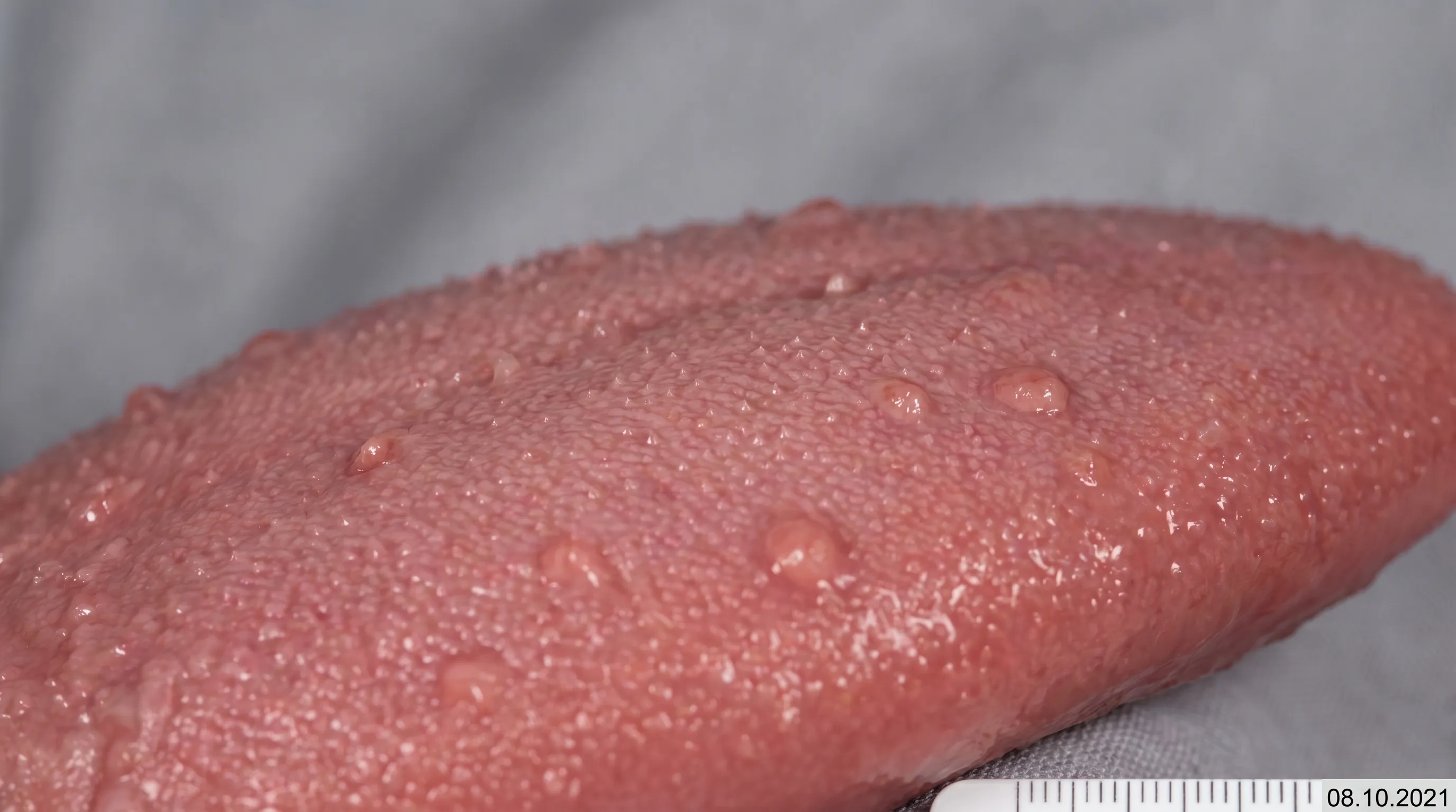
The human tongue is far more complex than it appears at first glance. Its surface is covered with thousands of microscopic projections called papillae, which serve distinct sensory, mechanical, and protective functions. Among these, the filiform papillae are the most numerous and play a crucial role in texture perception and food manipulation. Unlike the other papillae types, filiform papillae lack taste buds but possess keratinized epithelium that provides friction. Under normal physiological conditions, these structures grow, wear down, and shed at a predictable rate, maintaining a smooth and uniform appearance. When the natural desquamation process slows or stalls, however, the filiform papillae begin to elongate significantly, sometimes stretching up to fifteen times their normal length. This structural change creates a dense, carpet-like environment where dead skin cells, food debris, and microorganisms become trapped.
Free calculatorWater Intake CalculatorCalculate daily water needsOpen the calculatorThe Filiform Papillae Explained
Filiform papillae are arranged in parallel rows across the anterior two-thirds of the dorsal tongue. Their primary biological purpose is to facilitate mastication and bolus formation by providing grip on food surfaces. Each papilla consists of a connective tissue core surrounded by stratified squamous epithelium. Normally, mechanical abrasion from chewing fibrous foods, combined with natural enzymatic activity in saliva, keeps these projections trimmed and functional. When abrasive forces diminish, epithelial shedding slows dramatically. The retained keratin accumulates, creating an extended scaffold. This elongation does not represent tissue damage or disease in itself; rather, it reflects a temporary shift in the balance between cellular proliferation and mechanical exfoliation. Understanding this anatomy is essential because the physical structure of these elongated papillae directly enables the subsequent discoloration process.
The Chromogenic Process
Once the filiform papillae elongate, they create an ideal anaerobic microenvironment. The trapped debris and reduced oxygen flow allow chromogenic, or color-producing, microorganisms to thrive. These bacteria and fungi naturally inhabit the oral cavity but typically remain invisible in low numbers. In the elongated papillae environment, however, they proliferate rapidly. Certain species produce porphyrins and other pigmented metabolic byproducts that stain the keratinized surface. The resulting hue can vary widely depending on the dominant microbial strains, dietary intake, tobacco exposure, and medication use. While black and brown are frequently discussed, a gray tongue is equally common and follows the exact same pathophysiological pathway. The gray shade often indicates a mixed microbial colonization, sometimes involving Candida species alongside bacterial flora, or reflects specific chemical reactions from ingested compounds. WebMD's comprehensive guide on oral health outlines how these microbial interactions drive visible changes, emphasizing that color variation alone does not alter the underlying benign nature of the condition.
Primary Medical Causes of a Gray Tongue
Identifying the precise etiology of tongue discoloration is fundamental to implementing the correct intervention. While lifestyle factors frequently contribute, several distinct medical and pharmacological pathways directly trigger or accelerate the process. Clinicians categorize these causes into structural syndromes, medication-induced reactions, and nutritional or systemic deficiencies. Each category operates through unique biochemical and microbiological mechanisms, yet they all converge on the same clinical endpoint: visible alteration of the dorsal tongue surface. Recognizing these distinctions prevents unnecessary anxiety and ensures targeted management.
Black Hairy Tongue Syndrome
Despite its dramatic name, Black Hairy Tongue (BHT), medically termed lingua villosa nigra, is the most prevalent structural cause of gray, brown, yellow, or black discoloration. Epidemiological data suggests it affects approximately 0.5% to 1% of the adult population, with a higher incidence reported in males. The condition is characterized by marked hyperkeratosis and failure of the filiform papillae to undergo normal desquamation. As the projections lengthen, they trap chromogenic bacteria such as Corynebacterium, Bacteroides, and various anaerobic streptococci. Fungal colonization, particularly by Candida albicans, may also contribute to pigment variation. BHT prevalence increases significantly among smokers, individuals living with HIV, patients undergoing radiation therapy, and those with chronic xerostomia. Importantly, the syndrome is entirely reversible. It does not indicate malignancy, tissue invasion, or systemic infection. The discoloration remains superficial, affecting only the keratinized surface layer, which responds predictably to mechanical debridement and environmental modification.
Medication-Related Reactions
Pharmaceutical agents represent another major category of triggers. Certain medications alter the oral microbiome, reduce salivary flow, or directly stain the mucosal surfaces. Bismuth subsalicylate, commonly found in over-the-counter gastrointestinal remedies, produces a well-documented gray-black coating. The mechanism involves a straightforward chemical reaction: bismuth ions bind with trace sulfides in saliva, precipitating as bismuth sulfide, which adheres tightly to the tongue's surface. This staining is entirely transient and resolves within days of discontinuation. Antibiotics, particularly broad-spectrum agents like penicillins, cephalosporins, and clarithromycin, disrupt the delicate balance of oral flora by eliminating commensal bacteria, allowing resistant or pigmented organisms to overgrow. Antifungals, antipsychotics, and medications containing chlorhexidine digluconate similarly alter microbial equilibrium or directly deposit pigments. Iron supplements are another frequent culprit, as unabsorbed iron salts can interact with sulfur-containing compounds in the oral cavity, producing a darkened appearance that mimics a gray tongue. Patients initiating new regimens should be aware of these potential cosmetic side effects, which rarely necessitate medication cessation unless discomfort becomes pronounced.
Nutritional Deficiencies and Systemic Links
In less common but clinically significant cases, gray tongue discoloration may reflect underlying nutritional imbalances. Pellagra, a condition caused by severe deficiency of vitamin B3 (niacin), classically presents with a thick, grayish fur coating the dorsal tongue. This manifestation occurs alongside mucosal atrophy, glossitis, and the characteristic dermatological, gastrointestinal, and neurological symptoms historically known as the "3 Ds": dermatitis, diarrhea, and dementia. While pellagra is rare in developed nations with fortified food supplies, it remains a consideration in patients with severe malabsorption syndromes, chronic alcoholism, or restrictive diets. Other micronutrient shortfalls, particularly involving iron, folate, or vitamin B12, can indirectly contribute by altering epithelial turnover rates or compromising immune regulation in the oral cavity. Addressing the nutritional deficit typically restores normal papillae function and resolves discoloration, underscoring the importance of comprehensive dietary assessment when structural and environmental causes are ruled out.
| Condition | Primary Mechanism | Typical Presentation | Reversibility | Clinical Notes |
|---|---|---|---|---|
| Black Hairy Tongue | Filiform papillae elongation + chromogenic microbe accumulation | Diffuse gray/black/brown/yellow coating on dorsal surface | Highly reversible with hygiene | Most common cause; affects 0.5-1% of adults |
| Medication Staining (Bismuth) | Chemical reaction forming bismuth sulfide | Transient gray-black surface layer | Resolves in 24-72 hours post-discontinuation | Harmless; frequently seen with Pepto-Bismol use |
| Pellagra (Niacin Deficiency) | Epithelial atrophy and metabolic disruption | Thick grayish fur + glossitis + systemic symptoms | Reversible with B3 supplementation | Rare; accompanied by dermatitis, diarrhea, neurological changes |
| Antibiotic-Induced Dysbiosis | Flora disruption allowing pigmented overgrowth | Patchy or diffuse discoloration, possible halitosis | Reversible post-course or with probiotics | Common with broad-spectrum prescriptions |
Environmental and Lifestyle Triggers
Daily habits and environmental exposures exert a profound influence on oral mucosal health. The tongue exists in constant contact with ingested substances, temperature variations, and mechanical forces that collectively shape its structural integrity and microbial composition. When certain lifestyle patterns become entrenched, they can inadvertently create the exact conditions necessary for papillae elongation and subsequent gray tongue development. Recognizing these modifiable triggers empowers individuals to implement immediate, effective changes without requiring medical intervention.
Tobacco, Caffeine, and Staining Agents
Tobacco use remains one of the most significant lifestyle contributors to oral discoloration. The heat, chemical compounds, and particulate matter from cigarettes, cigars, and smokeless tobacco products directly irritate the lingual epithelium, accelerating keratin production while simultaneously impairing normal shedding mechanisms. Additionally, tobacco introduces exogenous pigments that bind readily to the porous, elongated papillae structure. Similarly, frequent consumption of heavily pigmented beverages like black coffee, dark teas, and red wines provides a continuous supply of staining agents. These compounds do not cause harm to the underlying tissue but readily accumulate in the retained keratin matrix, intensifying the visual appearance. Patients who consume multiple cups daily without adequate oral rinsing frequently notice progressive darkening, which may initially manifest as gray before transitioning to deeper brown or black hues if left unaddressed.
Oral Hygiene Practices and Mouthwash Misuse
While diligent cleaning prevents most oral conditions, excessive or inappropriate hygiene practices can paradoxically trigger discoloration. Overuse of aggressive oxidizing mouthwashes containing hydrogen peroxide, chlorhexidine, witch hazel, or high concentrations of menthol disrupts the natural microbial ecology and can induce chemical irritation. These agents strip away protective mucosal layers and alter pH balance, prompting compensatory hyperkeratosis. Patients who rinse multiple times daily or swish for extended periods essentially create a low-grade inflammatory state that slows desquamation. Furthermore, neglecting routine tongue cleaning while focusing exclusively on teeth allows debris to accumulate unchecked. A comprehensive hygiene protocol must include gentle lingual debridement to maintain papillae length within physiological limits and prevent the trapped buildup that leads to a gray tongue.
Dehydration and Xerostomia
Saliva serves as the oral cavity's primary cleansing and buffering agent. It continuously washes away dead cells, neutralizes acids, delivers antimicrobial peptides, and maintains optimal hydration of the mucosal surfaces. When saliva production declines due to dehydration, mouth breathing, stress, or medication-induced xerostomia, the protective washing mechanism fails. A dry oral environment promotes rapid keratin accumulation, reduces mechanical clearance of food particles, and creates a stagnant environment where bacteria flourish. Individuals who work long hours without adequate water intake, exercise intensely without rehydrating, or sleep with their mouths open frequently experience transient morning discoloration that progresses with chronic dryness. Restoring optimal hydration status directly supports epithelial turnover and significantly reduces the likelihood of pigment trapping.
Clinical Presentation and Associated Symptoms
Recognizing the precise characteristics of gray tongue helps differentiate it from more serious mucosal pathologies and guides appropriate management strategies. The clinical presentation follows a predictable pattern, with specific localization, texture variations, and accompanying sensory changes that clinicians use to establish a working diagnosis.
Visual Characteristics and Distribution
Discoloration typically appears as a diffuse, velvety coating concentrated on the dorsal midline of the tongue, extending from the posterior third forward to the circumvallate papillae. The lateral borders and ventral surface usually remain unaffected, preserving their normal pinkish hue. The affected area exhibits a distinctly elongated, hair-like texture when examined closely, though the projections are microscopic. The color palette ranges from light gray to charcoal, brown, yellow, or green, depending on the dominant chromogenic organisms and external staining factors. The coating is superficial and does not bleed or ulcerate when gently probed. Unlike true mucosal lesions, which integrate into the tissue architecture, the discoloration sits atop the keratin layer and can be partially or fully removed with mechanical scraping, revealing healthier tissue beneath.
Sensory Symptoms and Quality of Life Impact
Despite its striking appearance, a gray tongue is predominantly asymptomatic. Most individuals experience no pain, inflammation, or functional impairment. However, the altered surface topography and microbial overgrowth can produce secondary sensory phenomena. Patients frequently report a persistent metallic or foul taste due to bacterial metabolic byproducts. Halitosis, or oral malodor, commonly accompanies the condition as anaerobic bacteria produce volatile sulfur compounds. Some individuals describe a tickling sensation against the soft palate during swallowing, a gag reflex triggered by elongated papillae contact, or mild nausea related to taste alterations. When Candida overgrowth coincides with the structural changes, a burning sensation known as glossopyrosis may develop. These symptoms rarely become severe but can significantly impact social confidence and dietary enjoyment. Addressing the underlying buildup typically resolves the sensory complaints rapidly, restoring normal taste perception and oral comfort.
Diagnostic Procedures and Professional Evaluation
Accurate diagnosis relies primarily on visual inspection and clinical history, eliminating the need for invasive testing in the vast majority of presentations. Healthcare professionals follow standardized protocols to confirm benign etiology, rule out differential conditions, and identify contributing factors that require targeted intervention.
How Clinicians Assess the Condition
Dentists and physicians evaluate the tongue under adequate lighting, noting the distribution, texture, and removability of the coating. A gentle tongue depressor or gauze swab confirms that the discoloration sits superficially and does not adhere to underlying tissue. Medical history review focuses on medication use, tobacco habits, hydration status, dietary patterns, and oral hygiene routines. Because the presentation is highly characteristic, histologic biopsy is exceptionally rare and reserved only for cases with atypical features, persistent ulceration, or suspected neoplastic transformation. When histology is performed, it reveals marked filiform papillae elongation, hyperparakeratosis, and dense colonization of chromogenic bacteria or fungi on the epithelial surface. This microscopic confirmation reinforces the benign, reversible nature of the condition and validates conservative management approaches.
Red Flags and When to Seek Immediate Care
While a gray tongue is overwhelmingly benign, certain accompanying symptoms warrant prompt professional evaluation. If discoloration persists beyond fourteen days despite rigorous hygiene improvements, clinicians should investigate potential nutritional deficiencies, medication interactions, or chronic systemic conditions. Painful lesions, bleeding, induration, or inability to scrape the coating away suggest alternative diagnoses such as oral lichen planus, leukoplakia, or early mucosal malignancies. Fever, unexplained weight loss, persistent diarrhea, or widespread dermatological changes may indicate pellagra, autoimmune disorders, or infectious processes requiring laboratory workup and systemic treatment. Patients undergoing head and neck radiation therapy or living with significant immunosuppression should maintain regular oral examinations, as altered mucosal immunity increases susceptibility to fungal overgrowth and delayed healing. Early intervention prevents complications and ensures appropriate management of underlying health conditions.
Differential Diagnosis
Distinguishing gray tongue from other lingual conditions prevents misdiagnosis and ensures correct treatment pathways. Wikipedia's detailed medical overview provides extensive references highlighting these distinctions. Hairy leukoplakia presents as firm white plaques on the lateral tongue margins and correlates with Epstein-Barr virus reactivation, particularly in immunocompromised individuals. Geographic tongue features migratory, erythematous patches bordered by white keratinized lines, driven by benign inflammatory changes rather than papillae elongation. Oral thrush manifests as creamy white curd-like patches that bleed slightly when scraped, indicating active Candida infection. Median rhomboid glossitis appears as a smooth, red, rhomboid area on the dorsal midline, associated with chronic candidiasis or developmental anomalies. Understanding these clinical variations allows practitioners and patients to navigate symptoms accurately without unnecessary concern.
Evidence-Based Treatment and Home Care Protocols
Management of gray tongue centers on mechanical debridement, environmental modification, and, when indicated, targeted pharmacological intervention. Conservative approaches achieve resolution in the majority of cases, while medical treatments address persistent or complicated presentations. A structured, stepwise protocol ensures optimal outcomes without disrupting the natural oral microbiome.
Step-by-Step Oral Hygiene Optimization
Effective treatment begins with establishing a consistent, gentle cleaning routine. Patients should brush the dorsal tongue twice daily using a soft-bristled toothbrush. Starting at the posterior third, apply light pressure and sweep forward in smooth strokes, rinsing the brush after each pass to prevent redepositing debris. Avoid vigorous scrubbing, which causes epithelial micro-tears and delays healing. Following brushing, utilize a dedicated tongue scraper, typically made of stainless steel or flexible plastic. Place the scraper at the back of the tongue, pull it forward gently, and rinse thoroughly. Repeat three to five times until minimal residue appears. Standard tooth brushing should continue twice daily with fluoride toothpaste, focusing on plaque removal along the gingival margin. Replacing toothbrushes every three months prevents bacterial buildup and maintains bristle effectiveness. This mechanical approach physically removes elongated papillae tips, trapped debris, and chromogenic microorganisms, directly addressing the root cause of discoloration.
Medical and Pharmaceutical Interventions
When conservative measures prove insufficient after several weeks, clinicians may recommend adjunctive therapies. If fungal colonization is suspected or confirmed through clinical assessment, topical or systemic antifungal medications such as nystatin suspension or fluconazole can restore microbial balance. In refractory cases, topical tretinoin (Retin-A) may be prescribed to normalize epithelial turnover and reduce keratin accumulation, though it requires careful application to avoid mucosal irritation. Antibiotics are rarely indicated unless a specific pathogenic bacterial overgrowth drives persistent symptoms. For extreme, recalcitrant cases unresponsive to all conservative and pharmacological options, procedural interventions including laser therapy or electrosurgery may be considered to selectively reduce papillae length. These procedures remain last-resort options due to cost, recovery time, and the proven efficacy of simpler approaches. Patients should always coordinate medication use with their healthcare provider to prevent interactions and ensure appropriate dosing.
Nutritional and Hydration Support
Complementing mechanical cleaning with systemic support accelerates recovery. Increasing dietary fiber and roughage introduces natural mechanical abrasion during mastication, promoting physiological papillae wear. Consuming fibrous fruits, raw vegetables, and whole grains provides continuous, gentle debridement while supporting overall digestive health. Maintaining optimal hydration ensures consistent salivary flow, washing away metabolic waste and preventing dry mucosa. Patients should aim for adequate daily water intake, adjusting for activity level, climate, and medication use. Reducing or eliminating oxidizing mouthwashes, tobacco products, and excessive caffeine minimizes further keratin stimulation and staining. Implementing these modifications alongside diligent hygiene creates a synergistic effect, typically resolving a gray tongue within one to three weeks. Consistent monitoring confirms progress and allows timely adjustment of the care plan.
Long-Term Prevention and Daily Maintenance
Sustaining a healthy tongue requires integrating preventive habits into daily routines. Once discoloration resolves, maintaining the improved oral environment prevents recurrence and supports long-term mucosal vitality. Prevention strategies mirror treatment protocols but emphasize consistency, moderation, and proactive monitoring rather than intensive intervention.
Building a Sustainable Routine
Daily tongue cleaning should remain a standard component of oral care, equivalent in importance to brushing and flossing. Establishing a consistent morning and evening schedule ensures debris removal before microbial overgrowth initiates. Patients should monitor coating thickness and color changes, addressing minor buildup promptly rather than waiting for significant discoloration. Regular dental checkups every six months provide professional cleaning, early identification of emerging issues, and personalized guidance tailored to individual risk factors. Educating family members on proper techniques fosters shared accountability and reinforces healthy habits across all age groups.
Dietary and Hydration Adjustments
Nutrition plays a foundational role in maintaining epithelial health. A balanced diet rich in vitamins A, B-complex, C, and iron supports cellular regeneration and immune function. Avoiding highly processed, low-fiber foods reduces the risk of prolonged papillae retention. Moderating intake of staining beverages and consuming water immediately after coffee or tea minimizes pigment deposition. Patients with chronic dry mouth should consider saliva-stimulating strategies such as sugar-free xylitol gum, adequate room humidification, and consultation regarding medication adjustments. Proactive hydration and mindful dietary choices create an inhospitable environment for chromogenic bacteria, effectively preventing a gray tongue from redeveloping.
Frequently Asked Questions
Is a gray tongue a sign of a serious medical condition?
In the vast majority of cases, a gray tongue is a benign and reversible condition linked to harmless causes like elongated papillae, medication side effects, or dietary habits. However, if the discoloration persists beyond two weeks despite improved oral care, or is accompanied by pain, fever, difficulty swallowing, or systemic symptoms, it warrants professional evaluation to rule out underlying deficiencies or infections.
Can taking Pepto-Bismol actually turn my tongue gray or black?
Yes, this is a well-documented and completely harmless side effect. Bismuth subsalicylate reacts with trace amounts of sulfur naturally present in saliva to form bismuth sulfide, a blackish-gray compound that temporarily coats the tongue. The discoloration typically fades within 24 to 48 hours after stopping the medication and requires no specific treatment.
How do I properly clean my tongue to reverse discoloration?
Begin by gently brushing your tongue twice daily with a soft-bristled toothbrush, starting from the back and sweeping forward. Follow up with a dedicated tongue scraper for thorough removal of debris and elongated papillae buildup. Avoid aggressive scrubbing, which can cause micro-trauma, and pair this routine with standard twice-daily tooth brushing and consistent hydration for optimal results.
Should I stop using my mouthwash if I notice tongue changes?
Certain mouthwashes, particularly those containing high concentrations of hydrogen peroxide, chlorhexidine, witch hazel, or menthol, can contribute to papillae elongation and discoloration when used excessively. If you suspect your mouthwash is causing the issue, discontinue use temporarily or switch to an alcohol-free, non-oxidizing formula recommended by your dentist.
What is the difference between gray tongue and hairy leukoplakia?
Gray tongue typically stems from black hairy tongue syndrome or environmental factors and appears as a diffuse coating on the top surface that can be gently scraped away. Hairy leukoplakia, however, is an Epstein-Barr virus-related condition that manifests as firm, white, corrugated patches primarily on the lateral (side) edges of the tongue. Unlike gray tongue, leukoplakia cannot be scraped off and requires clinical assessment, particularly in immunocompromised patients.
Conclusion
A gray tongue may initially appear alarming, but clinical evidence consistently demonstrates that it represents a benign, highly reversible condition driven by temporary changes in papillae length, microbial balance, or external exposures. By understanding the anatomical mechanisms behind filiform elongation and recognizing common triggers such as bismuth medications, tobacco use, oxidative mouthwashes, and inadequate hydration, individuals can implement targeted interventions with confidence. Consistent, gentle mechanical cleaning, combined with strategic lifestyle modifications, resolves discoloration in the vast majority of cases without requiring pharmaceutical intervention. Maintaining long-term oral health hinges on proactive hygiene, balanced nutrition, and regular professional monitoring. When persistent discoloration, pain, or systemic symptoms arise, consulting a dental or medical professional ensures accurate diagnosis and appropriate care. Ultimately, addressing a gray tongue reinforces broader principles of preventive health, empowering patients to take control of their oral microbiome and sustain lasting wellness.
How we work
Researched and drafted with AI assistance, then reviewed by a human editor against the cited sources.
This article is general health information, not medical advice. Consult a qualified healthcare professional about your own situation.