What Is Crick Neck? Causes, Symptoms & Evidence-Based Relief Strategies

Key points
- Monitor centered at eye level
- Keyboard/mouse within elbow reach
- Feet flat, knees slightly below hips
- Headset use for prolonged calls
- Regular micro-breaks scheduled
- Laptop riser for secondary screens
Waking up to a sudden, sharp restriction in your neck movement or feeling an unexpected tightness after hours of focused work can instantly derail your daily routine. That familiar, frustrating sensation often described as a crick neck leaves many searching for answers, wondering why their cervical spine suddenly feels locked in place. Understanding what is crick neck from a clinical perspective is the first step toward effective relief and long-term prevention. Medically, a crick neck represents an acute episode of cervical stiffness, usually driven by localized muscle spasm, ligament strain, or facet joint irritation in the upper spinal region. While it is rarely a sign of serious pathology, the discomfort can be surprisingly debilitating, interfering with driving, working, exercising, and even sleeping comfortably. The condition affects millions annually, often striking without warning after seemingly minor triggers like poor sleeping posture, prolonged desk work, or sudden head movements. By exploring the underlying anatomy, evidence-based treatment protocols, and lifestyle modifications, you can regain full mobility, reduce recurrence, and maintain optimal cervical health. For clinical context, refer to the Mayo Clinic's comprehensive overview of neck pain. This comprehensive guide breaks down the medical mechanisms, clinical presentations, and proven interventions so you can confidently manage acute stiffness and protect your spine for the future.
Understanding the Anatomy of a Crick Neck
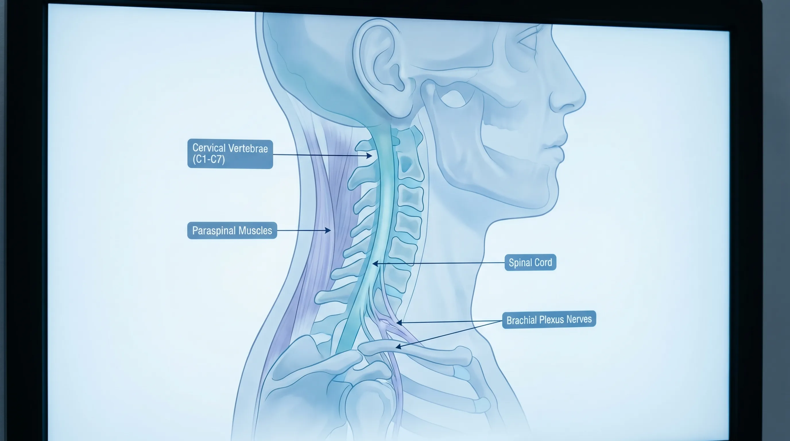
To fully grasp what is crick neck and why it occurs, we must first examine the complex biomechanical architecture of the cervical spine. Your neck is a remarkably versatile yet delicate structure designed to support approximately ten to twelve pounds of head weight while enabling a wide range of motion. When any component of this system becomes irritated or overloaded, the protective mechanisms of the body trigger localized inflammation and muscle guarding, resulting in that characteristic locked sensation.
Free calculatorWater Intake CalculatorCalculate daily water needsOpen the calculatorCervical Spine Structure
The cervical spine consists of seven vertebrae labeled C1 through C7, separated by intervertebral discs that act as shock absorbers. Unlike the thoracic and lumbar regions, the cervical vertebrae are smaller and more mobile, making them highly susceptible to repetitive stress and positional strain. The upper cervical region (C1-C2), known as the atlanto-axial joint, provides nearly half of the neck’s rotational movement, while the mid-to-lower segments (C3-C7) facilitate flexion and extension. When these segments are held in non-neutral positions for extended periods, facet joint capsules can become compressed, leading to micro-inflammation that manifests as acute stiffness. Understanding what is crick neck requires recognizing that it is often a localized joint capsule irritation rather than a disc injury. Detailed anatomical breakdowns are available through the Cleveland Clinic's cervical spine resources.
Muscle and Ligament Involvement
The cervical spine is stabilized by a sophisticated network of muscles and ligaments. The levator scapulae, trapezius, sternocleidomastoid, and splenius capitis work synergistically to control head positioning and stabilize the neck during movement. Ligaments such as the anterior longitudinal ligament, posterior longitudinal ligament, and ligamentum flavum maintain spinal alignment and prevent excessive motion. When a crick neck develops, the body’s natural protective response often involves involuntary muscle contraction or spasm. This myofascial response aims to immobilize the irritated area to prevent further damage, but it frequently results in the painful, restricted movement patients report. Trigger points can form within the muscle belly, creating localized knots that refer pain to surrounding regions and further limit mobility.
Joint Mechanics and Fascia
Fascia is a continuous, web-like connective tissue that surrounds muscles, nerves, and blood vessels throughout the cervical region. Healthy fascia glides smoothly during movement, allowing muscles to contract and relax efficiently. Prolonged static postures, dehydration, or sudden strain can cause fascial adhesions and increased tissue viscosity. When asking what is crick neck in relation to soft tissue mechanics, clinicians point to these fascial restrictions that limit gliding and create a sensation of internal tightness. The cervical facet joints, which are synovial joints containing lubricating fluid and a rich nerve supply, can also develop minor effusion or capsular tightening. This combination of muscular guarding, fascial stiffness, and joint irritation creates the classic clinical presentation of acute cervical stiffness.
Common Causes and Risk Factors
While a crick neck can appear out of nowhere, it almost always stems from identifiable mechanical, environmental, or lifestyle factors. Recognizing these triggers is essential for targeted intervention and long-term spine health.
Poor Posture and Ergonomics
Modern work environments have dramatically altered how humans use their spines. Forward head posture, where the ears shift ahead of the shoulders while looking at screens, increases the effective weight on the cervical spine exponentially. For every inch the head moves forward, the neck muscles must support an additional ten pounds of load. This constant eccentric contraction fatigues the posterior cervical muscles, leading to micro-tears, lactic acid buildup, and eventual spasms. Prolonged slouching, hunched shoulders, and improper monitor height compound the issue, creating a perfect storm for acute stiffness. Many patients first understand what is crick neck after realizing their workstation ergonomics have been compromised for months. The CDC’s workplace health guidelines emphasize proper alignment to prevent cervical overload and reduce musculoskeletal strain.
Sleeping Positions and Pillow Support
Sleep occupies a significant portion of daily spinal load time. The pillow you choose directly dictates whether your cervical spine maintains a neutral alignment or becomes strained during the night. Sleeping on the stomach forces the head into extreme lateral rotation, stretching the levator scapulae and facet joints for hours. Conversely, overly high or flat pillows disrupt the natural cervical lordosis, compressing anterior structures or straining posterior musculature. Temperature drops at night can also reduce muscle elasticity, making cold rooms a hidden risk factor for morning stiffness. Evaluating sleep hygiene is often the missing piece when patients ask what is crick neck and why it recurs frequently. The NIH’s Sleep Research Network notes that proper spinal alignment directly impacts musculoskeletal recovery during rest cycles.
Sudden Movements and Strain
Acute trauma or uncoordinated movements are classic triggers for cervical stiffness. Checking a blind spot while driving, lifting an object while twisted, or experiencing a sudden jolt (like a mild whiplash from a minor vehicle impact) can overstress the facet joint capsules and paraspinal muscles. The nervous system responds immediately with protective splinting. This acute guarding is the body’s way of creating a biological cast to allow healing. However, the resulting inflammation and restricted blood flow prolong discomfort and reduce flexibility temporarily.
Underlying Medical Conditions
While most crick neck episodes are mechanical, certain systemic conditions can predispose individuals to recurrent stiffness. Osteoarthritis of the cervical spine causes joint space narrowing and bone spur formation, which irritate surrounding tissues. Fibromyalgia amplifies pain signaling, making minor strains feel severe. Vitamin D and magnesium deficiencies impair muscle relaxation and nerve function. Thyroid dysfunction and dehydration also contribute to tissue irritability. When evaluating what is crick neck in a clinical setting, practitioners screen for these underlying factors to ensure treatment addresses the root cause rather than just masking symptoms.
Signs and Symptoms to Watch For
Recognizing the precise clinical presentation helps differentiate benign cervical stiffness from more serious neurological conditions. Accurate symptom identification guides appropriate self-care and determines when professional intervention is necessary.
Localized Pain vs. Radiating Discomfort
A classic crick neck presents with sharp or dull pain concentrated in one side of the upper cervical region, often near the base of the skull or alongside the trapezius ridge. The pain typically worsens with active movement in the restricted direction and eases with rest. Unlike nerve compression syndromes, the discomfort rarely travels past the shoulder blade or into the forearm. If you experience electric shock-like sensations, tingling, or numbness extending down the arm, this suggests cervical radiculopathy rather than a simple muscular crick. Understanding this distinction is crucial when determining what is crick neck versus nerve involvement.
Range of Motion Limitations
Reduced flexibility is the hallmark symptom. Patients frequently report an inability to turn their head fully to one side, look upward comfortably, or bring their chin to their chest. Passive movement (gently guiding the head with your hand) may be slightly easier but still reproduces a stretching or pulling sensation. The limitation is usually directional rather than omnidirectional, pointing to a specific strained muscle or irritated facet joint. Tracking these movement restrictions over several days helps gauge recovery progress.
Muscle Spasms and Stiffness
Involuntary muscle contractions often accompany acute stiffness. The affected area may feel firm, warm, or slightly swollen to the touch. Patients describe a sensation of internal tightness, as if the neck muscles are gripping themselves. Fatigue in the surrounding shoulder girdle is common due to compensatory overuse. Morning stiffness typically peaks after waking and gradually improves with gentle movement and warmth, though it may flare again after prolonged static postures later in the day.
Diagnosis and Medical Evaluation
Most cases resolve with conservative care, but clinical evaluation ensures safety and accuracy, especially when symptoms persist or present atypically.
Physical Examination Techniques
Healthcare providers conduct a systematic assessment beginning with posture analysis and active range of motion testing. Palpation identifies tender trigger points, muscle guarding, and joint line sensitivity. Neurological screening includes reflex testing, dermatomal sensation checks, and myotome strength assessment to rule out nerve compression. Special tests like the Spurling’s maneuver evaluate radicular symptoms, while the cervical distraction test assesses joint-mediated pain. These clinical findings help confirm what is crick neck versus more complex pathologies.
Imaging Studies and Diagnostic Tests
Routine imaging is unnecessary for uncomplicated cases. Radiographs (X-rays) are reserved for trauma, suspected fractures, or progressive neurological deficits. Magnetic resonance imaging (MRI) provides detailed visualization of discs, nerves, and soft tissues when conservative management fails after four to six weeks. Ultrasound-guided evaluations can assess muscle integrity and detect fascial thickening. Blood tests may be ordered if systemic inflammation, infection, or autoimmune conditions are suspected.
Differential Diagnosis
Distinguishing a crick neck from other cervical disorders prevents mismanagement. Cervical spondylosis involves degenerative disc disease and osteophyte formation. Cervical myelopathy affects the spinal cord, causing gait disturbances and hand clumsiness. Meningitis presents with neck stiffness alongside high fever, photophobia, and altered mental status. Torticollis involves sustained abnormal head posture, often congenital or drug-induced. Myofascial pain syndrome features widespread trigger points. Clinicians carefully evaluate these possibilities when determining what is crick neck and whether advanced intervention is warranted. For authoritative guidance on cervical conditions, consult resources like Mayo Clinic's Neck Pain Diagnosis & Treatment and Cleveland Clinic's Cervical Spine Care.
Evidence-Based Treatment Options
Effective management combines acute symptom control with progressive rehabilitation to restore full function and prevent recurrence.
Immediate First Aid and Acute Care
Within the first 48 hours, prioritize rest without immobilization. Avoid heavy lifting, sudden twisting, and high-impact activities. Apply ice packs wrapped in a cloth for 15-20 minutes every 2-3 hours to reduce localized inflammation. Gentle isometric contractions (pushing the head gently against your hand without moving the neck) maintain neuromuscular activation without straining tissues. Over-the-counter topical analgesics containing menthol, camphor, or diclofenac can provide targeted relief without systemic side effects. The NIH MedlinePlus guide to neck strain management recommends similar conservative first-aid protocols to safely reduce acute symptoms.
Physical Therapy and Targeted Stretching
Professional rehabilitation focuses on restoring mobility, strengthening stabilizers, and correcting biomechanical imbalances. Therapists employ joint mobilization, myofascial release, and progressive stretching protocols targeting the levator scapulae, upper trapezius, and deep cervical flexors. The McKenzie Method utilizes specific directional exercises to centralize pain and improve extension tolerance. Nerve gliding exercises address subtle neural tension that contributes to stiffness. Consistency is key; daily home exercises prescribed by a licensed therapist yield the most sustainable outcomes. For clinically vetted stretching demonstrations, refer to the Cleveland Clinic's recommended neck mobility exercises and the Mayo Clinic's physical therapy guidelines for cervical pain.
| Symptom / Feature | Crick Neck (Muscle/Joint Strain) | Cervical Radiculopathy (Nerve Compression) | Cervical Spondylosis (Degeneration) |
|---|---|---|---|
| Primary Pain Location | Localized upper neck, shoulder blade | Radiates down arm, forearm, hand | Central neck, stiffness with age |
| Neurological Signs | None or mild muscle fatigue | Tingling, numbness, weakness, reflex changes | Variable; may include mild nerve irritation |
| Onset | Sudden, often after sleep or strain | Gradual or sudden after disc injury | Progressive, chronic, worsens with time |
| Relief Strategies | Heat/ice, stretching, rest, NSAIDs | Postural correction, nerve glides, possible surgery | Activity modification, strength training, injections |
| Prognosis | Resolves in 1-2 weeks with self-care | Varies; may require PT or surgical intervention | Chronic management; focus on slowing progression |
Medications and Pain Relief Strategies
Non-steroidal anti-inflammatory drugs (NSAIDs) like ibuprofen or naproxen reduce prostaglandin-mediated inflammation. Acetaminophen targets pain perception without anti-inflammatory effects. Short-term prescription muscle relaxants may be indicated for severe spasms, though they carry drowsiness risks and should be used cautiously. Topical lidocaine patches provide localized analgesia without gastrointestinal concerns. Always consult a physician before combining medications or using them long-term.
Heat, Cold, and Massage Therapies
Thermal therapies modulate tissue response. Cold therapy constricts blood vessels, reducing edema and slowing nerve conduction speed to dull acute pain. Moist heat dilates vessels, increases oxygen delivery, and promotes collagen extensibility, making it ideal for subacute and chronic stiffness. Therapeutic massage, particularly trigger point release and Swedish techniques, breaks fascial adhesions and stimulates parasympathetic relaxation.
Home Remedies and Self-Care Strategies
Empowering patients with practical, daily habits transforms acute recovery into long-term spine resilience.
Ergonomic Adjustments
Optimizing your workspace prevents repetitive microtrauma. Position your monitor at eye level, approximately an arm’s length away. Keep elbows at 90 degrees, wrists neutral, and shoulders relaxed. Use a lumbar-supporting chair that encourages an upright posture. Implement the 20-8-2 rule: sit for 20 minutes, stand for 8, and move or stretch for 2 minutes every hour. Voice-to-text software and document holders reduce forward head positioning. Small adjustments compound into significant cervical relief.
Sleep Hygiene and Neck Support
Invest in a contour pillow that supports the natural cervical curve while allowing the shoulder to tuck comfortably beneath. Back sleepers benefit from thinner pillows, side sleepers require taller ones to maintain spinal neutrality, and stomach sleeping should be actively avoided. Maintain a bedroom temperature between 65-68°F to prevent muscle tension. Practice a 5-minute pre-sleep cervical mobility routine to release daily tension before lying down. Replace pillows every 18-24 months as foam degrades and support diminishes.
Stress Management and Tension Release
Chronic psychological stress elevates cortisol and sympathetic nervous system activity, directly increasing paraspinal muscle tone. Diaphragmatic breathing, progressive muscle relaxation, and mindfulness meditation downregulate the pain-spasm cycle. Yoga and tai chi integrate gentle movement with breath awareness, enhancing proprioception and tissue resilience. Journaling stress triggers and addressing them proactively reduces somatic manifestations. When stress is managed, the muscular guarding that contributes to what is crick neck naturally diminishes. The WHO guidelines on musculoskeletal conditions highlight the strong correlation between psychological stress management and reduced cervical muscle tension.

Prevention and Long-Term Neck Health
Sustainable cervical wellness requires proactive conditioning, nutritional support, and consistent posture awareness.
Daily Mobility Routines
Incorporate 5-10 minutes of cervical mobility daily, regardless of symptoms. Chin tucks strengthen deep neck flexors. Scapular retractions activate mid-back stabilizers. Thoracic spine extensions counteract desk-bound kyphosis. Resistance band exercises for the posterior shoulder chain balance anterior chest dominance. Consistency builds tissue tolerance and creates a protective neuromuscular buffer against sudden strain.
Workplace Ergonomics Checklist
- Monitor centered at eye level
- Keyboard/mouse within elbow reach
- Feet flat, knees slightly below hips
- Headset use for prolonged calls
- Regular micro-breaks scheduled
- Laptop riser for secondary screens
- Standing desk transitions throughout day
Nutrition and Hydration for Muscle Recovery
Muscles require adequate hydration, electrolytes, and micronutrients to contract and relax efficiently. Dehydration reduces tissue elasticity, increasing strain risk. Magnesium-rich foods (leafy greens, nuts, seeds, legumes) support neuromuscular relaxation. Vitamin D optimizes calcium metabolism and muscle function. Omega-3 fatty acids from fatty fish reduce systemic inflammation. Protein provides amino acids for tissue repair. Maintaining balanced blood sugar prevents energy crashes that lead to postural collapse and compensatory neck strain.
Frequently Asked Questions
How long does a typical crick neck episode last before resolving?
Most uncomplicated cases improve significantly within 24 to 72 hours and fully resolve within 1 to 2 weeks with consistent self-care, gentle movement, and proper rest. If stiffness persists beyond 14 days, worsens despite conservative measures, or disrupts daily activities, a clinical evaluation is necessary to rule out underlying structural issues.
Can I safely exercise with a crick neck, or should I rest completely?
Complete immobilization often prolongs recovery and increases muscle stiffness. Instead, practice active rest: avoid heavy lifting, overhead pressing, and sudden twisting, but continue gentle, pain-free movements. Walking, light cardio, and targeted cervical stretches maintain circulation, prevent adhesions, and promote tissue remodeling. Listen to your body; mild pulling is acceptable, but sharp or shooting pain means you should modify or pause.
Is cracking or popping my neck safe when I feel stiffness?
Self-manipulation, such as forcefully twisting your head to produce a popping sound, is strongly discouraged. It can overstretch ligaments, irritate facet joint capsules, and in rare cases, compromise vertebral arteries. The popping sensation does not correct misalignments and may temporarily mask symptoms while delaying proper healing. Seek guided mobilization from a licensed physical therapist or chiropractor trained in cervical care.
What is crick neck compared to a regular neck strain, and how do they differ?
A crick neck typically describes acute, localized stiffness often tied to sleeping posture or minor joint irritation, featuring prominent muscle guarding and limited directional movement. A broader neck strain usually results from heavier mechanical overload, affecting larger muscle groups with more diffuse pain, bruising, or weakness. Clinically, they overlap significantly, and management strategies largely align, focusing on inflammation reduction, graded mobility, and postural correction.
When should I stop home treatment and seek professional medical help?
Consult a healthcare provider if you experience trauma-related stiffness, fever, unexplained weight loss, persistent nighttime pain, neurological deficits (numbness, tingling, weakness), loss of bowel or bladder control, or pain that remains unchanged or worsens after two weeks of consistent self-care. Early evaluation prevents chronicity and ensures appropriate imaging or specialized therapy when needed.
Conclusion
Understanding what is crick neck demystifies a condition that, while intensely uncomfortable, is fundamentally the body’s protective response to localized mechanical stress. It rarely signals serious pathology, yet its impact on daily function demands respectful attention and evidence-based management. By recognizing the interplay between cervical anatomy, posture, sleep mechanics, and nervous system regulation, you can implement targeted interventions that restore mobility and prevent future episodes. Prioritize ergonomic optimization, maintain consistent mobility routines, and address underlying stressors that contribute to muscular tension. Remember that acute stiffness typically follows a predictable recovery timeline, but patience and consistency remain your greatest allies. When in doubt, or if symptoms deviate from the expected course, consult a qualified medical professional to ensure your cervical spine remains resilient, pain-free, and fully functional. Your neck supports your head, guides your vision, and enables countless daily movements; treating it with proactive care today guarantees unrestricted mobility for years to come.
How we work
Researched and drafted with AI assistance, then reviewed by a human editor against the cited sources.
This article is general health information, not medical advice. Consult a qualified healthcare professional about your own situation.