Pain Under Right Scapula: Causes, Diagnosis, and Evidence-Based Relief

Pain under right scapula is a surprisingly common complaint that affects millions of individuals across diverse age groups and lifestyles. Whether you are a desk worker spending eight hours daily hunched over a keyboard, an athlete performing repetitive overhead movements, or someone who simply experienced a minor lifting mishap, this specific discomfort can significantly disrupt daily function, sleep quality, and overall well-being. The shoulder blade region is a complex anatomical crossroads where musculoskeletal, neurological, and visceral systems converge. Because the scapula serves as an attachment site for over seventeen distinct muscles and sits in close proximity to the thoracic spine, rib cage, lungs, gallbladder, and liver, pinpointing the exact origin requires careful clinical reasoning (Cleveland Clinic shoulder anatomy). Many patients assume the discomfort is purely muscular, only to discover that postural imbalances, nerve compression, or even organ-related referral patterns are driving the symptoms. Fortunately, evidence-based medicine offers a clear diagnostic pathway and highly effective treatment protocols. Understanding the underlying mechanisms, recognizing warning signs, and implementing targeted rehabilitation strategies can transform a frustrating, chronic issue into a fully manageable condition. In this comprehensive guide, we will explore the anatomical foundations, differential diagnoses, clinical evaluation methods, and proven management strategies for pain under right scapula, ensuring you have the knowledge needed to navigate your recovery safely and effectively.
Understanding the Anatomy Behind Right Shoulder Blade Discomfort
The scapula, commonly known as the shoulder blade, is a large, triangular flat bone located on the posterior aspect of the thoracic ribcage. Unlike many other skeletal structures that connect primarily through direct joints, the scapula relies heavily on muscular stabilization and dynamic positioning. It is suspended by a complex web of seventeen muscles that work synergistically to control arm movement, maintain spinal alignment, and absorb mechanical stress during daily activities. Key players include the trapezius (upper, middle, and lower fibers), rhomboids (major and minor), levator scapulae, serratus anterior, latissimus dorsi, and the rotator cuff muscles. Each of these muscles has a specific biomechanical role, but they all share a common vulnerability: chronic overuse, prolonged shortening, or sudden eccentric overload can lead to microtears, inflammation, and the formation of hyperirritable myofascial trigger points.
Free calculatorBMI CalculatorCalculate Body Mass IndexOpen the calculatorThe periscapular region is also densely innervated. The dorsal scapular nerve (C5), long thoracic nerve (C5-C7), and suprascapular nerve traverse this area to provide motor and sensory function. Compression, stretch, or irritation of any of these neural pathways can generate localized discomfort, burning sensations, or radiating symptoms down the arm. Additionally, the scapulothoracic articulation is cushioned by several bursae, including the scapulothoracic bursa, which reduces friction during gliding motions. When repetitive trauma, poor posture, or muscle atrophy alters the natural kinematics of the shoulder blade, bursitis can develop, leading to painful crepitus or snapping sensations during arm elevation.
Crucially, the scapula shares neurological pathways with deeper visceral organs. The phrenic nerve (originating from C3-C5) and lower cervical/upper thoracic intercostal nerves create shared referral patterns that allow internal organ distress to manifest as superficial musculoskeletal pain. This neuroanatomical overlap is why clinicians never treat scapular discomfort in isolation. A thorough understanding of this biomechanical and neurological matrix is essential for distinguishing benign postural strain from clinically significant pathology.
Common Causes of Pain Under Right Scapula
Clinical evaluation of pain under right scapula typically begins by categorizing symptoms into three primary domains: musculoskeletal/biomechanical, neurological, and visceral/referred. Each category presents distinct clinical features, risk factors, and response patterns to treatment. Recognizing these distinctions empowers patients to communicate more effectively with healthcare providers and avoid unnecessary interventions.
Musculoskeletal and Biomechanical Triggers
Musculoskeletal etiology accounts for the vast majority of periscapular complaints in primary care and orthopedic settings. Rhomboid and levator scapulae strains are exceptionally common, particularly among individuals with sedentary occupations or repetitive manual labor demands. Forward head posture and rounded shoulders dramatically alter the biomechanical leverage of the upper thorax. Research indicates that this postural deviation can increase scapular muscle strain by up to 300%, forcing the rhomboids and middle trapezius to work overtime while the serratus anterior becomes inhibited. This muscular imbalance creates a cascade of dysfunction: the shoulder blade protracts and elevates excessively, compressing the surrounding soft tissues and generating localized tenderness along the medial border of the scapula.
Scapular dyskinesis, defined as abnormal resting position or motion of the shoulder blade, is another prevalent contributor. Rather than being a standalone disease, dyskinesis is typically a secondary adaptation to underlying issues such as rotator cuff weakness, tight pectoralis minor, or poor thoracic mobility. According to systematic reviews, scapular dyskinesis is present in 61 to 100% of patients presenting with shoulder and upper back injuries. When the scapula fails to upwardly rotate and posteriorly tilt during arm elevation, compensatory forces shift to the levator scapulae and upper trapezius, leading to fatigue, stiffness, and chronic ache beneath the blade (Mayo Clinic guidelines on shoulder pain and physical therapy).
Myofascial pain syndrome frequently accompanies these biomechanical faults. Repetitive strain or unresolved microtrauma causes sarcomeres within muscle fibers to contract and remain locked, forming trigger points. These hyperirritable nodules refer pain locally or predictably to surrounding regions. Trigger points in the infraspinatus, teres major/minor, or lower trapezius can mimic joint pathology, yet respond exceptionally well to targeted myofascial release, dry needling, and progressive loading.
Neurological Origins
Nerve-related pain under right scapula requires a different clinical approach. Cervical radiculopathy involving the C5-C8 nerve roots is a frequent culprit. Degenerative disc disease, osteophyte formation, or foraminal stenosis in the lower cervical spine can compress exiting nerve roots, sending pain, tingling, or weakness along the posterior shoulder and medial scapular border (NIH MedlinePlus on cervical radiculopathy). Patients typically report symptoms that worsen with neck extension or lateral bending toward the affected side. Thoracic outlet syndrome presents similarly but involves compression of the brachial plexus or subclavian vessels between the clavicle, first rib, and scalene muscles. This can cause scapular discomfort combined with vascular symptoms like cold hands, swelling, or color changes.
Herpes zoster (shingles) deserves specific mention. Before the characteristic dermatomal rash appears, patients may experience intense burning, itching, or shooting pain in the T2-T5 nerve distributions, which directly overlay the scapular region. Early recognition is critical, as antiviral therapy initiated within 72 hours of symptom onset significantly reduces the risk of postherpetic neuralgia.
Referred Visceral Pain
Visceral referral patterns represent the most clinically challenging subset of right-sided scapular discomfort. The gallbladder is the classic example. Cholelithiasis (gallstones) or cholecystitis can irritate the phrenic nerve and adjacent diaphragmatic peritoneum, triggering Boas' sign: referred pain localized to the right infrascapular region (Mayo Clinic patient information on gallstones and symptoms). This pain is typically colicky, occurs after fatty meals, and is accompanied by nausea, bloating, or right upper quadrant abdominal tenderness. Hepatic conditions, including hepatitis, liver abscess, or capsular stretching from hepatic congestion, follow similar referral pathways via the C3-C5 dermatomes.
Pulmonary and pleural pathologies must also be considered. Right-sided pneumonia, pleurisy, pulmonary embolism, or pneumothorax can cause sharp, pleuritic pain exacerbated by deep inspiration or coughing. Though less common, peptic ulcer disease and atypical cardiovascular events can occasionally refer pain to the right upper back. Clinicians emphasize that pain under right scapula without clear musculoskeletal tenderness warrants careful evaluation of the hepatobiliary and pulmonary systems.
When to Worry: Recognizing Red Flags
Not all scapular discomfort is benign, and knowing when to escalate care is essential for patient safety. While most cases respond well to conservative management, certain clinical features signal potentially serious underlying conditions. Sudden, severe, or tearing pain radiating to the back or shoulder should immediately raise suspicion for aortic dissection, a life-threatening emergency requiring rapid transport to an emergency department. Pain accompanied by shortness of breath, dizziness, profuse sweating, chest pressure, or unexplained palpitations demands urgent cardiac and pulmonary evaluation, regardless of whether the discomfort localizes to the right side (CDC warning signs for heart attack and cardiovascular emergencies).
Systemic symptoms such as persistent fever, chills, unexplained weight loss, night sweats, or jaundice strongly suggest infectious, inflammatory, or malignant processes. Neurological deficits, including progressive arm weakness, grip strength loss, sensory numbness, bowel or bladder incontinence, or gait disturbances, indicate significant spinal cord or nerve root compromise. A history of recent trauma, falls, motor vehicle accidents, or prior cancer diagnoses further elevates clinical concern. Patients experiencing any of these warning signs should bypass self-management protocols and seek immediate professional assessment.
Diagnostic Approach: How Professionals Evaluate the Pain
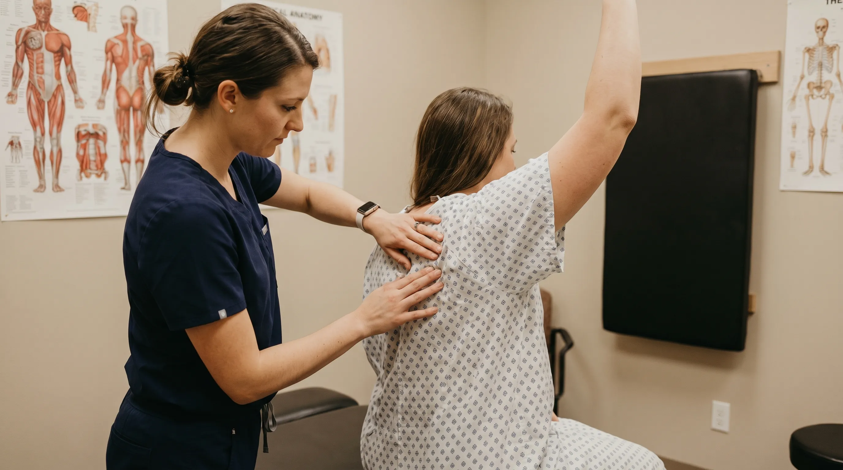
Clinical diagnosis of pain under right scapula relies on a systematic evaluation combining patient history, physical examination, and targeted diagnostic imaging. A thorough history captures onset, duration, quality of pain, aggravating and relieving factors, occupational exposures, and associated systemic symptoms. The physical examination includes observation of resting scapular position, assessment of active and passive shoulder range of motion, muscle strength testing, neurological screening (deep tendon reflexes, sensation, myotome strength), and palpation for localized tenderness, trigger points, or structural asymmetry.
Special tests help isolate the primary driver. The scapular assistance test and scapular retraction test evaluate whether correcting scapular positioning alleviates shoulder or periscapular pain, confirming dyskinesis or muscular imbalance. Spurling's test and upper limb tension tests assess for cervical radiculopathy or thoracic outlet syndrome. Visceral screening may involve abdominal palpation, Murphy's sign assessment, and cardiopulmonary auscultation.

Diagnostic imaging is reserved for cases with trauma, neurological deficits, persistent symptoms, or red flags. Plain radiographs (X-rays) serve as the initial imaging modality to rule out fractures, degenerative joint changes, or bony abnormalities. Magnetic resonance imaging (MRI) is the gold standard for evaluating soft tissue pathology, including rotator cuff tears, labral injuries, herniated cervical or thoracic discs, and nerve root compression. Diagnostic ultrasound offers dynamic, real-time assessment of the gallbladder, rotator cuff tendons, and scapulothoracic bursae, making it highly valuable for distinguishing musculoskeletal from visceral etiologies. In select cases, nerve conduction studies or electromyography (EMG) clarify peripheral nerve involvement, while CT scans with contrast may be ordered for suspected pulmonary embolism or complex thoracic pathology.
Conservative Management and Home Care Strategies
For the majority of patients, conservative management yields excellent outcomes. The American College of Physicians and leading orthopedic societies strongly recommend physical therapy, activity modification, and targeted self-care as first-line interventions for non-traumatic scapular pain (NIH MedlinePlus on physical therapy and non-invasive pain management). A structured rehabilitation program addresses muscle imbalances, restores joint mobility, and retrains optimal movement patterns.
Postural correction forms the foundation of conservative treatment. Ergonomic workstation adjustments are non-negotiable for desk workers: monitors should sit at eye level, keyboards and mice should allow elbows to rest at 90-degree angles, and chairs must provide adequate lumbar support. Implementing the 20-20-20 rule (every 20 minutes, look 20 feet away for 20 seconds) and incorporating micro-breaks reduces sustained muscle contraction. Chin tuck exercises and thoracic extension over a foam roller counteract forward head posture and restore mid-back mobility.
Targeted strengthening focuses on the muscles responsible for scapular stability. Serratus anterior activation, typically trained through push-up plus progressions or wall slides, ensures proper upward rotation and prevents winging. Prone Y-T-W raises engage the lower and middle trapezius and rhomboids, promoting posterior tilt and retraction. Rows using resistance bands or cables strengthen the periscapular stabilizers while improving posture. Rotator cuff external rotation exercises maintain glenohumeral balance and reduce compensatory strain on the shoulder blade muscles.
Myofascial release techniques provide rapid symptomatic relief. Trigger point massage, lacrosse ball self-massage, or professional myofascial therapy release taut muscle bands and improve local blood flow. Pectoralis minor doorway stretches, levator scapulae neck stretches, and cat-cow thoracic mobility drills complement strengthening work. Short-term use of NSAIDs and alternating heat or ice therapy can manage acute inflammation, though they do not replace movement-based rehabilitation.
Comparison Table: Musculoskeletal vs. Visceral Scapular Pain
| Feature | Musculoskeletal/Biomechanical | Visceral/Referred |
|---|---|---|
| Pain Quality | Aching, stiff, localized, worsens with movement | Burning, colicky, sharp, constant or episodic |
| Aggravating Factors | Poor posture, prolonged sitting, overhead activity, palpation | Deep breathing, fatty meals, systemic illness, lying flat |
| Associated Symptoms | Muscle tightness, clicking/crepitus, posture changes | Nausea, jaundice, fever, shortness of breath, abdominal tenderness |
| Response to PT | Excellent improvement within 2-6 weeks | Minimal or no improvement without treating underlying organ issue |
| Typical Onset | Gradual or acute after strain | Sudden or insidious, often unrelated to mechanical stress |
Incorporating evidence-based exercise routines accelerates recovery. Watching guided demonstrations from licensed professionals can ensure proper form and progression. For instance, comprehensive upper back mobility sequences help patients safely implement daily stretching protocols.
Additionally, understanding scapular movement faults is crucial for long-term prevention. Clinical breakdowns of dyskinesis assessment and rehabilitation provide deeper insight into why certain exercises are prescribed and how to track progress objectively.
Medical Interventions and Procedural Options
When conservative measures fail to produce meaningful improvement after 4 to 6 weeks, or when specific pathologies are identified, medical interventions become necessary. Corticosteroid or trigger point injections offer targeted anti-inflammatory effects for refractory myofascial pain, scapulothoracic bursitis, or facet joint inflammation. These procedures are typically ultrasound-guided to ensure precision and minimize complication risks.
For visceral etiologies, treatment shifts entirely to addressing the primary condition. Symptomatic gallstones or chronic cholecystitis often require laparoscopic cholecystectomy, a minimally invasive surgical procedure with high success rates. Antiviral medications manage herpes zoster infections, while proton pump inhibitors and H. pylori eradication protocols treat peptic ulcer disease. Gastroenterologists or hepatologists coordinate care for complex liver pathology.
Surgical intervention for isolated periscapular pain remains exceptionally rare. It is generally reserved for structural abnormalities such as snapping scapula syndrome unresponsive to rehabilitation, which may necessitate arthroscopic bursectomy or partial resection of a prominent scapular spine angle. Severe cervical or thoracic nerve compression causing progressive neurological deficits may warrant spinal decompression surgery. All surgical decisions require comprehensive imaging, functional assessments, and multidisciplinary consultation to ensure appropriate patient selection and realistic outcome expectations.
Frequently Asked Questions
What causes a dull ache under the right shoulder blade?
A dull ache under the right scapula is most commonly linked to musculoskeletal strain, particularly in the rhomboid, levator scapulae, or trapezius muscles. Poor posture, prolonged desk work, repetitive overhead motions, and scapular dyskinesis frequently trigger these symptoms. However, if the discomfort persists despite rest, it may indicate cervical nerve root irritation, myofascial trigger points, or even referred visceral pain from the gallbladder or liver.
Can pain under the right scapula be a sign of a heart problem?
While cardiac issues typically manifest as left-sided chest, arm, or jaw pain, atypical presentations can occasionally cause discomfort on the right side. Conditions like atypical myocardial ischemia or aortic dissection may refer pain to the right upper back or scapula, especially if accompanied by shortness of breath, sweating, dizziness, or sudden tearing sensations. Any unexplained, severe upper back pain with systemic symptoms warrants immediate medical evaluation (CDC guidance on heart attack symptoms).
How long does it take for right shoulder blade pain to heal?
Healing timelines depend heavily on the underlying cause. Simple muscle strains or postural imbalances often improve within 2 to 6 weeks with targeted stretching, strengthening, and ergonomic modifications. Myofascial pain or chronic scapular dyskinesis may require 6 to 12 weeks of structured physical therapy. If the pain originates from nerve compression or visceral pathology, resolution depends on treating the primary condition, which can range from weeks to several months.
What is the best sleeping position for pain under the right scapula?
The optimal sleeping position minimizes scapular compression and maintains spinal neutrality. Sleeping on your back with a thin pillow under your head and a small support under your knees is generally ideal. If you prefer side sleeping, avoid the painful right side; sleep on your left side with a supportive pillow between your knees and hug a pillow to prevent your right shoulder from collapsing forward. Stomach sleeping should be avoided as it forces cervical and thoracic rotation, increasing scapular muscle tension.
When should I see a doctor for right shoulder blade pain?
You should consult a healthcare provider if the pain persists beyond 2 to 4 weeks of conservative care, interferes with daily activities, or worsens progressively. Seek immediate medical attention if you experience red flags such as sudden severe tearing pain, shortness of breath, dizziness, unexplained weight loss, fever, jaundice, neurological deficits like arm weakness or numbness, or if the pain follows significant trauma. A thorough clinical evaluation is essential to rule out serious neurological or visceral pathology.
How can I prevent recurring pain under the right scapula?
Prevention hinges on consistent ergonomic habits, targeted strengthening, and movement awareness. Maintain neutral posture during work and rest, incorporate daily thoracic mobility drills, and strengthen the lower trapezius, rhomboids, and serratus anterior three to four times weekly. Avoid prolonged static positions, stay hydrated, manage stress levels, and progressively increase physical activity loads to allow connective tissues to adapt. Annual musculoskeletal screenings can identify biomechanical imbalances before they develop into symptomatic pain under right scapula.
Conclusion
Navigating pain under right scapula requires a balanced approach that respects both the mechanical complexity of the shoulder girdle and the potential for systemic referral patterns. While musculoskeletal strain, postural dysfunction, and scapular dyskinesis represent the most frequent culprits, clinicians must remain vigilant for neurological compromise and visceral pathology that demand entirely different management pathways. The cornerstone of successful recovery lies in early intervention, accurate differential diagnosis, and consistent participation in targeted rehabilitation. By integrating ergonomic modifications, progressive strengthening, myofascial release, and professional medical guidance when necessary, most individuals achieve complete functional restoration and long-term symptom prevention.
If you are experiencing persistent or worsening discomfort, prioritize a comprehensive evaluation from a licensed healthcare provider, physical therapist, or orthopedic specialist. Self-management is highly effective for mild, posture-related strain, but should never replace professional assessment when red flags are present or conservative care stalls. Your scapula functions as the structural foundation of the upper extremity kinetic chain; protecting its biomechanical integrity through mindful movement, proper recovery, and evidence-based interventions will serve you well for decades. Prioritize your posture, listen to your body's warning signals, and commit to a proactive approach to musculoskeletal health. With patience, consistency, and the right clinical guidance, relief is not only possible but highly achievable.
Disclaimer: This article is intended for educational purposes only and does not constitute medical advice, diagnosis, or treatment. Always consult a qualified healthcare professional for personalized evaluation of your symptoms. Individual conditions vary, and professional guidance ensures safe, effective management of pain under right scapula and related musculoskeletal or systemic concerns.
How we work
Researched and drafted with AI assistance, then reviewed by a human editor against the cited sources.
This article is general health information, not medical advice. Consult a qualified healthcare professional about your own situation.