Bilateral Foraminal Stenosis: Causes, Symptoms, Diagnosis, and Treatment Guide

Living with persistent back pain, radiating discomfort, or unexplained numbness in your limbs can feel overwhelming, especially when the root cause remains unclear. Bilateral foraminal stenosis is a complex yet increasingly common spinal condition that quietly alters how you move, rest, and function day to day. Unlike sudden injuries, this condition typically develops gradually, making early recognition and informed management essential for preserving mobility and quality of life. By understanding the anatomical mechanisms, recognizing the distinct symptom patterns across different spinal regions, and exploring evidence-based treatment pathways, you can take proactive steps toward lasting relief. This comprehensive guide breaks down everything you need to know about bilateral foraminal stenosis, drawing from clinical research, orthopedic best practices, and patient-centered rehabilitation strategies to help you navigate your spinal health with confidence and clarity.
Understanding the Anatomy and Pathophysiology
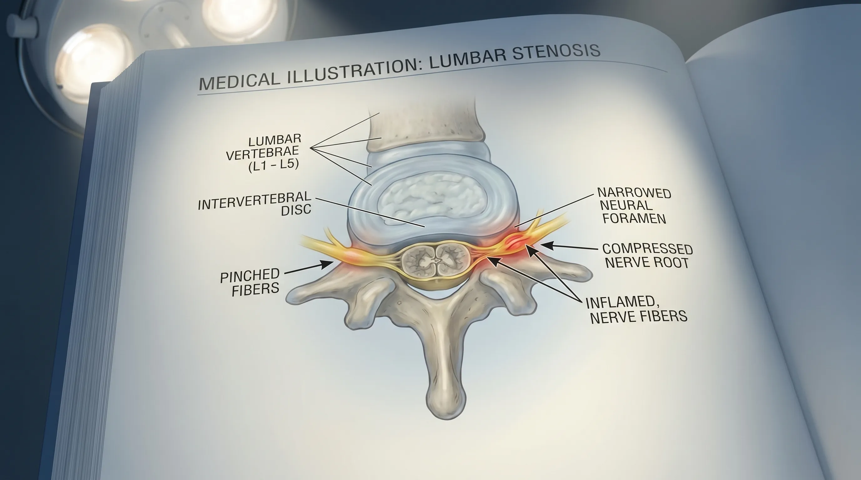
The human spine is a marvel of structural engineering, composed of thirty-three vertebrae stacked in precise alignment to support the torso, protect the spinal cord, and facilitate movement. Between each pair of vertebrae lie small, naturally curved openings known as the neural foramina. These bony tunnels serve as critical gateways, allowing spinal nerve roots to exit the spinal column and branch outward to innervate muscles, joints, skin, and internal organs throughout the body. When these openings remain unobstructed, nerve transmission occurs smoothly, and pain-free movement is maintained. However, when anatomical changes cause the neural foramina to narrow on both the left and right sides simultaneously, the condition is medically classified as bilateral foraminal stenosis. This bilateral compression disrupts normal nerve signaling and triggers a cascade of inflammatory and neuropathic responses.
Free calculatorBMI CalculatorCalculate Body Mass IndexOpen the calculatorThe Neural Foramina: Gateways for Nerve Roots
Each neural foramen is bordered anteriorly by the intervertebral disc, posteriorly by the facet joints and ligamentum flavum, and laterally by the pedicles of adjacent vertebrae. This delicate architectural balance requires precise spacing to prevent mechanical impingement. In a healthy spine, the foramina maintain enough clearance to accommodate nerve roots, blood vessels, and lymphatic channels. Over time, repetitive biomechanical stress, postural habits, and natural aging processes gradually alter this spacing. Disc height decreases, facet joint cartilage wears down, and supporting ligaments lose elasticity. These incremental changes reduce the available cross-sectional area of the foramina, setting the stage for nerve compression. Understanding this structural context is vital because it explains why symptoms often correlate directly with specific activities or postures that further compromise the narrowed pathways.
What Happens in Bilateral Foraminal Stenosis?
When bilateral foraminal stenosis develops, nerve compression occurs symmetrically at a specific spinal level. Unlike unilateral narrowing, which may cause symptoms predominantly on one side, bilateral involvement often produces more widespread neurological signs. The compression interferes with axonal transport, the microscopic process by which nutrients and signals travel along the nerve fibers. Prolonged mechanical pressure leads to localized ischemia, where blood flow to the nerve is restricted, triggering inflammation, demyelination, and eventually neuropathic pain. Patients frequently describe this sensation as a burning, electric, or deep aching discomfort that follows the dermatomal distribution of the affected nerve. Early intervention focuses on reducing inflammation and restoring mechanical space before irreversible nerve changes occur.
Cervical vs. Thoracic vs. Lumbar Involvement
Bilateral foraminal stenosis can manifest at any level of the vertebral column, though symptom profiles vary dramatically depending on the affected region. Cervical foraminal stenosis impacts the seven vertebrae in the neck. This area experiences tremendous mechanical demand from head weight and multi-directional mobility, making it highly susceptible to degenerative narrowing. Thoracic foraminal stenosis involves the twelve mid-back vertebrae and remains the least common variant due to the rib cage providing additional structural stability. When it does occur, it typically affects intercostal nerves, causing band-like pain around the chest or abdomen. Lumbar foraminal stenosis affects the five lower back vertebrae and represents the most prevalent form. The lumbar spine bears the majority of the body’s weight and undergoes constant flexion and extension during daily activities, accelerating wear and tear on the lower foramina. Recognizing regional differences is crucial for accurate diagnosis and targeted treatment planning.
Recognizing the Signs and Symptoms
The clinical presentation of bilateral foraminal stenosis is highly dependent on the spinal level affected, the degree of narrowing, and the patient’s unique neurological sensitivity. Symptoms rarely appear suddenly. Instead, they develop insidiously over months or years, often starting as mild stiffness or intermittent aches that gradually progress into persistent pain, tingling, or weakness. Many individuals dismiss early signs as normal aging or minor muscle strain, which can delay appropriate intervention until functional limitations become pronounced.
Cervical Symptom Profile
When bilateral foraminal narrowing occurs in the cervical spine, nerve compression primarily affects the brachial plexus and cervical nerve roots. Patients typically report neck pain that may radiate into the shoulders, upper back, or arms. A hallmark feature is bilateral paresthesia, characterized by numbness, pins-and-needles sensations, or burning pain in the hands, fingers, or forearms. As compression worsens, fine motor skills deteriorate, making tasks like buttoning shirts, typing, or holding utensils increasingly difficult. In advanced cases, spinal cord involvement may occur alongside foraminal compression, leading to gait instability, balance deficits, and generalized limb weakness. Early recognition of these upper extremity changes is critical, as cervical nerve damage can progress rapidly if left unaddressed.
Lumbar Symptom Profile and Neurogenic Claudication
Lumbar bilateral foraminal stenosis predominantly affects the lower spinal nerves that travel down the legs. The most recognizable symptom is bilateral sciatica, a radiating pain that originates in the lower back or buttocks and extends down the posterior or lateral thighs, often reaching the calves or feet. Unlike musculoskeletal back pain that remains localized, neuropathic pain follows distinct nerve pathways and frequently worsens with specific movements. A defining characteristic is neurogenic claudication, where patients experience cramping, heaviness, or fatigue in both legs after walking or standing for extended periods. Interestingly, symptoms typically improve rapidly when the patient sits down, leans forward, or bends at the waist. This positional relief occurs because spinal flexion temporarily widens the narrowed foramina and reduces nerve compression, a clinical clue that strongly differentiates bilateral foraminal stenosis from vascular claudication or muscular strain.
Progression Patterns and When to Seek Care
Symptom progression in bilateral foraminal stenosis is rarely linear. Patients often experience flare-ups triggered by prolonged activity, poor posture, or heavy lifting, followed by periods of relative remission. However, without proper management, the frequency and intensity of these episodes tend to increase. Warning signs that warrant prompt medical evaluation include persistent numbness that does not resolve with rest, progressive muscle weakness causing frequent tripping or foot drop, loss of coordination in the hands, and severe pain that disrupts sleep or daily routines. Rare but serious complications such as bowel or bladder dysfunction, sexual impairment, or sudden leg paralysis require immediate emergency care, as they indicate severe neurological compromise that may necessitate urgent decompressive intervention.
Underlying Causes and Risk Factors
Bilateral foraminal stenosis is predominantly a degenerative condition, meaning it develops gradually as spinal structures undergo wear and tear over decades. While aging is the most significant contributing factor, a combination of genetic predisposition, biomechanical stress, and underlying medical conditions determines the onset and severity of nerve compression.
Age-Related Degenerative Changes
As the spine ages, several structural transformations occur simultaneously that narrow the neural foramina. Intervertebral discs lose hydration and elasticity, causing them to thin and bulge outward into the foraminal space. The facet joints develop osteoarthritis, leading to cartilage breakdown and the formation of bone spurs, or osteophytes, that protrude into the nerve pathways. Additionally, the ligamentum flavum, a thick elastic band that stabilizes the spinal canal, becomes hypertrophic and calcified with age, further encroaching on the foramina. These cumulative changes reduce the available cross-sectional area for nerve roots, making older adults particularly vulnerable to symptomatic compression. Research from the National Institute of Arthritis and Musculoskeletal and Skin Diseases (NIAMS) confirms that aging-related structural deterioration is the primary driver of spinal stenosis in the majority of cases.
Disc Herniation, Spondylolisthesis, and Other Triggers
Beyond natural degeneration, acute or structural abnormalities can precipitate or exacerbate bilateral foraminal stenosis. A herniated disc occurs when the tough outer annulus fibrosus tears, allowing the gel-like nucleus pulposus to protrude into the foramen and directly compress the nerve root. Spondylolisthesis, a condition where one vertebra slips forward over the one below it, misaligns the spinal column and mechanically narrows the exit pathways for nerves on both sides. Less common causes include spinal tumors that occupy foraminal space, metabolic bone disorders like Paget’s disease that trigger abnormal bone remodeling, and traumatic fractures that displace vertebral segments. Understanding these underlying mechanisms helps clinicians differentiate stenosis from other pain syndromes and tailor treatment accordingly.
Congenital and Lifestyle Risk Factors
While degeneration dominates the clinical picture, some individuals are born with a naturally narrower spinal canal or smaller foramina, predisposing them to earlier symptom onset. Congenital spinal anomalies often remain asymptomatic until adulthood, when age-related changes compound the anatomical limitation. Lifestyle factors significantly influence disease progression. Prolonged sedentary behavior weakens core musculature, reducing spinal support and increasing disc compression. Obesity places excessive mechanical load on the lumbar spine, accelerating joint degeneration. Occupational hazards such as repetitive heavy lifting, prolonged vibration exposure, or sustained poor posture further stress the foramina. Additionally, a history of previous spinal surgery or trauma increases susceptibility to secondary stenosis due to scar tissue formation or altered biomechanics. Addressing modifiable risk factors through targeted lifestyle interventions plays a crucial role in slowing disease progression.
Diagnostic Pathway and Clinical Evaluation
Accurate diagnosis of bilateral foraminal stenosis requires a systematic clinical evaluation that combines patient history, physical examination, and advanced imaging. Because symptoms often mimic other spinal and neurological conditions, a thorough workup ensures appropriate treatment selection and rules out mimicking pathologies like peripheral neuropathy, vascular disease, or herniated discs without stenosis.
Physical Examination and Neurological Testing
The diagnostic process begins with a comprehensive physical and neurological assessment. A healthcare provider will evaluate posture, spinal alignment, and range of motion in all directions. Specific provocative tests help identify nerve root involvement. The straight leg raise test, for example, stretches the sciatic nerve and reproduces radiating pain if lumbar nerve compression is present. Neurological screening assesses muscle strength, reflexes, and sensory perception across multiple dermatomes. Diminished knee or ankle reflexes, reduced sensation in specific nerve distributions, and measurable weakness in key muscle groups provide critical localizing information that points directly to the affected spinal level.
Imaging Modalities: X-Rays, MRI, CT, and Myelography
Imaging is indispensable for confirming bilateral foraminal stenosis and visualizing the exact anatomical cause of nerve compression. Plain X-rays provide an initial overview of spinal alignment, disc height, and bony abnormalities like osteophytes or spondylolisthesis, though they do not visualize soft tissue or nerves. Magnetic Resonance Imaging (MRI) remains the gold standard for diagnosing foraminal stenosis because it clearly displays disc degeneration, nerve root impingement, ligament thickening, and inflammatory changes without radiation exposure. Computed Tomography (CT) scans offer superior bone detail and are particularly useful for surgical planning when evaluating facet joint hypertrophy or bony stenosis. In complex cases or when MRI is contraindicated, a CT myelogram may be performed, where contrast dye is injected into the spinal canal to highlight fluid flow and pinpoint exact compression sites.
Evidence-Based Treatment Approaches
Management of bilateral foraminal stenosis follows a stepwise approach, prioritizing conservative, non-invasive strategies before considering surgical intervention. The primary goals are to reduce nerve inflammation, alleviate pain, restore functional mobility, and prevent neurological deterioration. Treatment plans are highly individualized, accounting for symptom severity, functional limitations, patient age, and comorbid conditions.
Conservative Management and Pain Relief Strategies
First-line treatment focuses on symptom modulation and activity modification. Over-the-counter nonsteroidal anti-inflammatory drugs (NSAIDs) reduce nerve root inflammation and provide temporary pain relief. For more severe discomfort, physicians may prescribe short-term oral corticosteroids, neuropathic pain modulators like gabapentin or pregabalin, or targeted muscle relaxants. Epidural steroid injections deliver anti-inflammatory medication directly around the compressed nerve roots, offering rapid and often prolonged symptom relief that facilitates participation in physical therapy. Activity pacing and ergonomic adjustments are equally important. Patients are advised to avoid prolonged standing, heavy lifting, and repetitive spinal extension. Instead, frequent position changes, supportive seating, and the use of lumbar rolls during sitting help maintain optimal spinal alignment and reduce mechanical stress on the foramina.
Targeted Rehabilitation and Physical Therapy
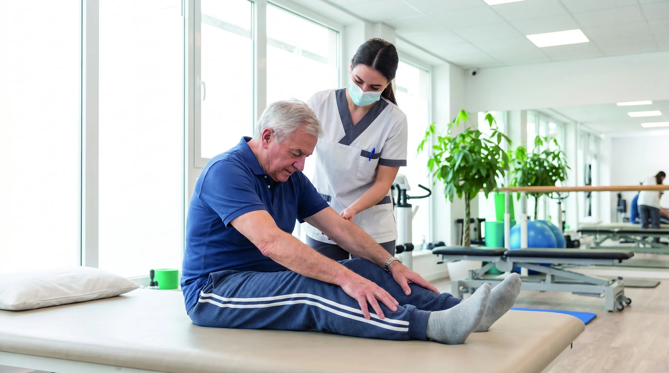
Physical therapy forms the cornerstone of long-term bilateral foraminal stenosis management. A licensed physical therapist designs a progressive exercise program tailored to the patient’s tolerance and spinal region involvement. Core stabilization exercises strengthen the deep abdominal and back muscles, creating a natural corset that unloads the spine and reduces disc compression. Flexion-based stretching routines, including knee-to-chest stretches, pelvic tilts, and cat-cow mobilizations, mechanically open the neural foramina and relieve nerve pressure. Aerobic conditioning through stationary cycling, elliptical training, or aquatic therapy improves cardiovascular health and promotes endorphin release without jarring the spine. Postural retraining and body mechanics education teach patients how to move safely during daily activities, minimizing future flare-ups. Consistent adherence to rehabilitation protocols significantly delays disease progression and enhances overall functional independence.
Surgical Interventions and Postoperative Expectations
When conservative measures fail to provide adequate relief, or when progressive neurological deficits develop, surgical decompression may be recommended. Common procedures include foraminotomy, which involves removing bone spurs, thickened ligaments, or disc fragments to widen the neural foramina and free the compressed nerve. In cases of spinal instability or severe spondylolisthesis, a spinal fusion may be performed to stabilize the affected segment. Minimally invasive techniques have revolutionized spinal surgery, allowing for smaller incisions, reduced muscle trauma, shorter hospital stays, and faster recovery timelines. It is important to understand that surgery addresses mechanical compression but does not cure underlying arthritis or reverse degenerative changes. Postoperative rehabilitation remains essential to rebuild strength, restore mobility, and prevent adjacent segment degeneration. Patients who follow structured recovery protocols and maintain spinal hygiene typically experience substantial long-term symptom resolution and improved quality of life.
Lifestyle Modifications and Long-Term Management
Living with bilateral foraminal stenosis requires a proactive, holistic approach that extends beyond clinical treatments. Sustainable lifestyle adaptations empower patients to manage symptoms, reduce flare-up frequency, and maintain an active, fulfilling life despite anatomical changes.
Ergonomic Optimization and Daily Habits
Small adjustments to daily routines yield significant spinal benefits. Workplace ergonomics should prioritize neutral spinal alignment, with computer monitors positioned at eye level to prevent forward head posture in cervical stenosis patients. Lumbar support cushions and standing desks with adjustable height settings allow periodic position changes that prevent static loading. When lifting objects, patients should practice proper biomechanics by bending at the knees, keeping the load close to the body, and engaging the core before movement. Sleep posture matters immensely; cervical stenosis sufferers benefit from supportive cervical pillows, while lumbar patients often find relief sleeping on their sides with a pillow between the knees to maintain pelvic neutrality and reduce disc pressure.
Weight Management and Anti-Inflammatory Nutrition
Maintaining a healthy body weight reduces the mechanical burden on weight-bearing spinal joints and slows degenerative progression. Excess adipose tissue also promotes systemic inflammation, which exacerbates nerve sensitivity and pain perception. An anti-inflammatory dietary pattern rich in omega-3 fatty acids, leafy greens, colorful vegetables, lean proteins, and whole grains supports tissue repair and modulates inflammatory pathways. Staying well-hydrated maintains disc hydration and elasticity, preserving some natural shock absorption capacity. Limiting processed foods, refined sugars, and excessive alcohol intake further reduces systemic inflammation and supports metabolic health, which is crucial for optimal nerve function and recovery.
Psychological Resilience and Pain Coping Techniques
Chronic spinal pain frequently impacts mental health, contributing to anxiety, depression, and sleep disturbances. Integrating mind-body practices enhances pain tolerance and improves coping mechanisms. Mindfulness meditation, guided breathing exercises, and progressive muscle relaxation activate the parasympathetic nervous system, lowering stress hormones and reducing central pain sensitization. Cognitive behavioral therapy (CBT) equips patients with strategies to reframe pain-related catastrophizing, set realistic activity goals, and maintain social engagement despite physical limitations. Building a strong support network, joining patient education groups, and maintaining open communication with healthcare providers fosters emotional resilience and promotes adherence to long-term management plans.
Frequently Asked Questions
What is the primary difference between bilateral foraminal stenosis and central canal stenosis?
Bilateral foraminal stenosis specifically involves the narrowing of the neural foramina on both sides of a spinal segment, compressing the exiting nerve roots. Central canal stenosis, in contrast, involves narrowing of the main spinal canal itself, which puts pressure on the spinal cord or the cauda equina bundle. While both can cause similar pain and neurological symptoms, foraminal stenosis typically presents with more localized radicular pain, numbness, or weakness along a specific nerve pathway, whereas central stenosis often causes broader symptoms like neurogenic claudication.
Can bilateral foraminal stenosis resolve on its own without surgery?
In many cases, conservative management can significantly reduce symptoms and improve quality of life without requiring surgery. Physical therapy, targeted exercises, anti-inflammatory medications, activity modification, and epidural steroid injections often provide meaningful relief. The condition is largely degenerative, so the anatomical narrowing itself typically does not reverse spontaneously, but symptom management is highly effective for most patients.
Why does bending forward or sitting relieve bilateral foraminal stenosis symptoms?
Flexing the spine by bending forward or sitting physically opens the intervertebral spaces and enlarges the neural foramina. This positional change reduces mechanical compression on the exiting nerve roots and temporarily restores normal blood flow to the affected nerves. Conversely, spinal extension, standing upright, or leaning backward compresses these spaces further, which is why symptoms typically worsen during prolonged standing or walking.
What are the warning signs of severe nerve damage requiring immediate medical attention?
Red flag symptoms that require urgent evaluation include loss of bowel or bladder control, saddle anesthesia (numbness in the groin, buttocks, and inner thighs), progressive and severe weakness in the legs, sudden onset of partial or complete paralysis, and unrelenting pain that does not respond to rest or medication. These may indicate severe nerve compromise or cauda equina syndrome, which are medical emergencies requiring prompt surgical consultation.
Is physical therapy safe and effective for bilateral foraminal stenosis?
Yes, when properly prescribed and supervised by a licensed physical therapist, physical therapy is a cornerstone of conservative treatment. Programs typically focus on core stabilization, gentle spinal flexion stretches, aerobic conditioning like stationary cycling or water therapy, and postural retraining. These interventions help maintain mobility, reduce inflammation, strengthen supporting musculature, and create more functional space around the compressed nerves, often delaying or eliminating the need for surgical intervention.
Conclusion
Bilateral foraminal stenosis is a manageable spinal condition that demands informed attention, consistent self-care, and professional guidance. By recognizing early symptoms, understanding the degenerative processes at play, and embracing a structured treatment plan, patients can effectively control pain, preserve neurological function, and maintain an active lifestyle. Whether through conservative rehabilitation, targeted lifestyle adjustments, or advanced surgical options when necessary, the path to relief is highly accessible. Prioritize proactive spine health, stay engaged with your healthcare team, and remember that small, consistent daily habits yield profound long-term benefits. For further reading and evidence-based resources on spinal health, explore the comprehensive guidelines provided by the Mayo Clinic, the NIAMS, and WebMD’s detailed foraminal stenosis overview. Your spine supports your entire life; investing in its care today ensures greater mobility and independence for years to come.
How we work
Researched and drafted with AI assistance, then reviewed by a human editor against the cited sources.
This article is general health information, not medical advice. Consult a qualified healthcare professional about your own situation.