Stabbing Pain Under Left Shoulder Blade: Causes, Relief & When to Seek Help
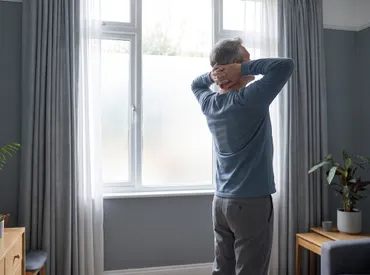
Experiencing a sudden, sharp discomfort in your upper back can be deeply unsettling, especially when it disrupts daily activities, impairs mobility, and interferes with restorative sleep. Many individuals first notice this symptom after lifting heavy objects, sleeping in an awkward position, or spending prolonged hours hunched over a computer screen. However, when you feel a stabbing pain under left shoulder blade, it is essential to look beyond simple muscle tension and understand the complex network of musculoskeletal, neurological, and visceral structures that could be involved. The upper thoracic and cervical regions house intricate musculature, branching spinal nerves, and critical internal organs, meaning that sharp discomfort in this area often stems from multiple overlapping physiological pathways. While the majority of cases resolve effectively with conservative care, targeted movement therapies, and postural correction, ignoring persistent, severe, or progressively worsening symptoms can delay appropriate medical intervention and prolong recovery timelines. This comprehensive, evidence-based guide explores the anatomical mechanisms, common triggers, red-flag warning signs, clinical diagnostic pathways, and proven pain relief strategies to help you navigate this uncomfortable symptom safely and confidently. By understanding the underlying causes and implementing structured self-management techniques, you can take informed, proactive steps toward lasting comfort, improved biomechanics, and sustainable upper back health.
Understanding the Anatomy of the Upper Back and Scapular Region
Musculoskeletal Structures and Nerve Pathways
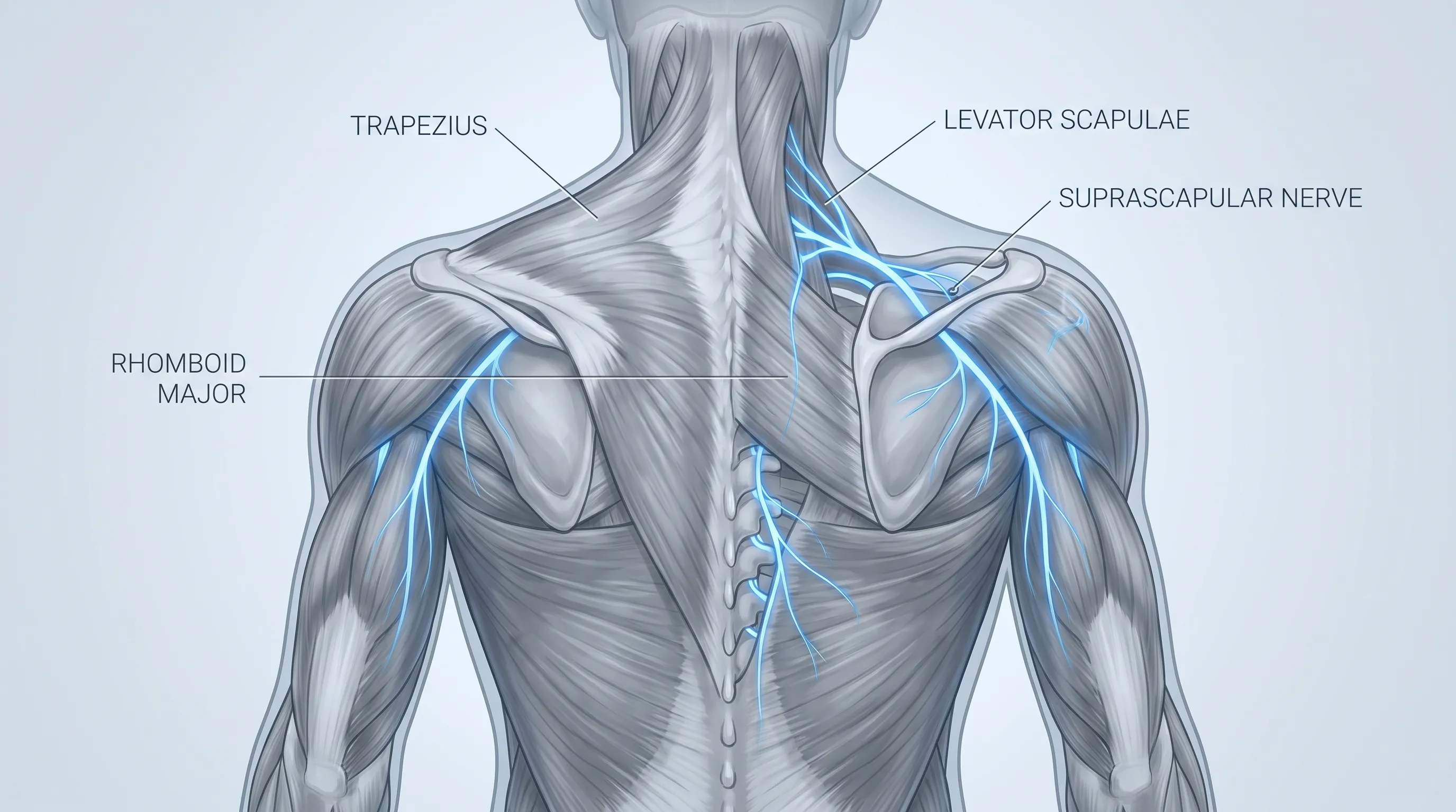
The scapula, commonly referred to as the shoulder blade, serves as a crucial anchor point for the upper extremity and upper back. It articulates with the humerus at the glenohumeral joint and connects indirectly to the rib cage through a complex web of stabilizing muscles, including the rhomboids, trapezius, levator scapulae, and serratus anterior. When you experience a stabbing pain under left shoulder blade, it often reflects irritation, microtrauma, or neuromuscular dysfunction within these supporting tissues. The dorsal scapular nerve, which innervates the rhomboid and levator scapulae muscles, frequently becomes a source of sharp, localized discomfort when compressed or irritated due to poor posture, repetitive strain, or cervical spine misalignment. Additionally, the intercostal nerves running between the ribs can refer sharp sensations to the scapular border when inflamed or compressed. The thoracic spine itself, though naturally less mobile than the cervical or lumbar regions, plays a vital role in load distribution. Prolonged sitting, forward head posture, and asymmetrical movement patterns increase mechanical stress on the facet joints and surrounding ligaments, creating a fertile environment for acute muscular spasms or joint capsule irritation.
Free calculatorBMI CalculatorCalculate Body Mass IndexOpen the calculatorHow Referred Pain Manifests
Referred pain occurs when a sensory signal originating from one anatomical location is perceived by the brain in a different, often distant, region. This phenomenon is governed by the convergence-projection theory of pain pathways. Nerves from various internal organs and deep musculoskeletal structures often share common spinal cord segments. When irritation or pathology occurs in the heart, lungs, spleen, or gastrointestinal tract, the afferent nerve signals can cross pathways in the dorsal horn of the spinal cord and be misinterpreted as originating from the upper back or shoulder blade region. This explains why visceral conditions frequently present as a stabbing pain under left shoulder blade without any direct injury to the musculoskeletal system itself. Recognizing referred pain patterns is clinically significant because treating only the superficial muscles will not resolve symptoms originating from deeper systemic sources. Clinicians use detailed pain mapping, symptom timing, and associated physiological markers to differentiate between localized musculoskeletal dysfunction and systemic referred pain. Understanding these neurological pathways empowers patients to track symptom triggers more accurately and seek appropriate diagnostic evaluation when standard musculoskeletal interventions fail to provide lasting relief.
Common Causes of Sharp Discomfort Near the Left Scapula
Musculoskeletal and Mechanical Triggers
Muscle strains and ligament sprains remain the most frequent culprits behind acute scapular pain. Overexertion during weightlifting, improper lifting mechanics, sudden twisting motions, or prolonged static postures can lead to microtears within the trapezius, rhomboids, or latissimus dorsi. When these tissues are overloaded, inflammatory mediators are released, stimulating nociceptors and generating sharp, localized aching or a stabbing pain under left shoulder blade during specific movements. Myofascial trigger points represent another highly prevalent musculoskeletal source. These hyperirritable nodules within taut muscle bands develop from chronic tension, dehydration, repetitive strain, or inadequate recovery. When pressed or activated during shoulder elevation or neck rotation, trigger points can produce intense, radiating discomfort that mimics nerve impingement. Costochondritis, an inflammation of the cartilage connecting the ribs to the sternum, can also refer sharp discomfort to the posterior thoracic region, particularly on the left side. While primarily an anterior chest condition, the biomechanical coupling of the rib cage means that breathing deeply or coughing often exacerbates posterior scapular tension.
Nerve Compression and Cervical Spine Involvement
The cervical spine, specifically levels C4 through C7, plays a dominant role in upper back and shoulder sensation. Cervical radiculopathy occurs when a herniated disc, bone spur, or degenerative joint narrowing compresses a cervical nerve root as it exits the spinal column. This compression disrupts normal signal transmission, leading to shooting, electric-like, or stabbing pain under left shoulder blade, frequently accompanied by numbness, tingling, or weakness radiating into the arm and fingers. Thoracic outlet syndrome (TOS) represents another neurovascular compression disorder where nerves or blood vessels become restricted between the collarbone and first rib. Patients with TOS often report aching, sharp sensations near the medial border of the scapula, especially when raising the arms overhead or carrying heavy loads. Postural abnormalities such as rounded shoulders, forward head posture, and excessive thoracic kyphosis significantly reduce the space within the intervertebral foramina and thoracic outlet, perpetuating nerve irritation and muscle guarding. Early recognition of neurogenic pain patterns is essential, as prolonged compression can lead to chronic neuropathy, reduced grip strength, and irreversible structural changes.
Cardiovascular and Internal Organ Referred Pain
While less common, visceral pathology can manifest prominently in the left upper back. The heart and lungs share autonomic innervation pathways that converge near the C3 to T5 spinal segments. Angina, myocardial ischemia, or pericarditis may present as a sudden, sharp, or pressure-like stabbing pain under left shoulder blade, often worsening with exertion or stress. Pleurisy, an inflammation of the lining surrounding the lungs, typically causes sharp pain that intensifies with deep inhalation or coughing and can radiate posteriorly. The spleen, located beneath the left rib cage, can refer pain to the scapular region when enlarged, inflamed, or traumatically injured. Pancreatic inflammation or biliary tract dysfunction may also produce mid-back discomfort, though it more commonly localizes slightly lower and often correlates with digestive disturbances. Because these conditions carry significant health implications, clinicians emphasize thorough cardiovascular and pulmonary screening when patients report atypical upper back pain, particularly if accompanied by systemic symptoms.
Gastrointestinal and Diaphragmatic Irritation
The diaphragm serves as the primary respiratory muscle and shares phrenic nerve innervation (C3 to C5) that refers sensation to the shoulder and upper back. Diaphragmatic irritation from acid reflux, gastric ulcers, or visceral fat displacement can generate sharp, intermittent sensations that patients describe as a stabbing pain under left shoulder blade. Additionally, gas entrapment in the splenic flexure of the colon, known as splenic flexure syndrome, can cause intense localized pressure and referred discomfort. While gastrointestinal causes rarely present as isolated musculoskeletal symptoms, they should be considered when pain correlates with meals, fasting periods, or changes in bowel habits. Functional gastrointestinal disorders often respond to dietary modifications, stress reduction, and postural optimization, highlighting the interconnected nature of digestive health and upper back biomechanics.

Warning Signs That Require Immediate Medical Attention
Differentiating Between Muscle Strain and Cardiac Events
One of the most critical challenges in managing upper back discomfort lies in accurately distinguishing benign musculoskeletal strain from life-threatening cardiovascular events. Musculoskeletal pain typically worsens with palpation, specific movements, or prolonged postures, and improves with rest, heat therapy, or gentle stretching. In contrast, cardiac-related discomfort often occurs spontaneously, remains relatively unaffected by movement, and may intensify during physical exertion or emotional stress. A stabbing pain under left shoulder blade that coincides with chest tightness, radiating jaw pain, profuse sweating, shortness of breath, nausea, or unexplained fatigue should never be dismissed as simple postural fatigue. Recognizing heart attack warning signs is critical, as women, older adults, and individuals with diabetes frequently experience atypical cardiac presentations, making clinical vigilance paramount. Emergency medical guidelines recommend immediate transport to a hospital or calling emergency services when any combination of cardiovascular symptoms accompanies acute upper back discomfort. Prompt electrocardiographic monitoring, cardiac enzyme testing, and clinical evaluation remain the gold standard for ruling out myocardial infarction or acute coronary syndrome.
Red Flags for Emergency Care
Beyond cardiovascular concerns, several red flags for back pain necessitate urgent medical evaluation for scapular pain. Unexplained weight loss, night sweats, fever, or persistent pain that awakens you from sleep may indicate underlying systemic infection, inflammatory conditions, or neoplastic processes. Neurological deficits such as progressive arm weakness, loss of fine motor coordination, bowel or bladder dysfunction, or bilateral sensory changes suggest spinal cord compression or severe radiculopathy requiring advanced imaging. Traumatic injuries involving falls, motor vehicle collisions, or direct blunt force to the back can cause scapular fractures, rib dislocations, or internal bleeding. If a stabbing pain under left shoulder blade follows significant trauma, immediate radiographic assessment is mandatory. Additionally, pain that rapidly escalates, becomes unresponsive to over-the-counter analgesics, or interferes with basic breathing and swallowing warrants prompt clinical intervention to prevent complications and establish an accurate diagnosis.
| Feature | Musculoskeletal/Postural Strain | Serious/Visceral or Neurological Cause |
|---|---|---|
| Onset | Gradual or activity-related, often after overuse | Sudden, unprovoked, or progressive worsening |
| Pain Triggers | Movement, palpation, prolonged sitting | Exertion, stress, breathing, or spontaneous |
| Relief Factors | Rest, heat, stretching, posture correction | Unresponsive to positional changes or analgesics |
| Associated Symptoms | Muscle stiffness, localized tenderness | Chest pressure, shortness of breath, numbness, fever |
| Neurological Signs | None or mild localized tingling | Weakness, radiating numbness, coordination loss |
| Duration | Days to 1-2 weeks with conservative care | Persistent, worsening, or recurrent beyond 2-3 weeks |
Diagnostic Approaches and Medical Evaluation
Physical Examination Techniques
A thorough clinical assessment begins with a detailed patient history and targeted physical examination. Physicians and physical therapists typically evaluate posture, spinal alignment, shoulder range of motion, and scapular tracking during overhead movements. Palpation along the medial scapular border, trapezius ridge, and levator scapulae insertion helps identify muscle spasms, trigger points, or localized tenderness. Orthopedic tests such as the Spurling maneuver assess cervical nerve root compression, while the Neer and Hawkins-Kennedy impingement tests evaluate shoulder joint mechanics. The clinician will also observe breathing patterns, diaphragmatic excursion, and thoracic spine mobility, as restricted mobility often contributes to compensatory scapular strain. Functional movement assessments, including scapular retraction strength, core stabilization endurance, and overhead squat mechanics, provide valuable insight into biomechanical deficits. By systematically reproducing or alleviating symptoms through specific movements, practitioners can isolate the primary pain generator and rule out secondary contributors.
Imaging Studies and Laboratory Diagnostics
When physical examination findings suggest structural pathology or when conservative management fails, diagnostic imaging becomes necessary. Plain radiographs (X-rays) effectively visualize scapular fractures, rib abnormalities, degenerative joint changes, and spinal alignment issues. If soft tissue pathology or nerve compression is suspected, magnetic resonance imaging (MRI) provides high-resolution visualization of intervertebral discs, nerve roots, spinal cord integrity, and surrounding musculature. Computed tomography (CT) scans may be utilized when detailed bone assessment or vascular evaluation is required. Ultrasound imaging offers dynamic, real-time assessment of muscle tears, tendon inflammation, and bursitis. In cases where systemic illness or visceral referral is suspected, clinicians order comprehensive blood panels, including complete blood count, inflammatory markers (CRP, ESR), cardiac enzymes, and metabolic panels. Evidence-based imaging guidelines ensure that electrocardiograms, pulmonary function tests, or abdominal ultrasounds are appropriately recommended to evaluate cardiac, respiratory, or gastrointestinal sources. A methodical diagnostic approach guarantees that a stabbing pain under left shoulder blade receives targeted, condition-specific management rather than generic symptom masking.
Evidence-Based Treatment and Pain Relief Strategies
Immediate Self-Care and First-Aid Measures
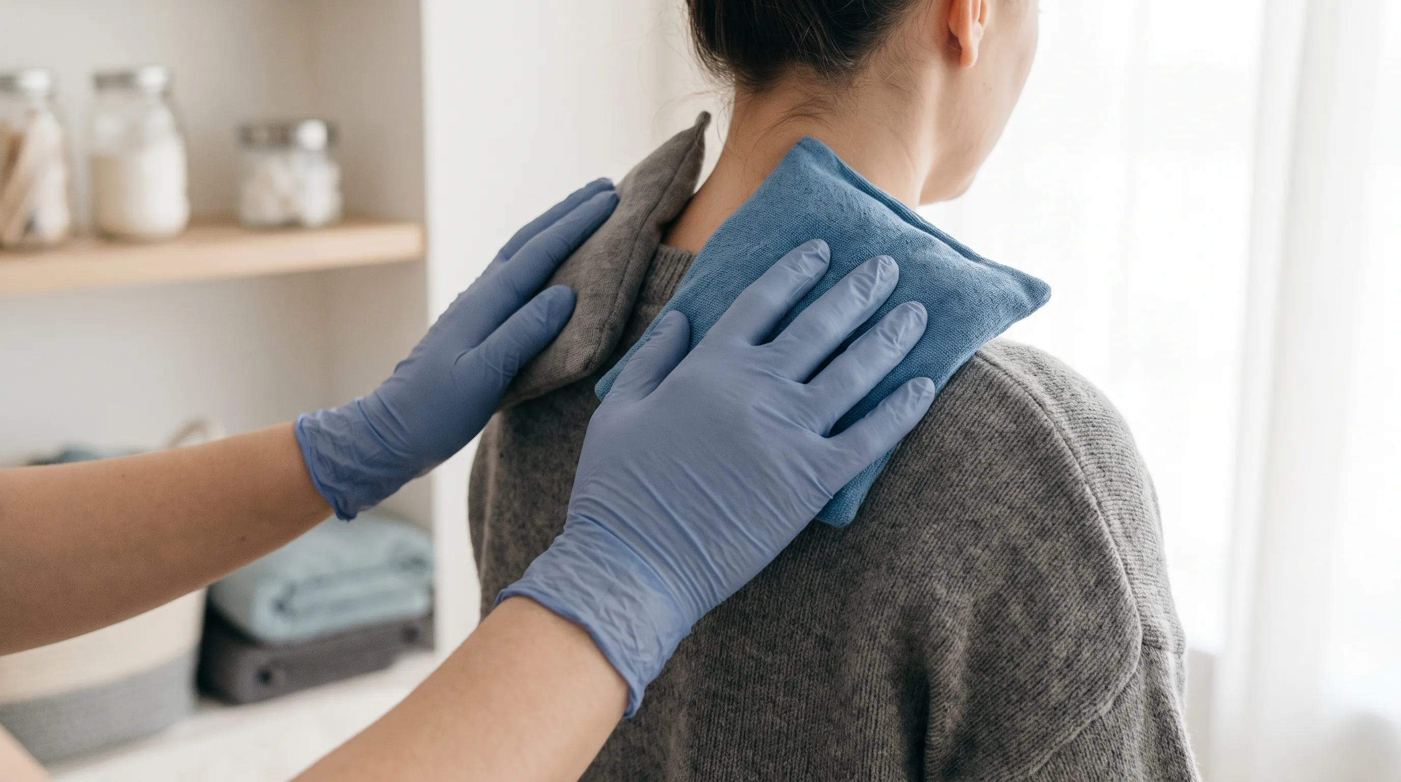
In the initial 48 to 72 hours following acute scapular strain, the priority shifts to reducing inflammation, minimizing further tissue stress, and restoring baseline mobility. Applying ice packs wrapped in a thin cloth for 15 to 20 minutes, three to four times daily, helps constrict blood vessels, decrease localized swelling, and numb nociceptive signaling. After the acute inflammatory phase transitions into a subacute stage, transitioning to moist heat therapy promotes vasodilation, increases tissue elasticity, and facilitates muscle relaxation. Proper use of ice and heat alongside over-the-counter nonsteroidal anti-inflammatory drugs (NSAIDs) such as ibuprofen or naproxen, when used according to manufacturer guidelines, can temporarily reduce pain and swelling, though they do not address underlying biomechanical dysfunction. Topical analgesic creams containing menthol, camphor, or diclofenac provide localized relief with minimal systemic absorption. Crucially, patients are advised to avoid complete immobilization, as prolonged rest can lead to joint stiffness, muscle atrophy, and delayed healing. Instead, engaging in gentle, pain-free range-of-motion movements such as shoulder rolls, chin tucks, and thoracic extensions encourages synovial fluid circulation and prevents compensatory movement patterns.
Physical Therapy, Mobilization, and Targeted Stretching
Once acute pain diminishes, structured physical therapy becomes the cornerstone of functional recovery. A licensed therapist designs a progressive rehabilitation protocol emphasizing scapular stabilization, thoracic mobility restoration, and neuromuscular re-education. Exercises such as scapular retractions, prone Y-T-W raises, and serratus punches activate weakened periscapular muscles while promoting proper joint centration. Thoracic spine foam rolling and extension over a stability ball counteract postural kyphosis and improve rib cage mechanics. Nerve gliding techniques, including cervical retraction and median nerve sliders, help restore healthy neural mobility when radicular symptoms are present. Manual therapy modalities such as myofascial release, joint mobilization, and dry needling effectively deactivate trigger points and restore normal tissue compliance. Research consistently demonstrates that patients who adhere to supervised exercise programs experience faster symptom resolution, lower recurrence rates, and improved long-term functional capacity. Home exercise programs should be performed consistently, typically 4 to 5 times weekly, with gradual progression in resistance, range, and movement complexity under professional guidance.
Medical Management and Pharmacological Options
When conservative measures prove insufficient, clinicians may recommend targeted medical interventions to disrupt the pain-inflammation-spasm cycle. Prescription muscle relaxants such as cyclobenzaprine or methocarbamol provide short-term relief for severe muscle guarding, though sedating side effects limit their use to brief durations. Corticosteroid injections into the facet joints, bursa, or around irritated nerve roots offer potent anti-inflammatory effects for patients with chronic radiculopathy or persistent joint inflammation. Epidural steroid injections are considered when imaging confirms significant nerve root compression. Neuropathic pain medications like gabapentin or duloxetine may be prescribed for chronic radicular symptoms that do not respond to standard analgesics. Trigger point injections using saline, anesthetic, or botulinum toxin can deactivate顽固性 trigger points and restore normal muscle function. In cases involving structural abnormalities, surgical intervention remains a last resort, reserved for severe spinal stenosis, refractory herniated discs, or traumatic fractures. Pharmacological management is always integrated with movement therapy, ensuring that medication serves as a bridge to functional recovery rather than a permanent dependency.
Prevention, Ergonomics, and Long-Term Musculoskeletal Health
Workspace Optimization for Desk Professionals
Prolonged sitting represents one of the most significant modifiable risk factors for developing a stabbing pain under left shoulder blade. Modern workstations frequently position monitors too low, keyboards too far forward, and chairs without adequate lumbar support, forcing the upper back into compensatory strain. Ergonomic workplace guidelines emphasize aligning the monitor's top third with eye level, positioning elbows at a 90-degree angle with forearms parallel to the floor, and maintaining a neutral wrist posture during typing. A chair with adjustable lumbar curvature, seat depth, and armrests reduces thoracic compression and encourages natural spinal curves. Implementing a sit-stand workstation allows periodic position changes, reducing static muscle loading and improving circulation. Micro-breaks every 30 to 45 minutes, featuring two to three minutes of standing, shoulder rolls, and gentle spinal twists, disrupt prolonged postural strain before it accumulates into tissue microtrauma. Investing in a supportive chair, monitor risers, and an ergonomic keyboard tray yields substantial long-term musculoskeletal benefits and significantly reduces the likelihood of recurrent upper back discomfort.
Strengthening Protocols and Movement Re-Education
Long-term resilience against scapular pain requires a balanced approach to strength, mobility, and motor control. The upper back muscles often become chronically lengthened and weakened due to dominant anterior chest musculature, creating a force couple imbalance that pulls the shoulders forward. Targeted strengthening of the lower trapezius, rhomboids, and middle deltoids restores proper scapulohumeral rhythm. Resistance band pull-aparts, inverted rows, and prone scapular retractions build endurance and promote postural correction. Core stabilization exercises, including planks, bird-dog movements, and diaphragmatic breathing drills, provide a stable proximal foundation for distal limb movement. Movement re-education focuses on retraining proper lifting mechanics, emphasizing hip hinge patterns over spinal flexion, and engaging the posterior chain before initiating heavy lifts. Incorporating mindful movement practices such as Pilates, yoga, or Tai Chi enhances body awareness, improves fascial glide, and promotes symmetrical muscle recruitment. Consistency remains the most critical factor; dedicating just 15 to 20 minutes daily to targeted mobility and strengthening yields measurable improvements within four to six weeks, significantly reducing the frequency and severity of future episodes.
Lifestyle Modifications and Stress Management
Chronic physiological stress exerts profound effects on musculoskeletal tension, directly influencing the frequency and intensity of a stabbing pain under left shoulder blade. Elevated cortisol levels and sympathetic nervous system activation increase baseline muscle tone, reduce pain thresholds, and impair tissue recovery. Mind-body interventions such as diaphragmatic breathing, progressive muscle relaxation, and guided meditation downregulate stress responses and promote parasympathetic recovery. Healthy sleep habits remain non-negotiable; seven to nine hours of uninterrupted, high-quality sleep facilitates tissue repair, hormone regulation, and neuromuscular reset. Nutritional strategies emphasizing anti-inflammatory foods, adequate hydration, and optimal protein intake support connective tissue resilience and muscle recovery. Avoiding repetitive unilateral loading, such as carrying heavy bags on one shoulder or cradling phones between ear and neck, prevents asymmetrical strain. Integrating stress management techniques into daily routines addresses the root physiological drivers of muscular tension, transforming short-term symptom relief into sustainable, long-term upper back health.
Frequently Asked Questions
What organ could cause stabbing pain under left shoulder blade?
Sharp discomfort in this region can sometimes stem from internal organs. The heart, lungs, spleen, and pancreas can all refer pain to the upper back through shared nerve pathways. Conditions such as pleurisy, splenic injury, pancreatitis, or even cardiac events may manifest as left-sided scapular discomfort. Always seek immediate medical evaluation if the pain is accompanied by chest tightness, shortness of breath, dizziness, or nausea.
How long does stabbing pain under left shoulder blade typically last?
If the discomfort originates from a simple muscle strain or postural fatigue, symptoms usually improve within three to seven days with appropriate rest, gentle movement, and conservative self-care. Chronic or recurrent pain lasting longer than two to three weeks may indicate an underlying structural issue, nerve involvement, or repetitive strain pattern requiring professional assessment and targeted rehabilitation.
Can a heart attack feel like a sharp pain near the left shoulder blade?
Yes, cardiovascular events can present as referred pain in the left upper back or shoulder blade region, particularly in women and older adults. While many heart attacks feature central chest pressure, atypical presentations may involve sharp, radiating discomfort, sweating, breathlessness, or unexplained fatigue. Any sudden, unexplained pain in this area with cardiovascular symptoms warrants emergency medical attention immediately.
What sleeping positions worsen pain near the scapula?
Sleeping directly on the affected side, using a pillow that is too high or too flat, or adopting a twisted spinal alignment can increase mechanical stress on the periscapular muscles. Experts recommend sleeping on your back with a supportive cervical pillow, or on your unaffected side with a firm pillow placed between your knees and arms to maintain neutral spinal alignment and reduce scapular compression.
When should physical therapy begin for scapular pain?
Physical therapy can typically begin within a few days of symptom onset once acute, severe inflammation subsides. Early intervention focuses on pain modulation, gentle range-of-motion restoration, and neuromuscular re-education. If symptoms persist beyond ten to fourteen days despite home management, a licensed physical therapist can design a progressive program emphasizing scapular stabilization, thoracic mobility, and postural correction.
Conclusion
Navigating a stabbing pain under left shoulder blade requires a balanced approach that respects both the mechanical and systemic origins of upper back discomfort. While most cases stem from manageable musculoskeletal strain, postural imbalance, or reversible trigger point activation, recognizing red-flag symptoms and understanding referred pain pathways ensures timely, appropriate intervention when necessary. By implementing structured self-care protocols, prioritizing ergonomic optimization, engaging in targeted strengthening routines, and addressing physiological stressors, you can effectively break the cycle of recurrent scapular tension and restore lasting upper back health. Always consult a qualified healthcare provider or licensed physical therapist for personalized evaluation, particularly when pain persists, worsens, or interferes with daily function. Proactive management, combined with evidence-based rehabilitation strategies, empowers you to reclaim mobility, eliminate chronic tension, and maintain a resilient, well-aligned upper back for years to come.
How we work
Researched and drafted with AI assistance, then reviewed by a human editor against the cited sources.
This article is general health information, not medical advice. Consult a qualified healthcare professional about your own situation.