Kyphosis vs Lordosis: Understanding Spinal Curvature and Treatment
The human spine is an architectural marvel, engineered to support weight, absorb shock, and protect vital neural pathways. Yet, when the delicate balance of its natural curves shifts, everyday movement can become painful, restrictive, and exhausting. Many individuals notice changes in their posture long before clinical symptoms emerge, often dismissing subtle rounding of the upper back or excessive lower back arching as mere cosmetic concerns. Understanding the distinction between kyphosis vs lordosis is the critical first step toward reclaiming pain-free mobility and optimizing long-term spinal health. While both conditions involve abnormal spinal curvatures, they differ significantly in anatomical location, underlying mechanisms, clinical presentation, and management strategies. This comprehensive guide explores the science behind these postural deviations, outlines evidence-based diagnostic approaches, and provides actionable rehabilitation protocols to help you restore proper alignment and enhance quality of life.
Understanding the Natural Curves of the Spine
Before examining pathological deviations, it is essential to appreciate how the spine functions in optimal alignment. A healthy vertebral column exhibits three primary physiological curves when viewed from the side, creating an S-shaped contour that distributes mechanical stress evenly across discs, ligaments, and facet joints. These curves are not random; they develop sequentially during infancy and childhood as the musculoskeletal system adapts to gravitational forces and upright posture.
Free calculatorBMI CalculatorCalculate Body Mass IndexOpen the calculatorThe Cervical, Thoracic, and Lumbar Regions
The cervical spine naturally curves inward (lordotic), supporting the head's substantial weight while allowing a wide range of motion for visual and auditory scanning. Below it, the thoracic region curves outward (kyphotic), anchoring the rib cage and protecting thoracic organs. The lumbar spine then curves inward again, serving as the primary weight-bearing segment that connects the axial skeleton to the pelvis and lower extremities. Together, these alternating curves act as biological springs, converting vertical compressive loads into manageable tensile forces that prevent premature disc degeneration.
How Spinal Alignment Affects Overall Health
Maintaining this physiological balance is crucial for far more than posture alone. Proper sagittal alignment ensures optimal lung expansion, digestive organ positioning, and cerebrospinal fluid circulation. When curves deviate beyond normal parameters, compensatory muscle activation patterns emerge, leading to chronic fatigue, restricted mobility, and systemic inflammation. Research consistently demonstrates that spinal misalignment correlates with reduced physical performance, impaired balance, and increased fall risk, particularly in aging populations. By recognizing the early warning signs of abnormal curvature, individuals can implement proactive interventions before structural remodeling occurs.
What Is Kyphosis?
Kyphosis refers to an exaggerated outward curvature of the thoracic spine, typically measured using the Cobb angle on lateral radiographs. While a normal thoracic curve ranges between 20 and 45 degrees, clinical kyphosis is diagnosed when the curvature exceeds 50 degrees. This condition manifests visually as a forward-rounded upper back, often accompanied by shoulder protraction and head-forward posture.
Types of Kyphosis
Postural kyphosis represents the most prevalent form, stemming from habitual slouching, prolonged screen time, and weakened thoracic extensor muscles. It is highly responsive to targeted exercise and ergonomic adjustments. Structural variants include Scheuermann's disease, an osteochondrosis affecting adolescent vertebral growth plates, and osteoporotic kyphosis, which occurs when compression fractures collapse anterior vertebral bodies. Congenital and degenerative forms exist as well, each requiring distinct clinical pathways for accurate management. The Mayo Clinic emphasizes that distinguishing between postural and structural etiologies dictates treatment intensity and prognosis.
Symptoms and Complications
Early-stage kyphosis often presents asymptomatically, but progressive curvature triggers localized upper back pain, muscle spasms, and stiffness. As the deformity advances, patients may experience reduced pulmonary capacity due to restricted rib cage expansion, gastrointestinal discomfort from abdominal compression, and neurological symptoms if spinal cord compression develops. Untreated severe kyphosis significantly diminishes quality of life, impeding basic activities like reaching overhead, driving, or maintaining eye contact during conversations. Early intervention remains the cornerstone of preventing irreversible vertebral remodeling.
What Is Lordosis?
Lordosis describes the natural inward curvature of the cervical and lumbar spine. When this curve becomes excessively pronounced, clinicians refer to it as hyperlordosis or swayback posture. This condition primarily affects the lumbar region, where the lower back arches dramatically forward, pushing the abdomen and pelvis anteriorly while tilting the buttocks posteriorly.
Common Causes of Hyperlordosis
Acquired hyperlordosis frequently originates from prolonged sitting, weak abdominal musculature, tight hip flexors, and inadequate gluteal activation. Pregnancy often induces temporary lordotic compensation as the growing uterus shifts the center of gravity forward. Obesity places sustained anterior load on the lumbar discs, while conditions like spondylolisthesis, discitis, or achondroplasia create structural drivers. The Cleveland Clinic notes that identifying the root mechanical imbalance is essential for designing effective corrective strategies that address muscle imbalances rather than merely masking symptoms.
Recognizing the Signs
Patients with hyperlordosis typically report dull, aching lower back pain that intensifies during prolonged standing, walking uphill, or wearing high heels. Tight hamstring and quadriceps muscles frequently accompany the condition, limiting forward flexion and contributing to compensatory pelvic tilt. In advanced cases, spinal stenosis or facet joint impingement may radiate pain into the hips and thighs. Visual assessment reveals a pronounced gap between the lumbar spine and a flat surface when lying supine, serving as a simple clinical screening tool for excessive anterior curvature.
Kyphosis vs Lordosis: Key Differences Explained
When evaluating kyphosis vs lordosis, the primary distinction lies in the anatomical location and directional vector of the abnormal curvature. Kyphosis involves an excessive posterior bowing of the thoracic vertebrae, creating a hunched silhouette, whereas lordosis features an exaggerated anterior arching of the lumbar spine, producing a swayback appearance. Both conditions disrupt sagittal balance, but they impose fundamentally different biomechanical stresses on adjacent tissues and require tailored therapeutic interventions.
| Feature | Kyphosis (Hyperkyphosis) | Lordosis (Hyperlordosis) | Natural Physiological Range |
|---|---|---|---|
| Primary Location | Thoracic spine | Lumbar spine | T1-T12 (outward), L1-L5 (inward) |
| Curve Direction | Excessive posterior arching | Excessive anterior arching | Balanced S-shape alignment |
| Visual Presentation | Rounded upper back, forward head | Swayback, anterior pelvic tilt | Smooth, proportional contour |
| Muscle Imbalance | Weak thoracic extensors, tight pecs | Weak abdominals/glutes, tight hip flexors | Balanced agonist-antagonist tone |
| Common Triggers | Poor sitting posture, osteoporosis | Prolonged sitting, pregnancy, obesity | N/A (baseline healthy state) |
Location and Direction of Curvature
The thoracic vertebrae naturally articulate with ribs, creating a semi-rigid structural unit that resists excessive flexion. When kyphotic deformation occurs, anterior vertebral bodies compress while posterior ligaments stretch, potentially altering intervertebral disc hydration. Conversely, lumbar hyperlordosis increases facet joint loading and posterior annulus fibrosus stress, accelerating degenerative disc disease progression. Understanding these directional forces clarifies why kyphosis vs lordosis demands distinct exercise prescriptions targeting specific muscle groups.
Impact on Mobility and Pain Levels
Kyphosis typically restricts overhead reach and spinal extension, while hyperlordosis limits flexion and rotational capacity. Pain patterns also diverge: upper back tension headaches and shoulder impingement frequently accompany thoracic deviations, whereas lumbar facet syndrome and radicular symptoms characterize excessive lordotic posture. Neither condition should be ignored, as chronic misalignment alters gait mechanics, increases energy expenditure during ambulation, and elevates the risk of secondary orthopedic injuries. Comparative analysis of kyphosis vs lordosis reveals that comprehensive management requires region-specific rehabilitation combined with global postural retraining.
Diagnosis and Clinical Evaluation
Accurate assessment begins with a thorough patient history, documenting symptom onset, progression rate, occupational demands, and previous injuries. Clinicians utilize standardized observation protocols to evaluate symmetry, shoulder height, scapular winging, and pelvic tilt. The Adam's forward bend test remains a cornerstone screening maneuver for identifying rotational deformities, though it primarily detects scoliosis rather than pure sagittal plane deviations. For kyphosis vs lordosis evaluation, plumb line analysis and lateral flexion/extension tests provide superior diagnostic sensitivity.
Physical Examinations and Postural Assessments
During the initial consultation, physical therapists measure thoracolumbar flexibility using Schober's test and assess core endurance through prone extension holds and plank variations. Muscle length testing identifies shortened hip flexors, hamstrings, and pectoral muscles that perpetuate abnormal curvatures. Functional movement screening reveals compensatory strategies, such as excessive lumbar extension during overhead squats or reduced thoracic rotation during golf swings. These objective measurements establish baseline data for tracking rehabilitation progress and modifying treatment intensity based on patient response.
Imaging and Advanced Diagnostic Tools
When clinical evaluation suggests structural involvement, full-length standing lateral radiographs provide definitive curvature quantification using the Cobb angle method. Magnetic resonance imaging becomes necessary if neurological deficits, suspected disc herniation, or inflammatory arthritis warrant further investigation. Dual-energy X-ray absorptiometry scans assess bone mineral density in osteoporotic kyphosis candidates. According to the American Academy of Orthopaedic Surgeons, imaging should never replace clinical correlation, as radiographic measurements must align with functional impairment and symptom severity to justify aggressive interventions.
Treatment Options and Management Strategies
Management of kyphosis vs lordosis follows a graduated approach prioritizing conservative rehabilitation before considering procedural interventions. The overarching goal involves restoring muscular equilibrium, improving joint mobility, and retraining neuromuscular control to maintain physiological alignment during dynamic activities. Early-stage conditions typically respond exceptionally well to structured exercise programs, whereas advanced structural deformities may require multimodal therapeutic coordination.
Conservative Rehabilitation Approaches
Evidence-based physical therapy forms the foundation of non-surgical management. Thoracic extension exercises, such as foam roller mobilizations, prone Y-T-W raises, and cat-cow variations, effectively reverse rounded upper back postures. For hyperlordosis, targeted core stabilization protocols featuring dead bugs, bird-dog progressions, and glute bridge variations reduce anterior pelvic tilt and decompress lumbar discs. Stretching regimens address tight myofascial structures, while proprioceptive neuromuscular facilitation enhances body awareness. Manual therapy techniques, including joint mobilizations and myofascial release, accelerate tissue pliability and pain reduction. Consistency proves paramount, with most patients experiencing measurable postural improvements after 8 to 12 weeks of supervised training followed by independent home programs.
Medical Interventions: Bracing, Medications, and Surgery
When conservative measures prove insufficient, orthotic bracing serves as a viable adjunct for adolescents with progressive structural kyphosis, particularly during skeletal growth phases. Custom-fitted thoracolumbosacral orthoses provide mechanical correction while remodeling forces guide vertebral alignment. Pharmacological management includes nonsteroidal anti-inflammatory drugs for acute inflammation, muscle relaxants for spasms, and bone-density medications for osteoporosis-related cases. Surgical intervention remains reserved for severe curves exceeding 75 degrees, progressive neurological compromise, or intractable pain unresponsive to rehabilitation. Spinal fusion procedures utilize instrumentation to stabilize segments and restore sagittal balance, though recovery demands extensive postoperative rehabilitation and lifelong lifestyle modifications.
Lifestyle Modifications for Spinal Health
Long-term spinal integrity depends heavily on daily habits, environmental design, and proactive self-care routines. Corrective exercises yield temporary benefits if immediately negated by poor workstation ergonomics or sedentary behavior. Integrating sustainable postural awareness into routine activities ensures lasting results and minimizes recurrence risk when comparing kyphosis vs lordosis management outcomes.
Ergonomics and Daily Posture Habits
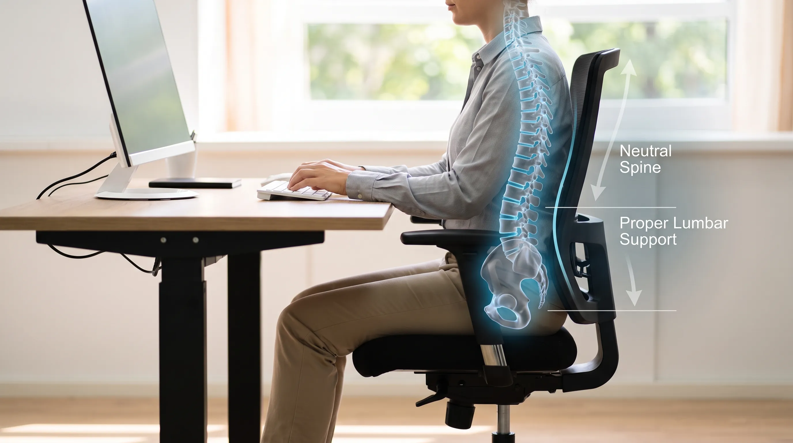
Optimizing your immediate environment dramatically reduces repetitive strain. Adjust monitor screens to eye level to prevent forward head posture, utilize lumbar support cushions to maintain natural lumbar lordosis, and position feet flat on the floor to distribute weight evenly. Implement the 20-20-20 rule: every twenty minutes, stand, stretch, and refocus your eyes for twenty seconds at a twenty-foot distance to interrupt prolonged static positioning. Standing desks, alternating with seated work, promote dynamic circulation and engage postural musculature. Additionally, wearing supportive footwear with adequate arch support prevents compensatory pelvic tilts that exacerbate both kyphotic and lordotic patterns.
Strengthening and Mobility Routines
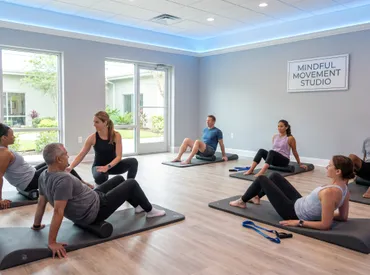
Incorporating daily movement practices sustains spinal resilience. Yoga and Pilates programs excel at integrating breath control with controlled spinal articulation, enhancing proprioceptive feedback and muscular endurance. Swimming and water aerobics provide low-impact resistance training that decompresses discs while building global stability. Mind-body disciplines like Tai Chi improve balance and reduce fall risk, particularly for older adults managing degenerative changes. Nutritional factors also play an underappreciated role; adequate protein intake supports connective tissue repair, while calcium, vitamin D, and magnesium optimize bone mineral density and muscle function. Hydration maintains intervertebral disc turgor, preventing premature desiccation and height loss.
Frequently Asked Questions
Can kyphosis and lordosis occur at the same time?
Yes, simultaneous presentation frequently occurs as compensatory mechanics. The human spine naturally attempts to maintain the center of gravity over the pelvis, meaning an exaggerated thoracic curve often triggers lumbar hyperextension to prevent forward falling. Clinicians refer to this pattern as postural swayback, requiring comprehensive assessment to address both regions concurrently.
Is kyphosis vs lordosis hereditary?
Structural variants exhibit genetic predispositions. Conditions like Scheuermann's disease or congenital vertebral segmentation defects follow familial patterns, whereas postural deviations primarily stem from environmental factors, occupational demands, and habitual movement patterns. Genetic screening remains unnecessary unless multiple family members develop severe structural deformities during adolescence.
At what age do spinal curvatures usually require medical intervention?
Adolescent growth spurts represent the critical window for intervention, as rapid skeletal development can accelerate curve progression. Adults over forty require evaluation when experiencing new neurological symptoms, progressive deformity, or pain disrupting sleep and daily function. Early adulthood represents an ideal phase for postural retraining before adaptive shortening becomes fibrotic.
How long does it take to correct postural kyphosis or lordosis?
Consistent adherence to targeted rehabilitation yields noticeable improvements within six to twelve weeks. Full neuromuscular reprogramming and sustained postural changes typically require six to twelve months of dedicated practice. Maintenance phases involve periodic reassessment and routine mobility sessions to prevent regression and sustain functional capacity.
Are there specific sleeping positions that help spinal alignment?
Back sleeping with a pillow beneath the knees maintains neutral pelvic tilt and reduces lumbar compression. Side sleeping requires a contoured pillow filling the space between the ear and mattress to prevent cervical lateral bending, alongside a knee pillow to maintain hip alignment. Stomach sleeping excessively rotates the neck and flattens natural lumbar curves, making it the least recommended position for spinal health.

Conclusion
Navigating the differences between kyphosis vs lordosis requires a clear understanding of spinal anatomy, early symptom recognition, and commitment to evidence-based rehabilitation. While both conditions disrupt the delicate equilibrium of physiological curves, they respond exceptionally well to targeted physical therapy, ergonomic optimization, and consistent lifestyle modifications. By addressing underlying muscle imbalances, improving postural awareness, and incorporating mobility practices into daily routines, individuals can restore functional alignment, reduce pain, and protect long-term spinal integrity. Proactive management not only enhances physical performance but also elevates confidence, breathing capacity, and overall well-being. Prioritizing spinal health today ensures decades of unrestricted movement, resilience, and vitality tomorrow.
How we work
Researched and drafted with AI assistance, then reviewed by a human editor against the cited sources.
This article is general health information, not medical advice. Consult a qualified healthcare professional about your own situation.