Pinched Nerve Scapula: Symptoms, Causes, and Evidence-Based Treatment
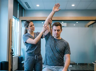
Living with unexplained upper back pain can be incredibly frustrating, especially when the discomfort wraps around your shoulder blade and refuses to fade despite weeks of self-massage, stretching, or over-the-counter pain relievers. A pinched nerve scapula is a surprisingly common yet frequently misunderstood condition that affects millions of individuals each year, spanning across athletes, office professionals, and aging adults alike (Mayo Clinic: Pinched Nerve Overview). Unlike typical muscular strain that resolves with simple rest and heat application, nerve compression in the shoulder girdle creates a distinct cascade of neurological symptoms, including sharp shooting pain, persistent numbness, muscle fatigue, and significant functional limitations that disrupt daily life. Many people initially dismiss these warning signs as simple postural tension or stress-related tightness, allowing the underlying neural irritation to progress unchecked. Understanding the precise anatomical mechanics behind this condition is the first critical step toward effective relief and sustainable recovery. When the delicate peripheral nerve pathways traveling beneath, through, or alongside the scapular region become compressed, inflamed, or mechanically entrapped, the nervous system responds with protective muscle guarding, altered movement compensation, and chronic discomfort that can radiate down the arm. By thoroughly exploring the structural roots, recognizing early clinical signals, and implementing medically verified management strategies, you can safely navigate the path from acute neural irritation to fully restored mobility. This comprehensive guide breaks down everything you need to know about identifying, treating, and preventing a pinched nerve scapula using evidence-based approaches, rehabilitation protocols, and lifestyle modifications designed to protect your nervous system long-term.
Understanding the Anatomy and Mechanics Behind a Pinched Nerve Scapula
The human shoulder complex represents one of the most sophisticated biomechanical systems in the body, engineered to deliver an extraordinary range of motion while simultaneously maintaining dynamic stability under heavy functional loads. However, this very architectural versatility renders the cervicothoracic junction highly vulnerable to mechanical stress, fascial restriction, and neural compromise. A pinched nerve scapula occurs when one or more peripheral nerves supplying the periscapular musculature become mechanically compressed, chronically irritated, or physically entrapped as they navigate through naturally narrow anatomical tunnels or beneath thickened myofascial bands. The primary neural structures typically implicated include the dorsal scapular nerve, the suprascapular nerve, and occasionally the long thoracic nerve (NIH: Brachial Plexus & Nerve Anatomy), each following a highly specific anatomical trajectory that leaves it uniquely susceptible to repetitive microtrauma or sudden structural shifts. Recognizing these pathways provides crucial context for why certain movements trigger disproportionate pain and why targeted decompression strategies prove far more effective than generalized stretching.
Free calculatorBMI CalculatorCalculate Body Mass IndexOpen the calculatorNerve Pathways Around the Scapular Region
The dorsal scapular nerve originates from the C5 cervical nerve root and travels deeply through the posterior neck and upper back before penetrating the middle scalene muscle to innervate the rhomboids and levator scapulae. This specific anatomical pathway requires the nerve to pierce muscular tissue early in its course, creating a natural pinch point where chronic scalene hypertrophy, forward head posture, or cervical facet joint dysfunction can easily trigger compression. Once past the scalene, the nerve runs parallel to the medial scapular border, making it highly sensitive to thoracic kyphosis and prolonged slouching. The suprascapular nerve follows a far more intricate and clinically significant route, originating from the upper trunk of the brachial plexus before descending to pass through the suprascapular notch beneath the rigid transverse scapular ligament, subsequently navigating the spinoglenoid notch to reach the infraspinatus. These anatomical tunnels are notoriously restrictive, and even minor soft tissue swelling, ganglion cyst formation, or repetitive overhead loading can drastically reduce available space. When available space decreases, the affected nerve loses its essential protective gliding ability, leading to localized ischemia, focal demyelination, and eventually axonal degradation if mechanical compression remains unaddressed. Understanding these precise pathways explains why a pinched nerve scapula often generates highly localized tenderness at specific bony landmarks while simultaneously referring discomfort along predictable distal trajectories.
Common Triggers and Risk Factors
Repetitive overhead activities, sustained poor postural alignment, acute traumatic incidents, and degenerative spinal changes represent the most frequent catalysts for scapular nerve entrapment. Individuals who participate in sports demanding repetitive arm elevation, including swimming, competitive volleyball, baseball pitching, tennis, and crossfit weightlifting, place continuous cyclical demand on the brachial plexus and periscapular nerves, gradually inducing microtrauma within the fascial tunnels. Similarly, remote workers and corporate employees who spend extended hours hunched over keyboards or staring at mobile devices frequently develop forward head posture, protracted shoulders, and thoracic stiffness (CDC: Ergonomics & Musculoskeletal Health), which systematically collapses the space within the cervicothoracic junction and compresses exiting neural structures. Additional risk factors encompass cervical spondylosis, where osteophytic bone spurs or bulging intervertebral discs directly impinge the nerve root at the spinal exit, radiating inflammatory signals outward toward the shoulder blade. Other significant contributors include systemic inflammatory conditions like rheumatoid arthritis, diabetes-induced peripheral neuropathy, chronic vitamin B deficiencies that impair myelin repair, and direct mechanical trauma from vehicular accidents, sports collisions, or improper heavy lifting mechanics. Identifying these predisposing factors enables patients and clinicians to implement targeted mechanical modifications and ergonomic interventions before acute compression progresses into chronic neurological dysfunction.
Recognizing the Signs: Symptoms and Clinical Presentation
The clinical manifestation of a pinched nerve scapula varies considerably depending on which specific peripheral nerve is compromised, the degree of mechanical obstruction, and the cumulative duration of tissue irritation. However, certain hallmark neurological symptoms consistently emerge across the majority of clinical presentations, providing highly reliable diagnostic indicators that distinguish neural compression from routine muscular fatigue. Unlike diffuse myofascial tension that typically responds favorably to massage therapy and heat application, nerve entrapment generates distinct sensory and motor disturbances that strictly follow established dermatomal distributions and myotomal activation patterns.
Localized Versus Radiating Pain Patterns
Patients frequently describe a deep, dull, and persistent aching sensation concentrated along the medial border or superior angle of the scapula, which often feels physically trapped beneath the bone and completely unresponsive to superficial stretching attempts. As the inflammatory cascade progresses and perineural edema accumulates, the discomfort frequently transitions into a sharp, electric, or intensely burning quality that radiates along the posterior shoulder capsule, down the lateral arm, and occasionally extends into the forearm, wrist, or fingers. This specific radiation pattern occurs because irritated peripheral nerves transmit ectopic electrical impulses to the central nervous system, creating pain referral trajectories that closely mimic rotator cuff tendinopathy, cervical disc herniation, brachial plexitis, or even cardiac angina when left-sided symptoms predominate. The pain intensity typically escalates dramatically during specific functional movements, including reaching across the body to buckle a seatbelt, lifting moderately heavy objects overhead, or maintaining static seated or standing postures for extended durations without positional shifts. Recognizing this distinctive pain architecture helps patients avoid counterproductive stretching that further irritates the compressed neural tissue.
Sensory Alterations and Neuromuscular Deficits
Beyond localized and radiating pain, mechanical nerve compression fundamentally disrupts the sensory afferent and motor efferent signaling pathways essential for coordinated, efficient shoulder function. Patients commonly report persistent paresthesia, characterized by continuous tingling, intermittent numbness, or a familiar pins-and-needles sensation wrapping around the posterior shoulder girdle or traveling distally along the arm. In more advanced clinical stages, objective motor weakness becomes readily apparent during functional testing. The affected musculature demonstrates markedly reduced contractile force, delayed neuromuscular firing sequences, and occasionally visible focal muscle atrophy if the compression persists untreated for several months. Scapular winging, a highly visible clinical sign where the medial border protrudes abnormally outward during forward arm elevation or push-up mechanics, strongly indicates long thoracic nerve compromise or severe periscapular stabilizer fatigue. Additional compensatory signs include persistent shoulder drooping, difficulty maintaining proper overhead alignment during lifting, and chronic upper cervical muscle hypertonicity as the body instinctively recruits accessory musculature to minimize traction on the entrapped nerve. Identifying these progressive neurological deficits during early clinical stages proves absolutely essential for preventing irreversible structural damage, optimizing targeted rehabilitation protocols, and accelerating functional restoration timelines.
Diagnostic Approaches: How Professionals Identify the Issue
Accurate clinical diagnosis requires a comprehensive, systematic evaluation that integrates detailed patient history, specialized orthopedic examination maneuvers, and targeted diagnostic imaging protocols. Because a pinched nerve scapula shares substantial symptomatic overlap with numerous cervical spine pathologies, shoulder joint injuries, and systemic neurological conditions, healthcare providers must carefully differentiate true neural compression from secondary musculoskeletal compensations, articular degeneration, or inflammatory arthropathies. A structured diagnostic workflow ensures that therapeutic interventions target the actual site of neural restriction rather than merely masking downstream pain symptoms.
Physical Examination and Orthopedic Testing
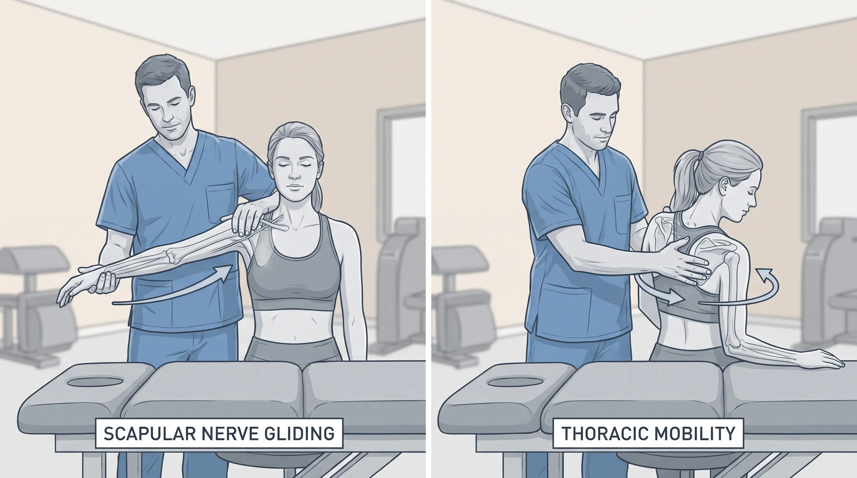
During a thorough clinical evaluation, physical therapists, sports medicine physicians, and orthopedic specialists perform specialized provocative tests designed to isolate specific peripheral nerve pathways and reproduce familiar patient symptoms under controlled conditions. The upper limb neurodynamic test (ULNT) or modified upper limb tension test systematically assesses neural mechanosensitivity by sequentially positioning the cervical spine, glenohumeral joint, elbow, and wrist into postures that gently elongate the entire brachial plexus pathway. Reproduction of the patient’s characteristic radiating pain, tingling, or electric sensations during ULNT strongly indicates primary neural hypersensitivity rather than isolated joint capsule dysfunction. Clinicians also perform scapular assistance and scapular retraction tests, which manually alter scapular positioning during active arm elevation to determine whether improved subacromial space and optimized nerve clearance temporarily alleviate symptoms. Manual muscle testing reveals asymmetric strength deficits, particularly in the infraspinatus, supraspinatus, major and minor rhomboids, and levator scapulae, helping pinpoint the exact nerve distribution affected. Furthermore, skilled palpation along the suprascapular notch, spinoglenoid fossa, and posterior cervical paraspinals identifies localized tenderness, palpable fascial bands, or abnormal tissue density that strongly suggests mechanical entrapment beneath fibrous structures or cystic formations.
Imaging and Electrodiagnostic Studies
When physical examination findings suggest moderate to severe neurological compromise, advanced diagnostic imaging becomes indispensable for confirming the diagnosis, mapping the exact compression site, and ruling out competing structural pathologies. Magnetic resonance imaging (MRI) of the cervical spine and shoulder girdle delivers high-resolution, multiplanar visualization of soft tissues, successfully revealing disc protrusions, nerve root impingement, paralabral ganglion cysts, hypertrophied ligaments, or inflammatory masses that physically compress the affected nerves. High-resolution musculoskeletal ultrasound provides a dynamic, real-time diagnostic alternative that allows clinicians to directly observe nerve swelling, altered longitudinal gliding mechanics, fascial tethering, and microvascular compromise during active shoulder movement sequences. Electrodiagnostic testing, specifically needle electromyography (EMG) and nerve conduction velocity (NCV) studies, objectively measures the electrical firing patterns of periscapular muscles and quantifies the conduction speed and amplitude of peripheral nerve signal transmission along compromised pathways. Abnormal EMG findings, including fibrillation potentials, positive sharp waves, or severely reduced motor unit recruitment patterns, definitively confirm active axonal injury and directly guide rehabilitation intensity and progression timelines. These objective diagnostic metrics effectively eliminate clinical guesswork, ensuring that targeted therapeutic interventions address the precise anatomical site of neural compression rather than secondary muscular adaptations.
Effective Treatment Options: From Conservative Care to Intervention
Successfully managing a pinched nerve scapula demands a phased, multimodal clinical approach that prioritizes acute inflammation reduction, mechanical neural decompression, tissue regeneration, and progressive functional restoration. Treatment selection depends heavily on symptom chronicity, compression severity, patient functional goals, and underlying anatomical etiology, with the overwhelming majority of acute and subacute cases responding favorably to structured conservative management protocols within a six to twelve-week rehabilitation window.
Rest, Activity Modification, and Ergonomic Adjustments
The foundational treatment phase focuses entirely on removing the repetitive mechanical stressors and postural triggers that continuously perpetuate neural irritation and perineural inflammation. Patients must temporarily modify daily activities that demand frequent overhead reaching, heavy unilateral lifting, or prolonged static postures that collapse the thoracic spine. Implementing structured microbreaks every thirty to forty-five minutes during desk work, utilizing ergonomic seating systems with integrated lumbar and mid-thoracic support, and adjusting computer monitor height to maintain a perfectly neutral cervical alignment drastically reduces cumulative neural traction and muscular fatigue. Sleep positioning requires equally careful attention; sleeping directly on the affected side compresses the brachial plexus against the rib cage, while sleeping flat without adequate cervical contouring chronically strains the upper trapezius and scalene attachments. Utilizing a memory foam contoured cervical pillow combined with placing a lightweight bolster beneath the upper arm prevents posterior shoulder collapse and maintains optimal nerve glide pathways throughout the entire sleep cycle.
Medication and Clinical Procedures
When mechanical and ergonomic modifications alone prove insufficient to interrupt the inflammatory cycle, targeted pharmacological interventions help rapidly reduce perineural edema and facilitate early participation in therapeutic exercise programs. Nonsteroidal anti-inflammatory drugs (NSAIDs) systematically decrease prostaglandin production and swelling, while short-term oral corticosteroids may be clinically indicated for severe acute exacerbations involving significant motor weakness or debilitating pain. Neuropathic pain modulators, including gabapentin, pregabalin, or duloxetine, selectively reduce central nervous system sensitization and decrease aberrant ectopic nerve firing, substantially improving sleep architecture and exercise tolerance during early recovery phases. For highly localized mechanical compression resistant to systemic medications, ultrasound-guided corticosteroid injections delivered precisely adjacent to the suprascapular notch or cervical intervertebral foramina provide potent, site-specific anti-inflammatory relief. Peripheral nerve hydrodissection utilizing sterile saline combined with localized anesthetic has gained significant clinical traction in regenerative medicine, offering a minimally invasive technique to mechanically separate entrapped nerves from dense fascial adhesions and restore independent gliding capacity. In highly refractory clinical cases where structural abnormalities like large ganglion cysts, severe foraminal osteophytes, or rigid fibrous bands cause persistent neurological compromise, surgical decompression, cyst aspiration, or fasciotomy becomes necessary to permanently restore adequate neural clearance.
Physical Therapy and Targeted Exercises
Structured physical rehabilitation forms the absolute cornerstone of long-term recovery for a pinched nerve scapula (Cleveland Clinic: Physical Therapy for Nerve Compression), focusing systematically on restoring joint mobility, rebuilding neuromuscular firing sequences, and retraining postural endurance across functional movement patterns. Evidence-based exercise programming progresses methodically from gentle neural mobilization to controlled strengthening integration, ensuring that recovering tissues adapt safely without triggering inflammatory symptom flare-ups.
| Exercise Modality | Recommended Sets & Reps | Primary Therapeutic Purpose | Key Progression Cues |
|---|---|---|---|
| Scapular Retraction Holds | 2-3 sets of 10-15 reps | Activate mid-trapezius and rhomboids | Maintain 3-second peak contraction, avoid shoulder elevation |
| Thoracic Wall Slides | 2 sets of 8-12 slow repetitions | Improve thoracic spine extension and motor control | Keep lower ribcage flat, move only at shoulder blades |
| Prone Y-T-W Raises | 2 sets of 10 reps each pattern | Strengthen lower trapezius and serratus anterior | Use light resistance bands, focus on scapular depression |
| Dynamic Serratus Punches | 3 sets of 12-15 controlled reps | Promote anterior scapular protraction and stability | Maintain straight elbow, exhale during full extension |
| Quadruped Rock-Backs | 2 sets of 8-10 rhythmic cycles | Enhance closed-chain proprioception and core control | Distribute weight evenly, avoid lumbar sagging |
Strengthening protocols must consistently prioritize movement quality over external load magnitude. The serratus anterior and lower trapezius function as primary dynamic stabilizers, actively anchoring the scapula against the thoracic rib cage and preventing excessive upward rotation, winging, or anterior tipping during arm elevation. Resistance band horizontal pull-aparts, prone cobra holds, and controlled scapular wall circles train these deep stabilizing muscles to fire synergistically rather than allowing the hyperactive upper trapezius and levator scapulae to dominate shoulder mechanics. As pain substantially diminishes and motor control improves, patients safely integrate closed-chain loading exercises like modified knee push-ups or quadruped alternating reaches, which dramatically enhance joint proprioception, compressive tolerance, and real-world functional capacity. Consistency consistently outperforms sporadic intensity; executing these targeted movements three to four times weekly with precise biomechanical alignment yields vastly superior neurological adaptation compared to occasional heavy resistance training.
Home Management and Self-Care Strategies
Daily symptom management plays a critical role in accelerating neurological recovery by maintaining optimal local tissue perfusion, regulating inflammatory mediators, and reducing compensatory muscular hypertonicity that restricts nerve mobility.
Heat, Cold, and Topical Therapies
Contrast hydrotherapy creates a highly effective physiological cycle that alternates between vasoconstriction and vasodilation to support perineural healing. Applying cold therapy for fifteen minutes immediately after activity or during acute inflammatory flare-ups effectively reduces localized tissue temperature and decreases nerve conduction velocity, temporarily numbing pain signals. Following this with moist heat application for twenty minutes stimulates local blood flow, delivers oxygen-rich blood to damaged tissues, and relaxes chronically tight fascial bands. Topical analgesic formulations containing menthol, camphor, capsaicin, or diclofenac provide highly concentrated localized pain relief without introducing systemic gastrointestinal side effects commonly associated with oral NSAIDs. These topical agents penetrate superficial myofascial layers, delivering targeted anti-inflammatory and counter-irritant effects directly at the symptom generation site.
Stretching and Mobility Routines
Safe mobility restoration requires gentle, sustained positioning that elongates restricted fascial lines without placing tensile load on the sensitized nerve. The pectoralis minor stretch, performed in a doorframe with elbows at ninety degrees, releases anterior fascial tension that chronically pulls the scapula forward and narrows the neural exit window. Thoracic spine extension over a foam roller with hands supporting the head effectively reverses compensatory kyphosis, restoring the natural thoracic curvature and increasing available space for the posterior scapular nerves. Diaphragmatic breathing drills performed in crook-lying positions simultaneously expand the posterior rib cage, mobilize restricted costovertebral joints, and reduce sympathetic nervous system dominance that perpetuates upper quarter muscle guarding. Patients must consistently avoid aggressive ballistic stretching, which triggers the stretch reflex and further tightens protective musculature around the compressed nerve.
Posture Correction and Workspace Setup
Long-term ergonomic sustainability requires proactive environmental redesigns that eliminate chronic mechanical compression triggers. Adjusting standing desks to maintain ninety-degree elbow flexion, utilizing split keyboard layouts that keep wrists neutral, and positioning document holders at direct eye level eliminates forward head compensation that systematically compresses exiting cervical nerve roots. Implementing lumbar support cushions, utilizing anti-fatigue mats during standing tasks, and scheduling deliberate posture reset alarms every forty minutes prevents cumulative microtrauma from compounding throughout the workday. When traveling or commuting, adjusting vehicle seat recline to support the mid-back, keeping the steering wheel within comfortable arm reach, and using small cervical rolls during flights maintains optimal neural alignment during prolonged sedentary periods.

Prevention and Long-Term Joint Health
Sustaining a pain-free, highly functional scapular region requires deeply integrated lifestyle habits that actively protect delicate peripheral nerves from cumulative microtrauma, postural degradation, and chronic inflammatory states. Prevention strategies seamlessly blend ergonomic awareness, consistent movement training, targeted fitness conditioning, and nutritional optimization that promotes rapid nerve myelination and fascial resilience.
Strengthening the Scapular Stabilizers
Proactive neuromuscular training focuses on building resilient periscapular musculature that naturally maintains optimal bone positioning and neural clearance without conscious postural effort. Integrating compound pulling movements like inverted rows, lat pulldowns, and single-arm dumbbell rows into weekly fitness routines strengthens the posterior shoulder chain, counteracting the chronic anterior dominance caused by prolonged sitting. Utilizing stability ball push-ups, bear crawls, and medicine ball wall throws develops dynamic shoulder girdle control and teaches the nervous system to distribute load evenly across multiple joints rather than concentrating stress on vulnerable neural pathways. Consistent incorporation of mobility flow sequences that combine cat-cow variations, thread-the-needle reaches, and controlled neck rotations maintains fascial pliability, prevents tissue adhesion formation, and ensures that peripheral nerves continue gliding freely through their anatomical tunnels under varying physiological demands.
Daily Habits for Nerve Protection
Long-term neural protection extends far beyond targeted exercise programming into foundational daily habits that regulate systemic inflammation, optimize hydration status, and support rapid cellular repair. Consuming a nutrient-dense diet rich in high-quality B vitamins, particularly pyridoxine (B6), folate (B9), and cobalamin (B12) (NIH Office of Dietary Supplements: Vitamin B12), directly supports robust axonal regeneration and maintains healthy homocysteine metabolism that protects microvascular circulation. Incorporating omega-3 fatty acids sourced from wild-caught salmon, sardines, chia seeds, and walnuts provides powerful systemic anti-inflammatory compounds that actively dampen perineural edema and accelerate tissue recovery. Maintaining consistent daily hydration prevents fascial desiccation that significantly increases mechanical friction during joint movement, while prioritizing seven to nine hours of uninterrupted sleep enables robust glymphatic clearance of metabolic neurotoxins and stimulates natural growth hormone release essential for structural tissue repair. Chronic stress management through daily meditation, extended nature exposure, progressive muscle relaxation, and structured breathwork protocols effectively lowers circulating cortisol concentrations, preventing prolonged sympathetic nervous system dominance that chronically elevates muscle tone and perpetuates pain sensitization cycles.
Frequently Asked Questions
What are the primary causes of a pinched nerve scapula?
A pinched nerve scapula typically develops from mechanical compression or chronic irritation of the dorsal scapular, suprascapular, or long thoracic nerves. Common causes include poor posture, repetitive overhead activities, cervical disc herniations, direct trauma, muscle hypertrophy in the neck and shoulder region, and systemic inflammation that reduces available space within anatomical nerve tunnels. Prolonged forward head positioning and thoracic stiffness significantly contribute to progressive neural entrapment over time.
How long does it take for a pinched nerve in the shoulder blade to heal?
Recovery timelines vary substantially based on compression severity, underlying etiology, and the extent of initial nerve damage. Mild irritation often resolves completely within two to four weeks with conservative management and strict activity modification. Moderate cases involving significant inflammation or measurable motor weakness typically require six to twelve weeks of structured physical therapy to fully restore neural gliding and muscular coordination. Severe axonal damage or structural entrapment can extend recovery to six months or longer, and in rare refractory instances, surgical decompression becomes necessary to permanently restore function.
Can a pinched nerve scapula resolve without medical treatment?
While exceptionally mild nerve irritation occasionally improves spontaneously with dedicated rest, ergonomic correction, and gentle mobility work, persistent or progressively worsening neurological symptoms rarely resolve independently without targeted clinical intervention. Ignoring active neural compression significantly increases the risk of chronic pain centralization, permanent muscle atrophy, irreversible demyelination, and long-term functional impairment. Consulting a qualified healthcare professional ensures accurate anatomical diagnosis and implementation of evidence-based therapeutic protocols that safely accelerate tissue healing and prevent permanent complications.
Is physical therapy effective for treating scapular nerve compression?
Physical therapy represents the undisputed gold standard for conservatively managing a pinched nerve scapula across all clinical severity stages. Certified therapists systematically utilize neural mobilization techniques, targeted myofascial release, progressive strengthening exercises, and postural retraining to mechanically decompress affected nerves and restore optimal shoulder biomechanics. Extensive peer-reviewed clinical studies consistently demonstrate that structured physical rehabilitation significantly reduces subjective pain scores, dramatically improves functional range of motion, accelerates return to daily activities, and substantially decreases the statistical likelihood of requiring invasive surgical intervention.
When should surgery be considered for a pinched nerve scapula?
Surgical intervention is strictly reserved for highly refractory clinical cases where comprehensive conservative treatment completely fails to produce meaningful improvement after three to six months, or when advanced diagnostic imaging definitively reveals space-occupying structural abnormalities such as large paralabral ganglion cysts, severe intervertebral foraminal stenosis, rigid calcified ligaments, or complete nerve transection. Experienced neurosurgeons and orthopedic shoulder specialists perform precise, minimally invasive decompression procedures to permanently release compressive fibrous bands, safely excise obstructive cystic formations, and restore adequate neural clearance. Comprehensive post-surgical rehabilitation remains absolutely essential for achieving optimal long-term functional outcomes and preventing recurrent compression.
Conclusion
Navigating the complex clinical landscape of a pinched nerve scapula demands a thorough understanding of peripheral anatomy, precise symptom recognition, and disciplined implementation of evidence-based management strategies. By accurately identifying the underlying causes of neural compression during early stages and consistently applying targeted interventions that prioritize mechanical decompression, fascial mobility restoration, and neuromuscular reeducation, individuals can effectively reclaim their functional range of motion and permanently eliminate debilitating discomfort. The transitional journey from acute nerve irritation to sustained neurological resilience absolutely requires patience, daily consistency, and professional clinical guidance, particularly when symptoms manifest as severe radiating pain or objective motor weakness. Seamlessly integrating structured therapeutic exercise, comprehensive ergonomic modifications, and nervous system-supportive nutritional habits constructs a highly resilient physiological defense against future inflammatory flare-ups and mechanical entrapment. Remember that enduring nerve health fundamentally depends on daily movement awareness, balanced posterior chain conditioning, and proactive lifestyle selections that actively protect the delicate neural pathways governing shoulder and arm function. With unwavering adherence to medically verified rehabilitation protocols, a commitment to progressive self-care practices, and consistent monitoring of postural mechanics, lasting pain relief, optimal scapular stability, and peak upper extremity performance remain completely achievable for patients of all ages and activity levels.
How we work
Researched and drafted with AI assistance, then reviewed by a human editor against the cited sources.
This article is general health information, not medical advice. Consult a qualified healthcare professional about your own situation.