Recovery Time for Herniated Lumbar Disc Surgery: A Complete Medical Guide

Living with the burning, shooting pain of a herniated lumbar disc can be profoundly debilitating. For many patients, the sensation of sciatica radiating down the leg disrupts sleep, limits mobility, and diminishes overall quality of life. When conservative treatments like targeted physical therapy, epidural injections, and anti-inflammatory medications no longer provide adequate relief, surgical intervention becomes a highly effective pathway to reclaim your health. Understanding the recovery time for herniated lumbar disc surgery is crucial for setting realistic expectations, preparing your home environment, and navigating the healing process with confidence. This comprehensive guide breaks down the physiological healing stages, clinical evidence, and evidence-based strategies to optimize your post-operative journey. By aligning with established spine surgery protocols and adhering to structured rehabilitation milestones, most patients experience dramatic improvements in leg pain and functional mobility within a predictable timeframe.
Understanding the Surgical Intervention
Before diving into rehabilitation timelines, it is essential to understand exactly what occurs during a lumbar decompression procedure and why it effectively addresses nerve root compression. The primary goal is to relieve pressure on the affected spinal nerve while preserving spinal stability.
Free calculatorSleep CalculatorCalculate sleep cyclesOpen the calculatorThe Most Common Surgical Approaches
Microdiscectomy remains the gold-standard surgical treatment for symptomatic lumbar disc herniations. This minimally invasive technique utilizes a small 1-to-1.5 inch incision, specialized retractors, and a high-magnification surgical microscope. The surgeon carefully navigates through paraspinal muscles to access the lamina, removes a small portion of bone or ligament if necessary, and extracts only the herniated disc fragment compressing the nerve root. By preserving the majority of the intact disc and surrounding structures, patients experience significantly reduced recovery time for herniated lumbar disc surgery compared to traditional open procedures.
Endoscopic discectomy has emerged as an advanced alternative, utilizing a 2-to-12 millimeter portal and a high-definition internal camera system. This ultra-minimally invasive approach minimizes muscle stripping, reduces intraoperative bleeding, and often allows for same-day discharge. Some patients report slightly faster initial pain relief, though long-term outcomes at six and twelve months closely mirror traditional microdiscectomy results according to comparative clinical studies. Open discectomy or laminectomy, involving a larger incision and more extensive tissue retraction, is generally reserved for complex multi-level pathologies, severe spinal stenosis, or cases requiring concurrent spinal fusion.
Determining Surgical Candidacy
Not every patient requires immediate intervention. The American Academy of Orthopaedic Surgeons (AAOS) recommends considering surgery when radicular symptoms persist beyond 6 to 12 weeks despite structured conservative management. Immediate surgical consultation is warranted for progressive neurological deficits, such as worsening muscle weakness, or the development of cauda equina syndrome, a rare but surgical emergency characterized by bowel or bladder dysfunction and saddle anesthesia. The landmark Spine Patient Outcomes Research Trial (SPORT Trial) demonstrated that while surgery yields faster and greater symptom relief at 3, 6, 12, and 24 months, many patients still experience meaningful improvement with prolonged non-operative care. Shared decision-making between patient and spine specialist remains the cornerstone of optimal treatment planning.
The Staged Recovery Timeline
The physiological healing process after nerve decompression follows a predictable biological sequence. Recognizing these stages prevents premature overexertion and reduces anxiety during periods of slower progress. The recovery time for herniated lumbar disc surgery is best understood through four distinct clinical phases.
Phase 1: Immediate Post-Operative Care (Days 0–7)
The first week focuses on acute pain management, incision protection, and safe early mobilization. Most microdiscectomies are performed on an outpatient basis, or require only a single overnight observation stay. Within 2 to 6 hours post-anesthesia, nurses and physical therapists encourage supervised walking. Early ambulation stimulates blood flow, prevents venous thromboembolism, and jumpstarts neuromuscular re-education.
Surgical site discomfort is normal and typically peaks around 48 to 72 hours before gradually subsiding. Residual nerve inflammation, often described as lingering tingling or mild aching in the previously affected leg, is expected because nerves heal slowly. Pain control strategies rely on a multimodal approach combining acetaminophen, NSAIDs, muscle relaxants, and occasionally a short, carefully monitored course of opioids for breakthrough pain. During this critical window, strict adherence to BLT (Bending, Lifting, and Twisting) restrictions is non-negotiable. Patients must avoid lifting anything heavier than 5 to 10 pounds, refrain from sitting for longer than 20 to 30 minutes consecutively, and maintain a neutral spine during transfers. Stool softeners and adequate hydration are routinely prescribed to prevent straining, which dramatically increases intra-abdominal and intradiscal pressure.
Phase 2: Early Recovery and Mobilization (Weeks 2–6)
As acute inflammation diminishes, patients enter the tissue remodeling phase. Staples or sutures are typically removed during a follow-up clinic visit at 10 to 14 days. Incision care involves keeping the area clean and dry, monitoring for signs of infection, and avoiding submerging the wound in baths or pools until fully sealed.
Activity progression during this window is guided by symptom tolerance rather than a rigid calendar. Patients gradually increase walking duration, aiming for multiple short walks daily that collectively reach 30 to 45 minutes by week four. Light household tasks such as wiping counters or folding laundry become feasible, but vacuuming, mopping, and heavy grocery shopping remain off-limits. For those with sedentary occupations, returning to desk work around weeks 2 to 4 is often achievable with ergonomic modifications like lumbar support cushions, sit-stand desk converters, and scheduled micro-breaks to prevent spinal stiffness.
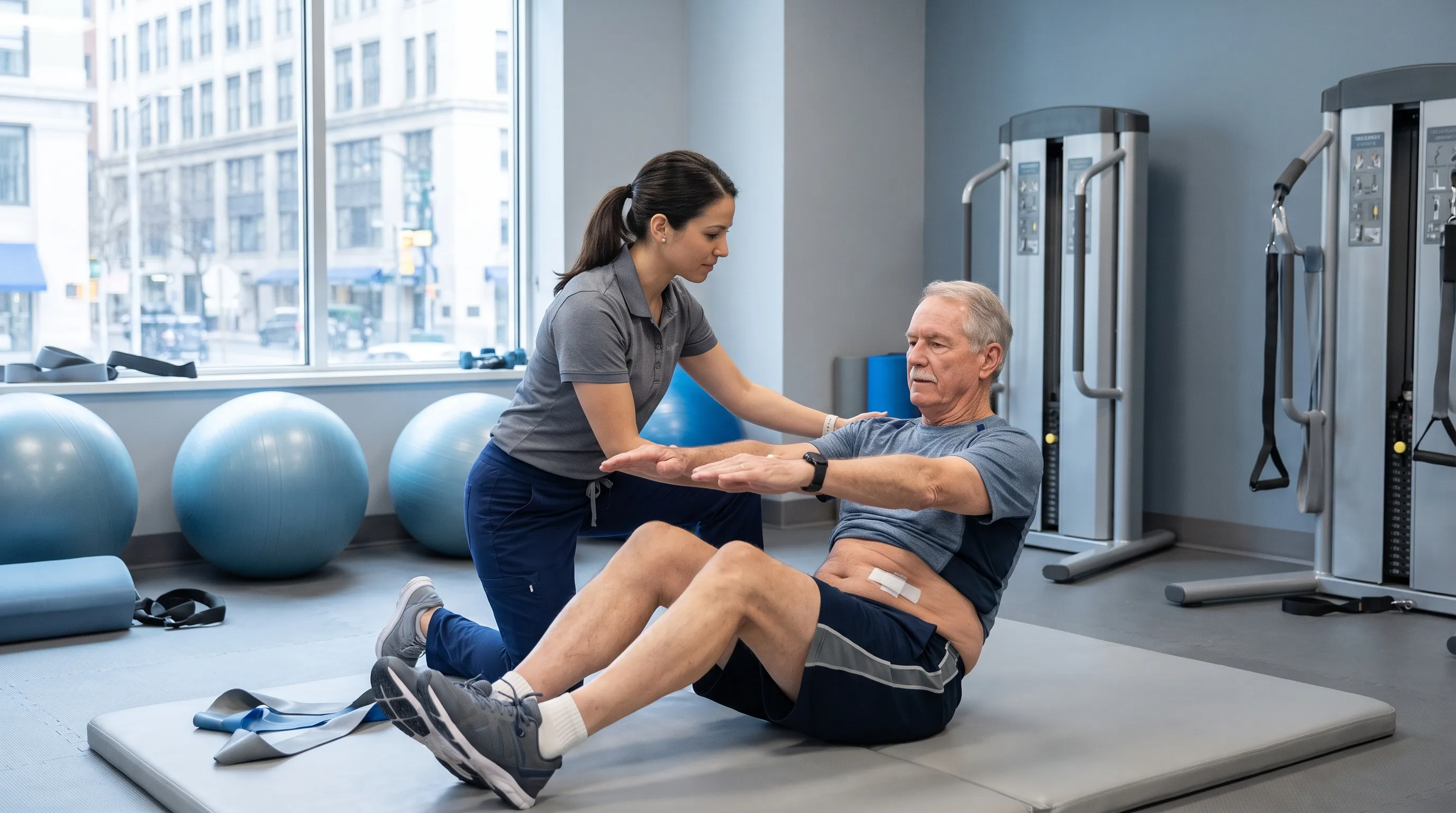
Formal physical therapy typically initiates around weeks 3 to 4. Early sessions prioritize gentle neural mobilization, hamstring and hip flexor stretching, and foundational core activation exercises that avoid spinal flexion. The focus remains on establishing movement patterns that protect the healing annulus fibrosus while restoring functional mobility. Driving is usually permitted once the patient is completely weaned off narcotic medications, can comfortably operate pedals without pain, and possesses adequate reaction time for emergency braking, which commonly occurs between weeks 2 and 4.
Phase 3: Intermediate Recovery and Strengthening (Weeks 6–12)
By the second month, the majority of surgical tissue healing is complete, though deep ligamentous and annular remodeling continues. Patients experience substantial freedom in daily activities and can gradually resume most household responsibilities. The recovery time for herniated lumbar disc surgery accelerates significantly during this phase as physical therapy transitions from protective mobilization to structured strengthening.
Exercise programming introduces low-impact cardiovascular conditioning, including stationary cycling, elliptical training, and aquatic walking or swimming. Water buoyancy reduces axial loading on the lumbar spine while allowing full range of motion and resistance training. Core stabilization intensifies with progressive loading of the transverse abdominis and multifidus muscles. Patients begin practicing safe hinge mechanics, learning to bend at the hips and knees rather than the lumbar spine. Moderate lifting restrictions gradually ease around weeks 8 to 12, but heavy compound lifts, repetitive overhead reaching, and contact sports remain contraindicated. Return to light physical labor is often cleared at the 6-week mark, provided the patient demonstrates proper body mechanics and lacks pain flare-ups.
Phase 4: Full Recovery and Athletic Reintegration (Months 3–6+)
The final phase represents the transition from rehabilitation to performance optimization. Between 3 and 6 months post-surgery, patients are typically cleared for heavy manual labor, high-intensity interval training, and return to recreational sports, contingent upon successful completion of functional capacity evaluations. The annulus fibrosus, the tough outer ring of the disc where the tear occurred, requires 6 to 12 months to fully scar and regain structural integrity. During this extended remodeling period, gradual progression is paramount to prevent tissue overload.
Athlete recovery data from systematic reviews indicate that 75% to 100% of competitive athletes successfully return to play, with average rehabilitation timelines ranging from 2.8 to 8.7 months depending on sport-specific demands. Professional athletes undergoing microdiscectomy often follow highly regimented, sport-specific progressions supervised by athletic trainers and spine biomechanics specialists. Long-term outcomes remain exceptionally favorable, with 85% to 95% of patients reporting significant, sustained relief from radicular leg pain. However, it is important to recognize that axial lower back pain may persist in some individuals if underlying degenerative disc disease or facet joint arthropathy exists, as decompression surgery specifically targets nerve compression rather than age-related spinal wear.
Factors That Influence Your Recovery Speed
While standardized timelines provide valuable benchmarks, individual healing trajectories vary based on several physiological and lifestyle variables. Understanding these modifiable and non-modifiable factors empowers patients to actively participate in optimizing their recovery time for herniated lumbar disc surgery.
Age, Baseline Health, and Tissue Quality
Younger patients generally exhibit accelerated cellular turnover and more robust inflammatory responses conducive to rapid healing. However, age alone is a poor predictor of surgical success. Baseline cardiovascular fitness, muscle mass, and metabolic health play far more significant roles. Patients with well-controlled diabetes, healthy body mass indices, and regular pre-surgical activity levels consistently outperform sedentary counterparts with identical surgical procedures. Chronic systemic inflammation from obesity or metabolic syndrome can delay soft tissue repair and prolong post-operative pain duration.
Pre-Existing Nerve Compression Duration
The length of time a nerve root remains compressed prior to surgery directly impacts neurological recovery. Acute herniations decompressed within weeks often result in near-instantaneous pain relief and rapid motor return. Chronic compression extending beyond 6 to 12 months can lead to demyelination or axonal degeneration. Nerve regeneration occurs at an approximate rate of 1 millimeter per day, meaning prolonged pre-operative deficits may take many months to fully resolve. Residual numbness or mild paresthesia can persist even after the disc fragment is removed, representing the nerve’s biological timeline rather than surgical failure.
Lifestyle Choices and Compliance
Behavioral adherence to post-operative protocols dramatically alters outcomes. Smoking or vaping introduces nicotine and carbon monoxide into the bloodstream, causing microvascular vasoconstriction that severely impairs oxygen delivery to healing spinal tissues. Research consistently demonstrates that smokers face significantly higher rates of delayed wound healing, disc degeneration acceleration, and symptomatic re-herniation. Complete cessation at least 4 weeks prior to surgery and throughout the rehabilitation window is strongly advocated by spine surgeons. Similarly, psychological readiness, stress management, and realistic expectation setting contribute substantially to perceived recovery speed and overall satisfaction.
Evidence-Based Recovery Tips and Lifestyle Modifications
Implementing clinically validated strategies accelerates tissue repair, minimizes complication risks, and enhances functional restoration. The following protocols synthesize recommendations from leading spine centers, rehabilitation medicine specialists, and peer-reviewed outcome studies.
Structured Walking Protocols
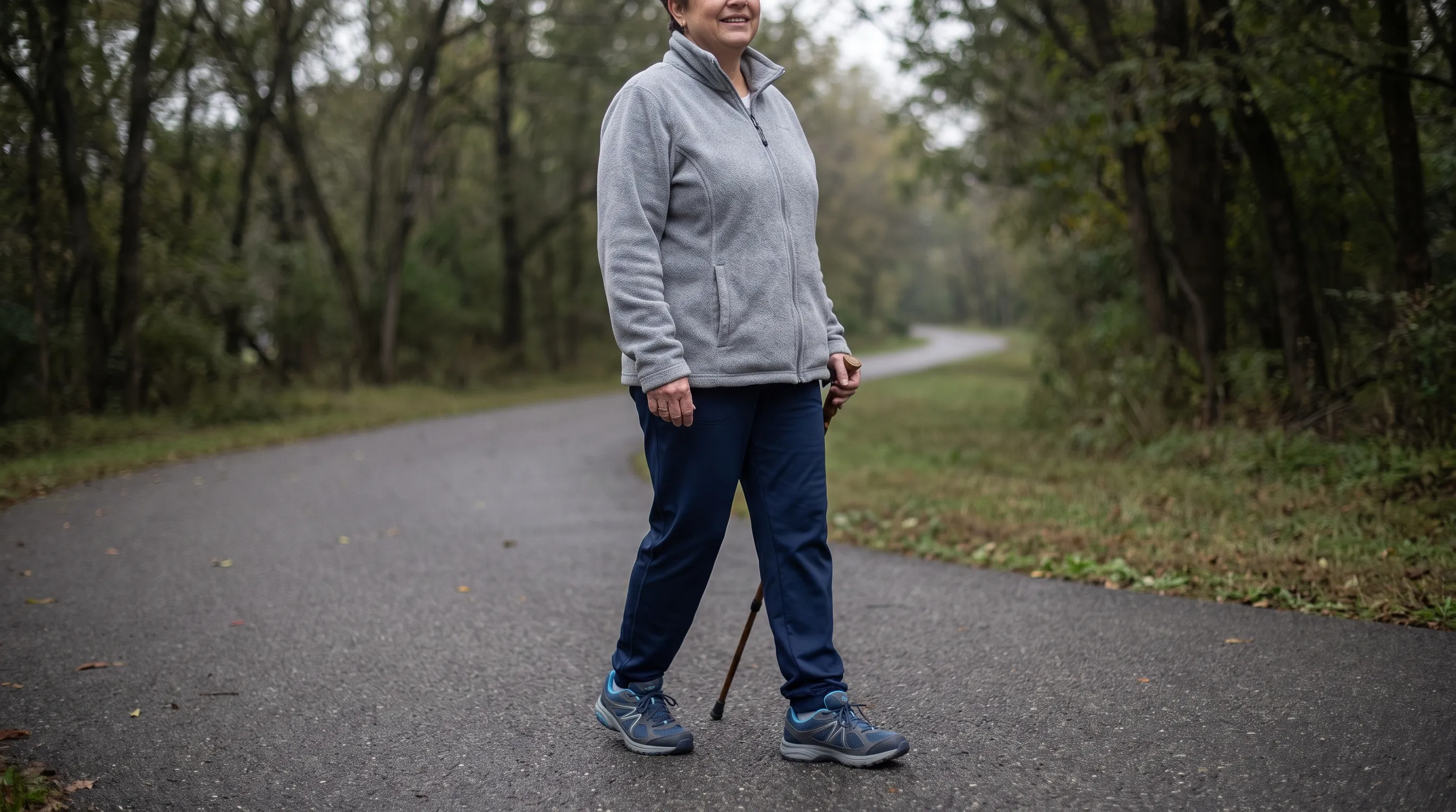
Walking is the most accessible, evidence-backed exercise for post-microdiscectomy recovery. It promotes lymphatic drainage, reduces epidural inflammation, and maintains cardiovascular conditioning without axial loading. Begin with 5-to-10-minute walks on flat, even surfaces within the first 24 to 48 hours. Increase frequency rather than duration initially, taking three short walks daily. By week four, progress toward a single continuous 30-minute walk. Utilize proper gait mechanics: engage your core lightly, maintain a neutral head position, and allow natural arm swing to counterbalance pelvic rotation. Avoid uneven terrain, steep inclines, or prolonged treadmill sessions until cleared by your physical therapist.
Mastering Physical Therapy and Core Stabilization
Physical therapy is not optional; it is a critical component of preventing recurrence. Early rehabilitation emphasizes motor control re-education over brute strength. The McGill Big 3 exercises—modified curl-ups, side planks, and bird-dogs—are frequently prescribed because they build isometric trunk endurance without placing the spine in flexion or excessive rotation. Neural gliding exercises, such as seated sciatic nerve flossing, prevent adhesions and maintain nerve root mobility through the newly decompressed space. Patients must communicate openly with their therapists regarding symptom response. Sharp, shooting pain indicates neural irritation requiring exercise modification, whereas mild muscular fatigue signifies appropriate training adaptation.
Optimizing Sleep Ergonomics and Daily Posture
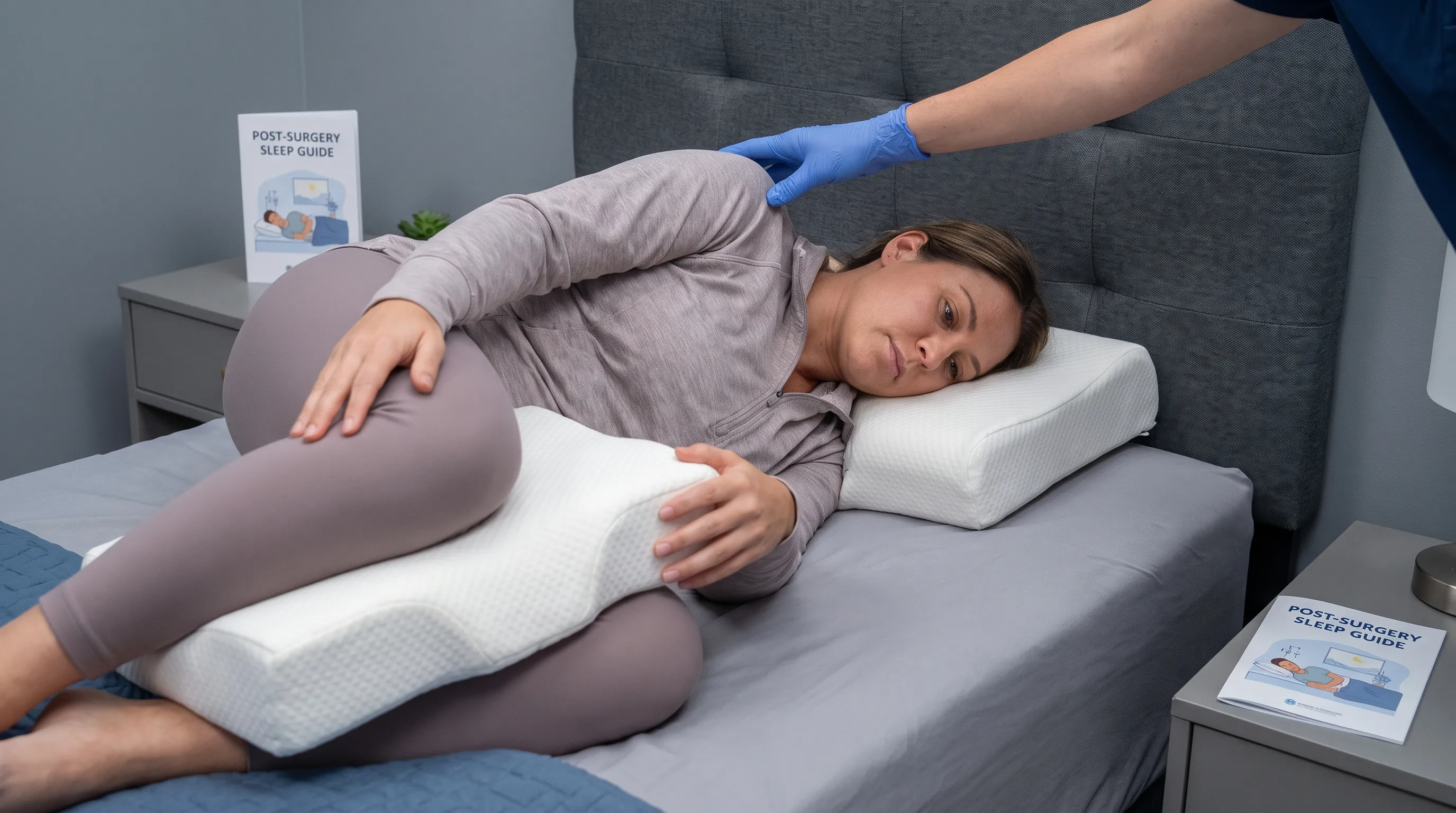
Quality sleep accelerates cellular repair and pain modulation, yet post-operative discomfort frequently disrupts rest. Sleep positioning drastically influences lumbar mechanics. When lying supine, place a firm pillow under your knees to flatten the lumbar lordosis and reduce discal pressure. Side-sleepers should position a thick pillow between the knees to prevent the upper leg from rotating the pelvis and twisting the lower spine. Avoid prone sleeping entirely, as it forces hyperextension of the lumbar facets and strains healing paraspinal musculature.
During waking hours, practice dynamic sitting. Limit continuous seated time to 20 to 30 minutes during the first two weeks. Stand, stretch gently, and walk for 2 to 3 minutes before resitting. Invest in lumbar roll cushions for chairs and vehicles. When picking up lightweight objects, utilize the golfer’s lift technique or squat with a straight back rather than rounding the lumbar spine. Consistent postural awareness prevents micro-traumas that delay healing.
Nutritional Support and Smoking Cessation
Healing demands specific macronutrients and micronutrients. Prioritize high-quality protein intake (aiming for 1.2 to 1.5 grams per kilogram of body weight daily) to supply amino acids for collagen synthesis and muscle repair, as supported by clinical wound healing guidelines. Vitamin C, zinc, and omega-3 fatty acids support connective tissue formation and modulate inflammatory pathways. Hydration maintains intervertebral disc hydration status and facilitates nutrient diffusion through the avascular disc. Eliminate processed sugars and excessive alcohol, which promote systemic inflammation and interfere with tissue regeneration. If you smoke, seek medical support for cessation programs; nicotine replacement therapy and prescription aids can facilitate quitting during this critical window.
Clinical Evidence, Success Rates, and Realistic Expectations
Understanding the data behind lumbar discectomy outcomes helps patients separate marketing hype from medical reality. The recovery time for herniated lumbar disc surgery aligns closely with robust clinical trial findings and long-term registry data.
Insights from the SPORT Trial
The Spine Patient Outcomes Research Trial (SPORT), a landmark NIH-funded randomized controlled trial published in the New England Journal of Medicine, compared surgical decompression against structured non-operative management for lumbar disc herniation. The study revealed that patients undergoing surgery experienced significantly faster and greater improvement in pain, disability, and quality-of-life metrics at 3, 6, 12, and 24 months. The lead authors noted that while surgery was associated with more rapid improvement than nonoperative treatment, by 2 years, the differences between groups were small, though the surgical cohort maintained slightly better functional outcomes. This data supports surgery as the optimal choice for patients seeking expedited relief and rapid return to work, particularly those with severe radicular symptoms.
Success Metrics and Re-Herniation Risks
Success is primarily defined by relief of leg pain rather than complete elimination of all back discomfort. Between 85% and 95% of patients achieve substantial radicular pain reduction and improved mobility. Return-to-work statistics indicate that approximately 70% to 80% of patients resume employment within 3 months, climbing to 90% by 6 months.
Re-herniation represents the most common post-operative complication, occurring in 5% to 15% of cases. The risk peaks within the first 6 months when the annular defect remains vulnerable. Contributing factors include heavy lifting violations, smoking, obesity, and multi-level disc degeneration. Recurrent herniations typically present with sudden return of severe sciatica and may require revision microdiscectomy, which generally follows a similar but slightly more cautious recovery protocol.
Comparative Recovery Milestones Table
The table below outlines typical progression markers to help patients track their healing trajectory against clinical expectations.
| Recovery Phase | Typical Timeframe | Activity Restrictions | Expected Symptom Progression | Clinical Milestones |
|---|---|---|---|---|
| Acute Inflammatory | Days 0–7 | Strict BLT avoidance, no sitting >30 mins | Surgical pain peaks then declines; leg tingling common | Outpatient discharge, first supervised walks, staple care |
| Early Mobilization | Weeks 2–6 | No lifting >10 lbs, avoid prolonged sitting | Incision heals; radicular pain improves significantly | Suture removal, driving clearance (off narcotics), sedentary work return |
| Tissue Remodeling | Weeks 6–12 | Gradual lifting, avoid heavy bending/twisting | Residual nerve sensitivity fades; strength improves | PT intensifies, low-impact cardio introduced, moderate labor clearance |
| Functional Optimization | Months 3–6+ | Sport-specific restrictions lifted | Full daily function restored; occasional weather-related stiffness | Heavy labor clearance, return to athletics, annual follow-up |
Warning Signs: Red Flags During Recovery
Vigilance during the post-operative period ensures timely intervention if complications arise. While mild fluctuating discomfort is normal, specific symptoms warrant immediate medical evaluation.
Neurological Red Flags
New or progressive lower extremity weakness, particularly foot drop or inability to stand on toes/heels, indicates potential nerve root compromise. Sudden, explosive return of severe shooting leg pain accompanied by neurological deficits suggests acute re-herniation. Cauda equina syndrome, though rare, constitutes a surgical emergency. Symptoms include new-onset bowel or bladder incontinence, urinary retention requiring catheterization, numbness in the perineal or saddle region, and bilateral severe leg weakness. Contact emergency services or your surgeon immediately if these occur.
Surgical Site and Systemic Concerns
Infection risk remains low but serious. Monitor for fever exceeding 101°F (38.3°C), increasing redness, warmth, or swelling around the incision, foul-smelling or purulent wound drainage, and escalating pain unresponsive to prescribed medications. Deep vein thrombosis presents with unilateral calf swelling, warmth, redness, and tenderness. Pulmonary embolism may manifest as sudden shortness of breath, chest pain, or unexplained tachycardia. Prompt communication with your healthcare team ensures appropriate imaging, laboratory work, and targeted treatment.
Frequently Asked Questions
How long after herniated lumbar disc surgery can I return to work?
Return-to-work timelines depend heavily on your job's physical demands. Patients with sedentary or desk-based roles typically return within 2 to 4 weeks, provided they can modify their workstation. Those involved in light physical labor generally need 4 to 6 weeks. For heavy manual labor or jobs requiring repetitive bending and lifting, clearance usually occurs between 8 and 12 weeks post-surgery, following physical therapy clearance and surgeon approval.
Is walking necessary after a lumbar discectomy?
Yes, walking is considered one of the most beneficial activities during early recovery. Surgeons typically recommend beginning short 5-to-10-minute walks within hours after surgery. Gradually increasing to 30 or more minutes of daily walking by week four improves circulation, reduces swelling, prevents blood clots, and accelerates tissue healing. Prolonged sitting or bed rest should be avoided in favor of frequent, gentle movement.
What are BLT restrictions and why are they important?
BLT stands for Bending, Lifting, and Twisting. Patients are instructed to strictly avoid bending at the waist, lifting objects heavier than 5 to 10 pounds, and twisting the lower spine for at least the first 6 weeks post-operatively. These movements place significant shear force and pressure on the healing annulus fibrosus and the surgical site, dramatically increasing the risk of re-herniation or delayed nerve recovery.
When does physical therapy typically begin after spine surgery?
Formal physical therapy usually commences between weeks 3 and 4, once initial incision healing has occurred and acute post-surgical pain has subsided. Early PT focuses on core stabilization, gentle neural gliding exercises, posture education, and gradual flexibility work. Programs like the McGill Big 3 exercises are often introduced to build trunk endurance without stressing the healing lumbar segments.
What is the success rate and re-herniation risk for microdiscectomy?
Clinical studies consistently show that microdiscectomy achieves an 85% to 95% success rate for relieving radicular leg pain and sciatica symptoms. However, there is a 5% to 15% risk of disc re-herniation at the same level, with the highest vulnerability occurring during the first 6 months while the annular defect fully scars over. Adhering to post-operative restrictions and completing rehabilitation significantly mitigates this risk.
Conclusion
Navigating the recovery time for herniated lumbar disc surgery requires patience, adherence to clinical guidelines, and proactive engagement with rehabilitation professionals. While the procedure itself typically lasts less than an hour, the biological healing of compressed nerve roots and compromised disc tissue unfolds over several months. By understanding the phased recovery trajectory, implementing evidence-based movement strategies, avoiding high-risk behaviors like smoking and heavy lifting violations, and maintaining open communication with your spine care team, you position yourself for optimal long-term outcomes. The vast majority of patients experience transformative relief from debilitating leg pain and successfully return to meaningful work, recreational activities, and an active lifestyle. Always consult your neurosurgeon or orthopedic spine specialist for personalized medical advice tailored to your specific anatomy, surgical approach, and rehabilitation milestones. With disciplined self-care and realistic expectations, your journey back to full spinal health is well within reach.
How we work
Researched and drafted with AI assistance, then reviewed by a human editor against the cited sources.
This article is general health information, not medical advice. Consult a qualified healthcare professional about your own situation.