Understanding and Managing Hip and Knee Pain: A Complete Guide
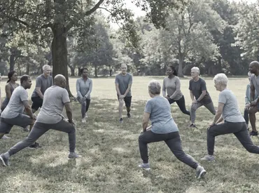
Living with persistent lower extremity discomfort can silently erode your independence, limit your favorite activities, and disrupt your daily routine. When mobility becomes a conscious effort rather than an automatic function, it is often time to investigate the underlying mechanics of your movement system. The phenomenon of hip and knee pain rarely occurs in isolation; these two powerhouse joints function as interdependent components of a complex kinetic chain. A disruption in one area invariably places compensatory stress on the other, creating a cycle of inflammation, muscle inhibition, and joint degradation that requires targeted intervention. As noted by the World Health Organization, musculoskeletal conditions remain a leading cause of disability worldwide, profoundly impacting mobility and quality of life. Millions of individuals navigate the frustrating reality of navigating stairs, standing for extended periods, or simply walking across a room with compromised comfort. Understanding why this happens, how it manifests, and what you can do about it is the first critical step toward reclaiming pain-free movement. This comprehensive guide explores the anatomical relationships, evidence-based diagnostic pathways, conservative and surgical management strategies, and actionable lifestyle protocols that healthcare professionals recommend for sustainable joint health. By examining both the biological mechanisms and the practical rehabilitation frameworks, you will gain the tools necessary to break the cycle of discomfort and restore functional mobility.
Understanding the Hip-Knee Connection and Biomechanical Interplay
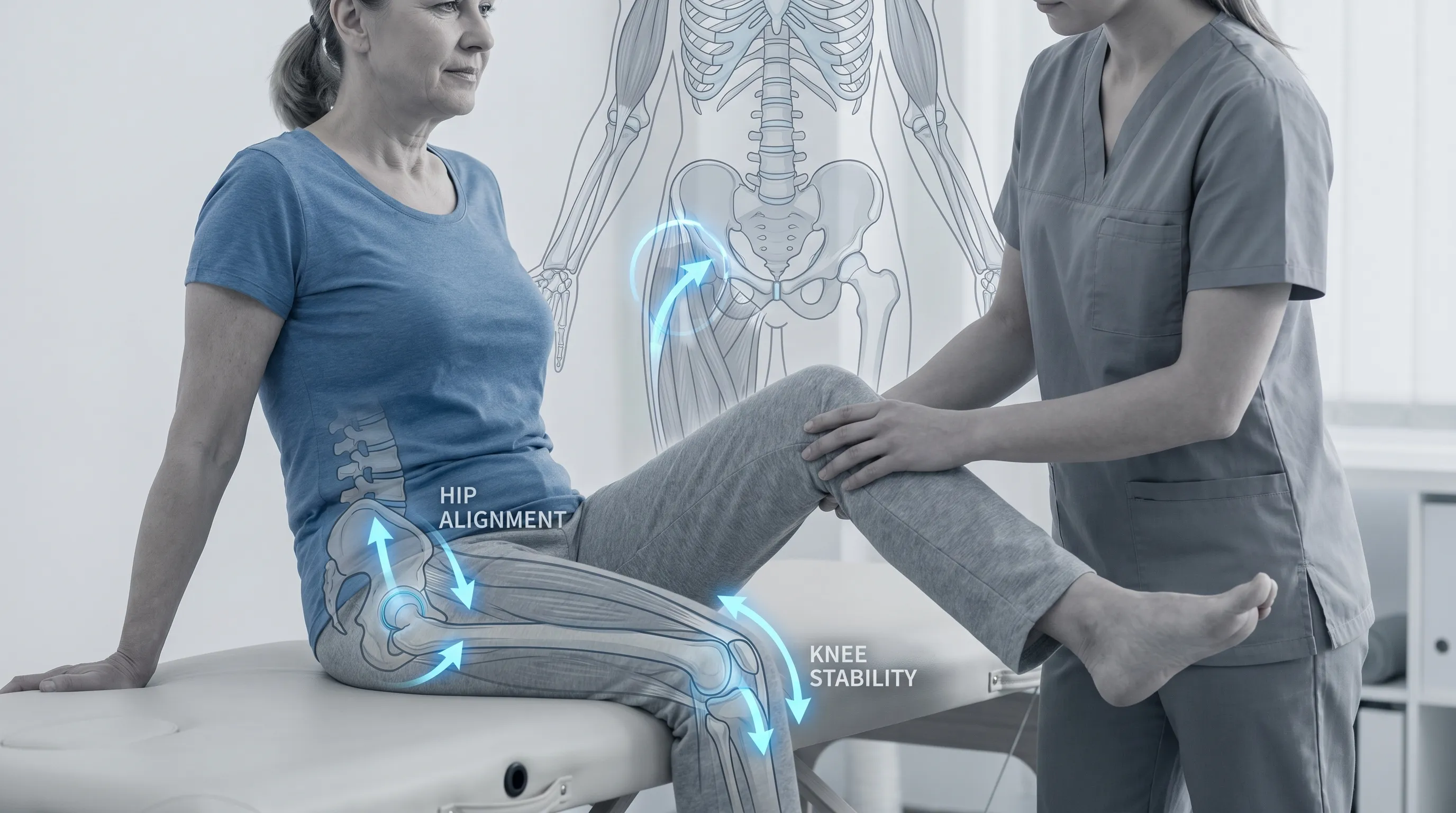
The lower extremity operates as a highly coordinated mechanical system designed to absorb impact, transfer weight, and generate propulsion during gait. The hip joint, a true ball-and-socket synovial articulation, provides multidirectional mobility while maintaining pelvic stability during weight-bearing activities. Meanwhile, the knee functions primarily as a hinge joint, relying heavily on surrounding musculature, ligaments, and meniscal cartilage to maintain alignment and absorb compressive forces. When these structures work in harmony, movement feels effortless. However, when one component falters, the entire system must compensate, often leading to accelerated wear and secondary discomfort.
Free calculatorKnee Pain Location ChartIdentify knee pain causes by locationOpen the calculatorThe Kinetic Chain in Lower Extremity Movement
Human locomotion is governed by sequential muscle activation patterns that travel from the distal foot to the proximal pelvis. During a normal gait cycle, the hip initiates femoral control while the knee stabilizes the tibia against ground reaction forces. The gluteal muscles, quadriceps, hamstrings, and calf complex coordinate in precise timing to prevent joint collapse. Research in biomechanical gait analysis consistently demonstrates that proximal weakness disrupts distal alignment. When the hip abductors and external rotators fail to adequately control femoral positioning, the knee experiences abnormal rotational and valgus stress. This phenomenon explains why individuals with gluteal inhibition frequently develop patellofemoral tracking issues or medial compartment knee degeneration over time.
Why Pain in One Joint Often Affects the Other
The nervous system responds to localized inflammation or structural compromise by altering motor recruitment patterns. This neuromuscular adaptation, while initially protective, quickly becomes maladaptive if sustained. Antalgic gait modifications shift weight-bearing loads away from the painful joint, overloading adjacent structures. For example, favoring a stiff hip forces the knee to absorb greater impact forces during heel strike and terminal stance phases. Similarly, chronic knee discomfort leads to reduced push-off force, compelling the hip flexors and lumbar spine to compensate during propulsion. This cross-joint referral pattern is well-documented in orthopedic literature and underscores the necessity of evaluating both articulations simultaneously. Comprehensive rehabilitation must address the entire lower kinetic chain rather than isolating a single symptomatic region.
Common Causes of Hip and Knee Pain: From Degeneration to Overuse
The etiology of lower extremity discomfort spans acute trauma, repetitive microtrauma, inflammatory processes, and age-related tissue degeneration. Clinicians categorize these conditions based on tissue involvement, symptom duration, and functional limitation patterns. Recognizing the specific pathological mechanism is essential for directing appropriate therapeutic interventions and preventing further structural compromise.
Osteoarthritis and Cartilage Degeneration
Osteoarthritis remains the most prevalent cause of progressive joint dysfunction worldwide, characterized by the gradual breakdown of articular cartilage and subchondral bone remodeling (NIH). It involves synovial inflammation and marginal osteophyte formation. Unlike autoimmune arthritis, which targets multiple systems, osteoarthritis stems from mechanical overload combined with genetic susceptibility and metabolic factors. As cartilage thins, joint lubrication decreases, and bone-on-bone friction increases, triggering localized pain, crepitus, and morning stiffness that typically resolves within thirty minutes. Weight-bearing joints experience accelerated degeneration due to cumulative gravitational loading. Studies indicate that every additional pound of body mass multiplies joint compressive forces by three to four times during ambulation. Managing osteoarthritis requires a multimodal approach combining load reduction, anti-inflammatory strategies, and targeted strengthening to preserve remaining cartilage and delay structural collapse.
Tendinopathies, Bursitis, and Soft Tissue Inflammation
Soft tissue structures frequently become the primary pain generators before joint surfaces deteriorate. The iliotibial band, patellar tendon, gluteus medius tendon, and hamstring insertions endure high tensile forces during repetitive movement. Tendinopathy develops from chronic overload without adequate recovery, leading to collagen disorganization, neovascularization, and localized nociceptor sensitization. Unlike acute tendon tears, tendinopathies often present with insidious onset, warmth during activity, and stiffness after periods of inactivity. Concurrent bursitis occurs when fluid-filled sacs designed to reduce friction become inflamed due to repetitive compression or direct trauma (Cleveland Clinic). Trochanteric bursitis manifests as lateral hip pain exacerbated by lying on the affected side, while pes anserine bursitis causes medial knee discomfort. Addressing soft tissue dysfunction requires load management, eccentric strengthening protocols, and temporary modification of aggravating activities to facilitate tissue remodeling and pain resolution.
Biomechanical Misalignments and Compensatory Gait Patterns
Structural deviations from optimal alignment significantly alter joint loading vectors. Excessive femoral anteversion, tibial torsion, genu varum, or pes planus change how ground reaction forces travel through the lower limb. Even minor discrepancies, such as a leg length difference exceeding five millimeters, can tilt the pelvis, compress one hip joint, and shift knee alignment into valgus or varus deviation. Over time, these asymmetries concentrate stress on specific cartilage zones, accelerate wear, and provoke chronic inflammation. Footwear selection, occupational posturing, and prior injuries all contribute to cumulative biomechanical strain. Corrective strategies include custom orthotics, gait retraining, manual therapy to restore joint arthrokinematics, and neuromuscular re-education to establish symmetrical weight distribution. Addressing foundational alignment issues often yields dramatic improvements in both hip and knee pain outcomes.
Recognizing Clinical Symptoms and Determining Severity
Symptom differentiation is critical for accurate self-assessment and timely clinical intervention. Lower extremity discomfort varies widely in presentation, intensity, and functional impact. Understanding how pain behaves under different conditions helps distinguish mechanical issues from systemic inflammation or neurological referral.
Differentiating Referred Pain Patterns
Pain localization does not always correspond to the primary pathological site. Hip joint pathology frequently refers discomfort to the anterior thigh, medial knee, or groin region due to shared innervation from the obturator and femoral nerves. Conversely, lumbar spine radiculopathy can mimic hip and knee pain, radiating down the lateral thigh and calf. True knee pain typically localizes around the patella, joint line, or posterior fossa. Mechanical symptoms such as catching, locking, or sudden giving way suggest intra-articular derangements like meniscal tears or loose bodies. Inflammatory pain presents as diffuse aching, prolonged stiffness, and warmth that worsens at night or during rest. Keeping a symptom diary tracking onset, duration, aggravating movements, and relieving factors provides clinicians with invaluable diagnostic information.
Red Flags That Demand Immediate Medical Evaluation
Certain clinical indicators require urgent orthopedic or emergency assessment. Sudden inability to bear weight following trauma suggests fractures or ligamentous rupture. Unexplained weight loss, fever, or severe night pain may indicate infectious processes or systemic disease. Progressive neurological deficits such as numbness, tingling, or bowel/bladder dysfunction point toward spinal cord compression. Rapidly expanding swelling with extreme tenderness warrants evaluation for deep vein thrombosis or septic arthritis. Joint effusions that feel warm, appear erythematous, or restrict range of motion within hours require immediate aspiration and laboratory analysis (Mayo Clinic). Ignoring these warning signs can lead to permanent structural damage, chronic instability, or life-threatening complications. Prompt clinical evaluation ensures timely intervention and optimal recovery trajectories.
Evidence-Based Diagnostic Pathways for Joint Pain
Accurate diagnosis relies on systematic clinical evaluation combined with selective imaging and laboratory testing. Overreliance on advanced imaging without clinical correlation frequently leads to incidental findings that do not align with symptom severity. A structured diagnostic algorithm ensures appropriate resource utilization and targeted treatment planning.
Comprehensive Physical Examination Techniques
Orthopedic assessment begins with detailed history taking and observational gait analysis. Clinicians evaluate posture, step length, heel strike symmetry, and pelvic tilt during ambulation. Specialized orthopedic tests isolate specific structures to reproduce symptoms and confirm tissue involvement. The FABER (Flexion, Abduction, External Rotation) test assesses hip joint integrity and sacroiliac function. The Trendelenburg maneuver identifies gluteus medius weakness through pelvic drop during single-leg stance. Knee examinations incorporate the Lachman and anterior drawer tests for anterior cruciate ligament integrity, McMurray and Thessaly tests for meniscal pathology, and patellar apprehension tracking for instability. Palpation identifies localized tenderness over the joint line, trochanteric region, or pes anserine bursa. Range of motion measurements establish baseline mobility and track rehabilitation progress.
Imaging Modalities and Laboratory Assessments
Radiography remains the gold standard for initial structural evaluation, revealing joint space narrowing, osteophyte formation, subchondral sclerosis, and malalignment. Weight-bearing X-rays accurately depict functional loading conditions. Magnetic resonance imaging provides superior soft tissue visualization, detecting meniscal tears, ligament sprains, bone marrow edema, and early cartilage defects before they appear on X-rays. Ultrasound offers dynamic assessment of tendons, bursae, and joint effusions, allowing real-time visualization during movement. Laboratory testing becomes essential when inflammatory, autoimmune, or infectious etiologies are suspected. Erythrocyte sedimentation rate, C-reactive protein, rheumatoid factor, and anti-CCP antibodies help differentiate systemic arthritis from mechanical degeneration. Synovial fluid analysis guides antibiotic therapy for septic joints or crystal identification for gout and pseudogout.
Comprehensive Management Strategies and Conservative Interventions
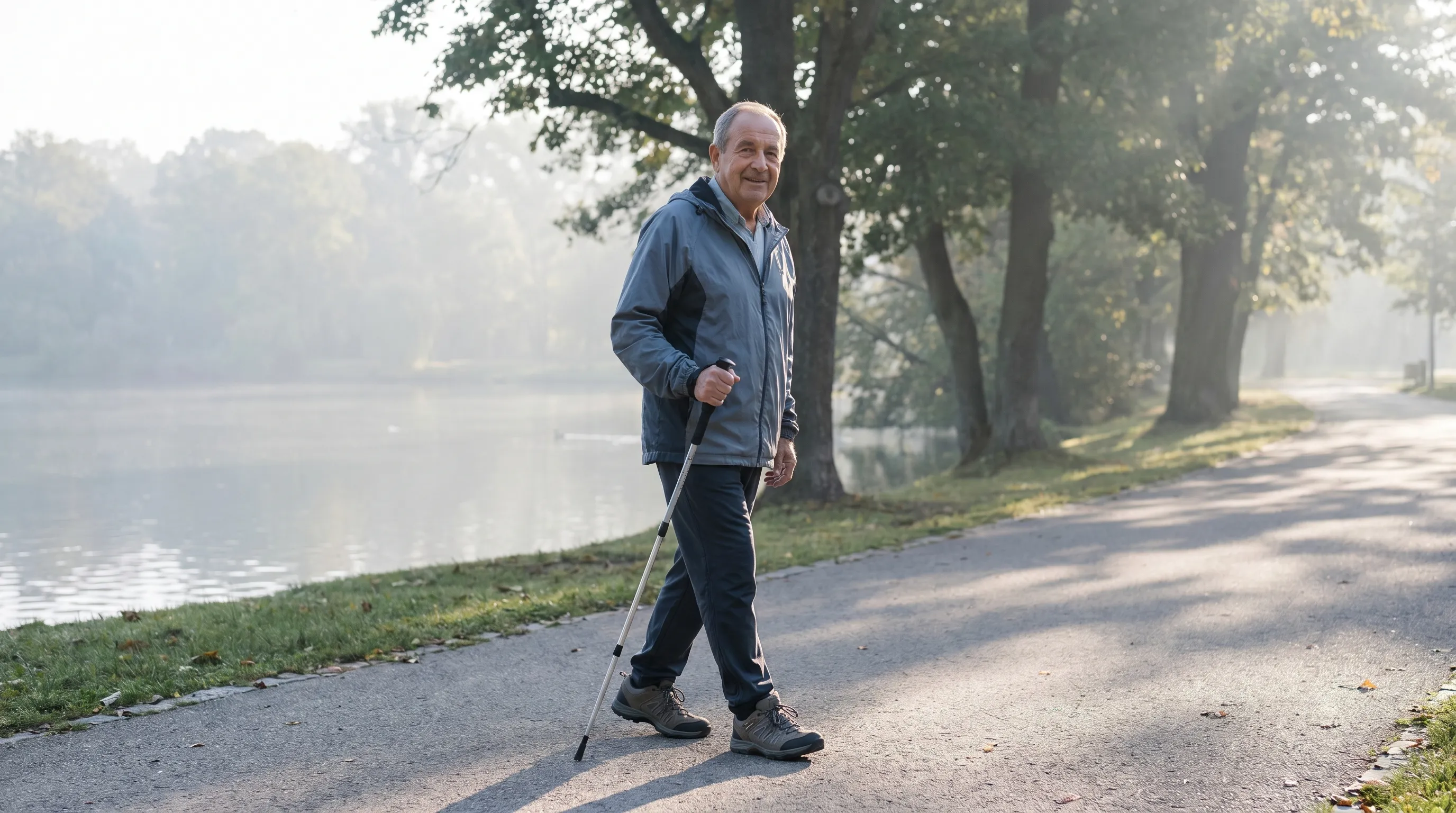
Treatment planning follows a stepwise approach prioritizing non-invasive, evidence-based interventions before considering surgical alternatives. Multimodal conservative care addresses inflammation, restores mobility, strengthens stabilizing musculature, and educates patients on sustainable joint preservation techniques.
Pharmacological Approaches and Topical Therapies
Medication management focuses on pain modulation and inflammation control while minimizing systemic side effects. Acetaminophen remains a first-line option for mild discomfort, though its efficacy varies among individuals. Nonsteroidal anti-inflammatory drugs reduce synovial swelling and provide functional improvement during acute exacerbations, but require cardiovascular and gastrointestinal risk assessment for long-term use. Topical formulations deliver concentrated analgesic effects directly to periarticular tissues with minimal systemic absorption, making them ideal for localized joint discomfort. Corticosteroid injections provide rapid relief during severe inflammatory flares but are limited to three to four administrations annually to prevent cartilage toxicity. Viscosupplementation with hyaluronic acid may offer temporary lubrication improvement in osteoarthritic knees, though clinical evidence shows mixed outcomes. Platelet-rich plasma and stem cell therapies continue to generate research interest, with emerging data suggesting potential cartilage-modulating effects, though regulatory approval and standardized protocols remain under development.
Lifestyle Modifications, Ergonomics, and Activity Pacing
Sustainable joint health requires daily habit optimization beyond clinical interventions. Weight management dramatically reduces compressive loading across both articulations. Nutritional strategies emphasizing anti-inflammatory foods, lean proteins, and adequate hydration support tissue repair and metabolic efficiency. Ergonomic workstation adjustments prevent prolonged static posturing that stiffens hip flexors and weakens gluteal activation. Activity pacing techniques distribute exercise volume throughout the day rather than concentrating stress during single prolonged sessions. Implementing the two-hour pain rule—evaluating symptom intensity two hours after activity—helps individuals identify safe thresholds and avoid boom-bust cycles. Proper sleep positioning with knee pillows between the legs or under the knees maintains neutral alignment and prevents nocturnal compression. Mind-body practices such as diaphragmatic breathing and guided relaxation reduce central sensitization and improve pain tolerance thresholds.
Targeted Exercise Protocols and Rehabilitation Frameworks
Movement is medicine when applied correctly. Rehabilitation programs prioritize progressive overload, neuromuscular control, and joint stability to restore functional capacity and prevent recurrence. Exercise selection must balance therapeutic benefit with tissue tolerance. Regular, guided physical activity is strongly recommended by public health authorities to maintain joint mobility and reduce pain severity (CDC).
Progressive Strengthening for Gluteal and Quadriceps Musculature
The foundation of lower extremity rehabilitation begins with restoring hip extensor and abductor strength. Gluteal activation drills include supine bridges with progressive single-leg progressions, side-lying clamshells emphasizing external rotation, and standing resisted abduction bands. Quadriceps re-education focuses on terminal knee extensions, isometric wall sits with proper patellar tracking, and controlled step-downs emphasizing eccentric control. Hamstring strengthening incorporates Nordic curls and Swiss ball leg curls to balance anterior-posterior thigh forces. Progression follows pain-free range of motion, proper movement patterns, and gradual resistance increases. Avoiding knee valgus collapse during lower body movements protects medial compartment structures. Consistency over intensity yields superior long-term outcomes, as neuromuscular adaptations require repetitive, high-quality repetitions to establish automatic motor patterns.
Low-Impact Conditioning and Mobility Workflows
Cardiovascular fitness maintains systemic health and supports joint nutrition through cyclic synovial fluid pumping. Stationary cycling, elliptical trainers, and aquatic therapy provide sustained movement without excessive ground reaction forces. Swimming engages the entire kinetic chain while buoyancy eliminates compressive loading, making it ideal for advanced degenerative conditions. Mobility routines target joint capsule elasticity and fascial glide. Hip flexor half-kneeling stretches restore anterior pelvic tilt, while ankle dorsiflexion mobilizations improve tibial progression during gait. Foam rolling addresses myofascial restrictions in the tensor fasciae latae, quadriceps, and gastrocnemius. Dynamic warm-ups incorporating leg swings, lateral shuffles, and controlled squats prepare tissues for higher intensity work. Integrating these components into a structured weekly schedule prevents deconditioning, maintains cartilage metabolism, and promotes lasting mobility.
Comparative Overview of Treatment Modalities
Selecting the appropriate intervention depends on symptom duration, structural severity, functional goals, and individual risk factors. The following comparison outlines conservative and surgical pathways to help patients and clinicians make informed decisions tailored to specific clinical presentations.
| Treatment Category | Indications | Expected Recovery Timeline | Risk Profile | Long-Term Efficacy |
|---|---|---|---|---|
| Physical Therapy & Strengthening | Mild to moderate degeneration, early tendinopathy, post-injury rehab | 6 to 12 weeks for initial improvement; ongoing maintenance required | Minimal; delayed onset muscle soreness common | High when adherence maintained |
| Pharmacological Management | Acute inflammatory flares, breakthrough discomfort during rehabilitation | Hours to days for symptom modulation | Gastrointestinal, renal, cardiovascular with long-term oral use | Temporary relief; does not reverse structural damage |
| Intra-Articular Injections | Moderate osteoarthritis, severe bursitis, refractory synovitis | Days to weeks depending on agent used | Infection risk, cartilage toxicity with frequent corticosteroid use | Variable; PRP shows promise, hyaluronic acid mixed evidence |
| Joint Replacement Surgery | Severe cartilage loss, intractable pain, failed conservative care | 3 to 6 months for functional independence; 12 months for full tissue maturation | Surgical risks: infection, DVT, nerve injury, implant loosening | Excellent durability; 90%+ survival rates at 15 years |
Frequently Asked Questions
Can weak hips actually cause knee pain?
Yes, proximal muscle weakness directly impacts distal joint mechanics. The gluteus medius controls femoral positioning during weight-bearing activities. When these stabilizers fatigue or become inhibited, the knee experiences abnormal rotational stress and inward collapse, accelerating patellofemoral compression and medial compartment wear. Strengthening hip musculature is a foundational element in knee rehabilitation protocols.
Is it normal to feel discomfort in both joints simultaneously?
Absolutely. The lower extremity functions as an integrated kinetic chain. Compensatory movement patterns from hip stiffness or injury transfer excessive load to the knee, while knee dysfunction forces the hip and lumbar spine to absorb impact forces. Additionally, systemic conditions like widespread osteoarthritis or inflammatory arthritis frequently affect multiple weight-bearing joints concurrently. Comprehensive evaluation ensures both regions receive appropriate attention.
What are the safest exercises for managing chronic joint discomfort?
Low-impact, controlled movements prioritize joint preservation while maintaining muscular support. Stationary cycling, swimming, and aquatic therapy provide cardiovascular benefits without excessive ground reaction forces. Within strength training, focus on isometric quadriceps sets, supine glute bridges, clamshells, and straight-leg raises. Avoid deep squats, plyometric jumping, and sudden pivoting until baseline strength and mobility improve. Always progress gradually and monitor symptom response.
When should I consider joint replacement surgery?
Surgical consultation becomes appropriate when conservative interventions no longer control pain, structural damage significantly restricts daily activities, and quality of life declines substantially. Radiographic confirmation of severe joint space narrowing, combined with persistent symptoms lasting beyond six to twelve months despite guided therapy, typically indicates advanced degeneration requiring orthopedic evaluation. Patient motivation, overall health status, and realistic expectations heavily influence surgical success.
Does footwear make a measurable difference in lower extremity pain?
Footwear profoundly influences biomechanical loading patterns. Shoes with proper arch support, adequate midsole cushioning, and stable heel counters reduce ground reaction forces transmitted upward through the kinetic chain. Individuals with excessive foot pronation or supination often benefit from custom orthotics that normalize tibial alignment and prevent femoral internal rotation. Replacing worn footwear every three hundred to five hundred miles maintains shock absorption properties and prevents compensatory strain on the hips and knees.
Conclusion
Navigating the complexities of hip and knee pain requires a shift from passive symptom suppression to active functional restoration. These joints do not operate independently; they respond as integrated components of a dynamic movement system. Understanding the biomechanical relationships, recognizing early warning signs, and implementing structured rehabilitation strategies empower individuals to take control of their joint health. Conservative management remains highly effective for the majority of patients, emphasizing targeted strengthening, activity modification, and sustainable lifestyle integration. Surgical interventions offer reliable outcomes for advanced structural damage when conservative pathways reach their limits. Consistency in exercise adherence, mindful pacing of daily activities, and proactive collaboration with healthcare professionals create the foundation for lasting mobility and comfort. By approaching hip and knee pain with evidence-based strategies and realistic expectations, you can preserve joint integrity, restore functional independence, and confidently return to the activities that bring meaning to your daily life. Commit to gradual progress, prioritize tissue recovery, and view movement as a vital component of long-term wellness.
How we work
Researched and drafted with AI assistance, then reviewed by a human editor against the cited sources.
This article is general health information, not medical advice. Consult a qualified healthcare professional about your own situation.