Understanding Broken Neck Symptoms: A Comprehensive Guide to Recognition and Recovery
The human cervical spine is a marvel of biomechanical engineering, designed to support the weight of the head, facilitate complex rotational and flexion movements, and protect the delicate spinal cord that transmits signals between the brain and the rest of the body. Despite its remarkable resilience, the cervical vertebrae remain highly vulnerable to traumatic forces. When a sudden impact, extreme deceleration, or severe mechanical overload occurs, the delicate bony architecture can fracture. Recognizing broken neck symptoms early and responding appropriately can be the difference between a full functional recovery and permanent neurological impairment. This comprehensive guide explores the clinical presentation, diagnostic pathways, treatment modalities, and evidence-based recovery strategies associated with cervical spine fractures. Whether you are seeking information for personal awareness, preparing for a medical consultation, or supporting a loved one through spinal trauma recovery, understanding the physiological and clinical realities of these injuries is essential.
Understanding Cervical Spine Fractures
Cervical Spine Anatomy and Structural Vulnerability
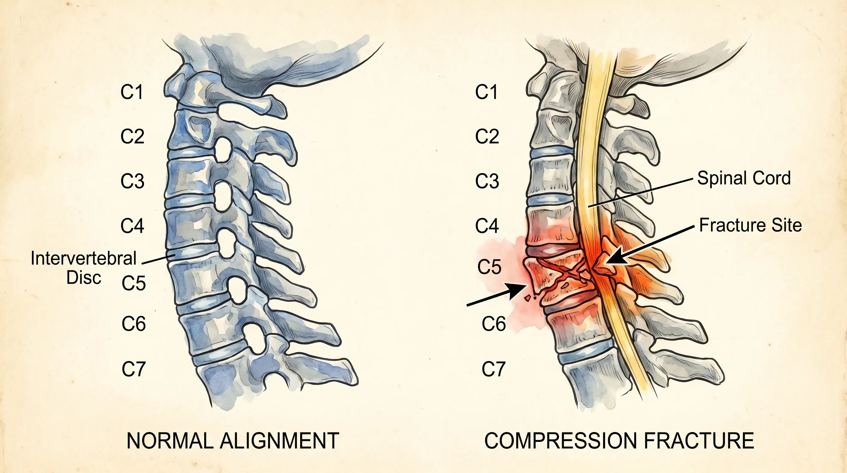
The cervical spine consists of seven vertebrae (C1 through C7), each uniquely shaped to balance mobility with structural stability. The atlas (C1) and axis (C2) form the craniovertebral junction, enabling the majority of head rotation and nodding movements. Lower cervical vertebrae (C3-C7) bear increasing mechanical loads and facilitate lateral flexion and extension. Between each vertebra lie intervertebral discs that act as shock absorbers, while a complex network of ligaments, tendons, and paraspinal muscles maintains alignment and restricts excessive motion. The spinal cord, encased within the vertebral foramen, is surrounded by cerebrospinal fluid and protected by the meninges. When a fracture occurs, the structural integrity of this system is compromised. Depending on the mechanism of injury, fractures may involve the vertebral body, pedicles, laminae, transverse processes, or odontoid process. The severity of tissue damage directly correlates with the risk of neurological compromise, making early recognition of clinical indicators absolutely critical. For more details on cervical spine anatomy and function, clinical resources provide comprehensive structural breakdowns.
Free calculatorBMI CalculatorCalculate Body Mass IndexOpen the calculatorCommon Mechanisms of Cervical Trauma
Cervical fractures typically result from high-energy trauma or compromised bone integrity. Motor vehicle collisions, particularly rear-end impacts causing hyperextension or head-on crashes inducing hyperflexion, remain the leading cause of acute cervical spine injuries. Falls from elevated positions, sports-related impacts, diving accidents into shallow water, and direct blows to the head or neck also frequently precipitate vertebral fractures. In older adults, osteoporosis and degenerative disc disease significantly lower the threshold for injury, meaning even minor falls or sudden twisting motions can result in compression fractures. NIH research on cervical spine trauma consistently highlights that approximately ten to fifteen percent of cervical spine injuries involve multiple vertebral levels, increasing complexity and treatment urgency. Understanding the mechanism behind the trauma helps clinicians anticipate fracture patterns and associated soft tissue damage, but for patients and caregivers, recognizing broken neck symptoms remains the most immediate priority regardless of how the injury occurred.
Recognizing Broken Neck Symptoms: A Critical Complete Guide
Immediate and High-Risk Warning Signs
Following acute cervical trauma, broken neck symptoms often manifest rapidly. The most prominent indicator is severe, localized neck pain that intensifies with any movement or attempted weight-bearing. Unlike muscular strains that may improve with rest or gentle stretching, fracture-related pain typically remains unrelenting and may be accompanied by profound mechanical instability, described by patients as a feeling of "giving way" or structural collapse in the neck region. Neurological indicators represent the most urgent red flags. These include radiating pain, paresthesia (tingling or burning sensations), numbness, or progressive weakness extending into the shoulders, arms, forearms, or fingers. When the spinal cord itself sustains compression or contusion, patients may develop myelopathic symptoms such as gait instability, loss of fine motor coordination, hyperreflexia, or sudden bowel and bladder dysfunction. In high cervical fractures (C1-C2), compromised phrenic nerve function can impair diaphragmatic movement, leading to shallow breathing, respiratory distress, or complete respiratory failure requiring immediate intubation and ventilatory support.
Delayed, Subtle, or Atypical Presentations
Not all cervical fractures present dramatically. Stable compression fractures, minor avulsion injuries, or isolated transverse process fractures may initially masquerade as severe whiplash or musculoskeletal strain. Patients might report intermittent stiffness, dull aching that fluctuates with posture changes, or mild headaches originating from the suboccipital region. Delayed onset broken neck symptoms frequently emerge hours or days after the initial trauma as localized inflammation increases, hematoma formation expands, or minor displacement occurs with routine activities. Older adults and individuals with neuropathic conditions like diabetes may experience blunted pain perception, masking fracture severity entirely. Medical professionals emphasize that any unexplained neck discomfort following significant trauma warrants diagnostic evaluation, regardless of symptom intensity. Assuming a minor strain without ruling out bony injury significantly increases the risk of secondary neurological damage through improper movement or delayed intervention.
Symptom Differentiation and Clinical Overlap
Distinguishing cervical fractures from other spinal or neurological conditions requires careful clinical correlation. Cervical disc herniations often produce identical radicular pain patterns and sensory deficits, but typically lack the profound structural instability seen in acute fractures. Ligamentous sprains and facet joint dysfunction cause localized pain and restricted range of motion but generally preserve neurological function and bony continuity on imaging. Occipital neuralgia, tension-type headaches, and temporomandibular joint disorders can refer pain to the cervical region, creating diagnostic confusion. Clinicians rely on standardized decision-making frameworks like the Canadian C-Spine Rule and NEXUS criteria to determine when radiographic evaluation is medically necessary. These protocols assess midline cervical tenderness, focal neurological deficits, altered mental status, intoxication, distracting injuries, and dangerous trauma mechanisms. When applied correctly, they safely identify patients requiring imaging while minimizing unnecessary radiation exposure. Understanding these clinical distinctions empowers patients to advocate appropriately for comprehensive evaluation when broken neck symptoms persist or worsen.
The Diagnostic Process: How Medical Professionals Assess Cervical Injuries
Physical Examination and Neurological Screening
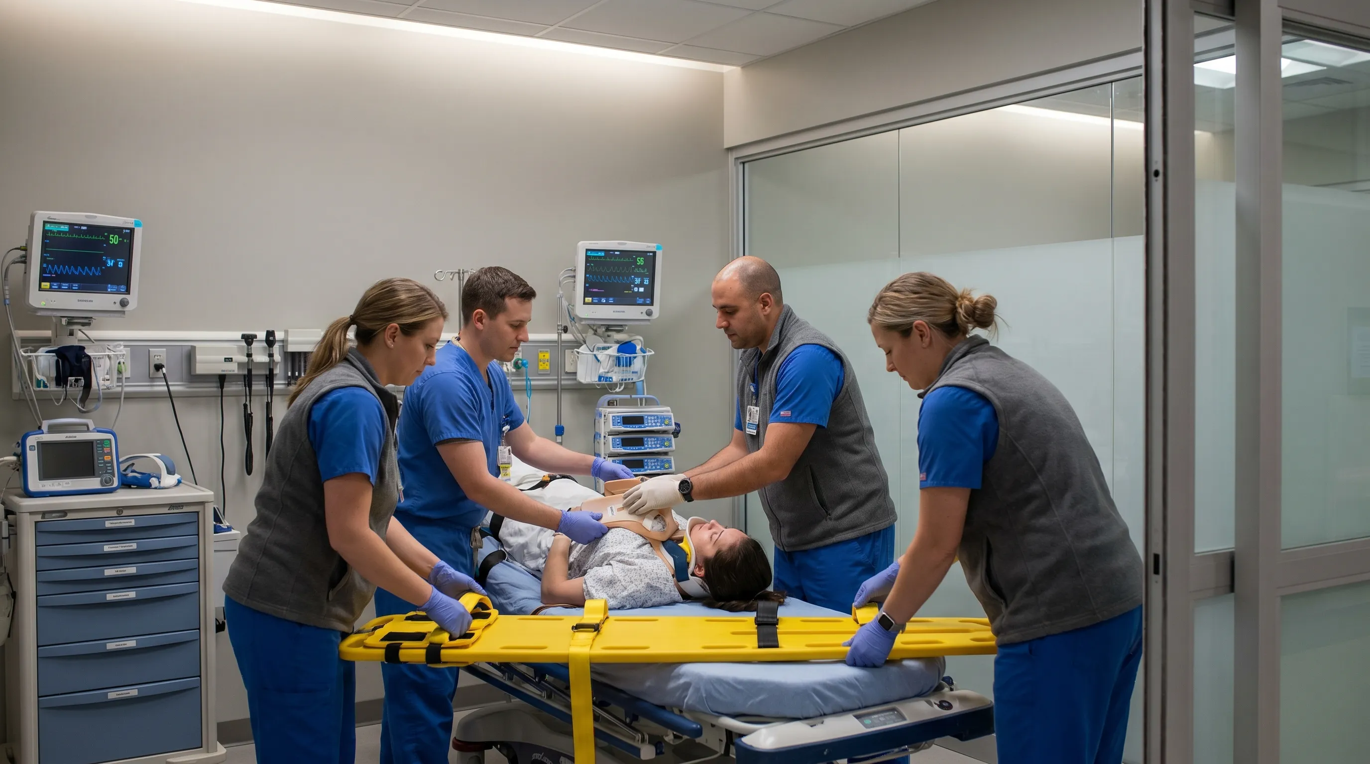
Upon presentation to emergency or urgent care settings, clinicians initiate a structured trauma assessment focusing on airway, breathing, and circulation before addressing the cervical spine. Strict spinal immobilization is maintained using rigid collars and vacuum mattresses until bony injury is definitively excluded. Physical examination begins with careful palpation of posterior bony prominences, paraspinal musculature, and anterior vertebral bodies to identify focal tenderness or step-offs indicating displacement. Motor strength, sensory perception, and deep tendon reflexes are systematically documented using the ASIA Impairment Scale for neurological assessment. Special attention is given to dermatomal sensory mapping and myotome testing to pinpoint nerve root involvement. Vascular assessment includes bilateral carotid and vertebral artery palpation to screen for dissection, which can accompany cervical trauma and precipitate posterior circulation stroke. A thorough neurological baseline establishes a reference point for monitoring clinical deterioration or improvement throughout the treatment continuum.
Advanced Imaging Modalities and Diagnostic Accuracy
Radiographic evaluation remains the cornerstone of cervical fracture diagnosis. While traditional cervical spine radiographs provide initial screening, their sensitivity for detecting subtle fractures ranges between eighty and eighty-five percent. Computed tomography has emerged as the diagnostic gold standard, offering multiplanar reconstructions that visualize complex fracture morphology, comminution, and spinal canal compromise with exceptional precision. CT protocols typically employ slice thicknesses of one millimeter or less, enabling three-dimensional surgical planning when intervention becomes necessary. Magnetic resonance imaging complements CT by visualizing ligamentous disruption, intervertebral disc herniation, spinal cord edema, hemorrhage, or contusion. MRI sequences including T1, T2, and STIR provide critical information regarding soft tissue integrity that directly influences surgical decision-making. Dynamic flexion-extension radiographs may be utilized cautiously in alert, cooperative patients to assess ligamentous stability, but are contraindicated in acute trauma due to the risk of neurological deterioration. Advanced CT and MRI diagnostic protocols interpretation requires specialized radiological expertise, as subtle alignment variations, prevertebral soft tissue swelling, or occult fracture lines can easily be overlooked without meticulous analysis.
Treatment Pathways and Intervention Strategies
Conservative Management and External Immobilization
Stable cervical fractures without neurological compromise, spinal canal stenosis, or ligamentous disruption are frequently managed conservatively. The primary objective is rigid external immobilization to facilitate osseous union while preventing secondary displacement. Rigid cervical orthoses including the Philadelphia, Miami J, or Aspen collars restrict flexion, extension, and lateral rotation through strategically placed mandibular and occipital supports. Halo-vest immobilization provides the highest degree of restriction and is typically reserved for upper cervical fractures involving the atlas or odontoid process that require strict rotational control. Conservative treatment mandates strict compliance with immobilization protocols, typically spanning six to twelve weeks depending on fracture location, patient age, and healing progression. Adjunctive pharmacological management includes acetaminophen, nonsteroidal anti-inflammatory drugs (NSAIDs), and occasionally short-course opioids for breakthrough pain, though prolonged opioid use is discouraged due to side effects and bone healing inhibition. Regular clinical and radiographic follow-up ensures appropriate alignment maintenance and timely identification of nonunion or hardware failure. Patient education regarding proper collar hygiene, skin breakdown prevention, and activity modification remains essential throughout conservative management phases.
Surgical Interventions and Structural Reconstruction
When fractures demonstrate instability, neurological compromise, significant displacement, or failure to respond to conservative measures, surgical stabilization becomes necessary. Modern cervical spine surgery employs anterior, posterior, or circumferential approaches depending on fracture pattern and anatomical location. Anterior cervical discectomy and fusion (ACDF) addresses fractures involving the vertebral body or disc space by removing compromised tissue, decompressing neural elements, and reconstructing anterior column stability with structural grafts or titanium cages secured by locking plates. Posterior cervical fusion utilizes lateral mass or pedicle screws connected to contoured rods to restore posterior tension band integrity, particularly effective for facet dislocations, lamina fractures, or ligamentous instability. Circumferential procedures combine both approaches when three-column disruption occurs, maximizing biomechanical stability across all spinal planes. Intraoperative neuromonitoring, including somatosensory and motor-evoked potentials, provides real-time neurological feedback to minimize iatrogenic injury. Surgical recovery protocols emphasize early mobilization within prescribed parameters, progressive physical therapy, and vigilant wound care to optimize fusion rates and minimize complications.
Pain Management and Supportive Care
Multimodal pain management forms the foundation of cervical fracture recovery, addressing both nociceptive and neuropathic pain pathways. Initial acute management emphasizes scheduled acetaminophen and NSAIDs to reduce prostaglandin-mediated inflammation and central sensitization. Neuropathic pain arising from nerve root compression or spinal cord irritation often responds to gabapentinoids, tricyclic antidepressants, or serotonin-norepinephrine reuptake inhibitors. Muscle relaxants like cyclobenzaprine or tizanidine alleviate paraspinal spasm but require careful dosing to prevent excessive sedation that could impair respiratory function or increase fall risk. Non-pharmacological interventions including transcutaneous electrical nerve stimulation (TENS), cryotherapy, gentle myofascial release techniques (once fracture stability is confirmed), and mindfulness-based stress reduction significantly enhance pain tolerance and functional recovery. Sleep hygiene optimization remains critical, as cervical fractures frequently disrupt restorative sleep due to positioning discomfort. Patients benefit from specialized cervical pillows, elevated bed head positioning, and structured relaxation protocols that promote tissue repair and psychological resilience during extended recovery periods.
Recovery, Rehabilitation, and Long-Term Care
Structured Physical and Occupational Therapy
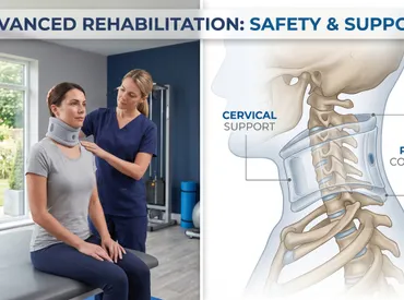
Once radiographic evidence confirms adequate fracture consolidation and clinical clearance is obtained, structured rehabilitation commences. Physical therapy protocols progress systematically from passive range-of-motion exercises to active strengthening, proprioceptive training, and functional movement reintegration. Initial phases focus on restoring cervical flexibility, reducing residual muscular tension, and reestablishing normal scapular mechanics. Progressive resistance training targets deep cervical flexors, extensors, and lateral stabilizers to compensate for prolonged immobilization-induced atrophy. Occupational therapy addresses activities of daily living that may have become compromised, emphasizing ergonomic body mechanics, proper lifting techniques, and adaptive equipment utilization when necessary. Neuromuscular reeducation exercises improve postural control, while vestibular rehabilitation addresses dizziness or balance deficits that frequently accompany upper cervical injuries or prolonged collar wear. Consistent home exercise compliance, guided by certified therapists, significantly reduces chronic pain development and accelerates return to occupational and recreational activities.
Lifestyle Modifications and Long-Term Monitoring
Long-term outcomes following cervical fractures depend heavily on sustained lifestyle modifications and vigilant monitoring. Weight management reduces mechanical stress on healing vertebrae and adjacent spinal segments. CDC guidelines on smoking and bone health confirm that smoking cessation is mandatory, as nicotine and tobacco byproducts significantly impair osteoblast function, delay bone union, and increase pseudarthrosis risk by up to four hundred percent. Nutritional optimization emphasizing calcium, vitamin D3, vitamin K2, magnesium, and high-quality protein supports ongoing skeletal remodeling. Regular follow-up imaging at three, six, and twelve months post-injury monitors fusion progression and screens for late complications including hardware migration, adjacent segment disease, or degenerative changes. Patients are educated to recognize warning signs requiring immediate reevaluation, including recurrent neurological symptoms, mechanical instability sensations, or sudden pain escalation. Psychological support remains integral, as cervical trauma frequently triggers anxiety, depression, or post-traumatic stress responses that can impede rehabilitation adherence. Multidisciplinary care coordination involving orthopedic specialists, neurologists, pain management physicians, and mental health professionals ensures comprehensive recovery addressing physical, emotional, and functional domains simultaneously.
Prevention and Safety Protocols
Ergonomic Practices and Daily Movement Optimization
Preventing cervical spine injuries begins with optimizing daily mechanical environments. Workplace ergonomics should prioritize monitor positioning at eye level, chair lumbar support, and neutral neck alignment to prevent chronic forward head posture that progressively weakens posterior musculature and increases disc pressure. Frequent micro-breaks every thirty to forty-five minutes interrupt sustained static loading and promote circulation to cervical soft tissues. Proper lifting mechanics utilizing lower extremity power, maintaining objects close to the body, and avoiding twisting under load significantly reduce sudden cervical torque. Sleep posture optimization using supportive mattresses and contour cervical pillows maintains neutral alignment throughout the night, preventing morning stiffness and cumulative strain. Regular strength training targeting the upper back, shoulders, and core musculature creates a protective muscular corset that absorbs mechanical forces before they reach the vertebral column. Consistent flexibility routines incorporating dynamic stretching, yoga, or Pilates improve tissue resilience and joint mobility, creating a more adaptable and injury-resistant cervical spine.
High-Risk Activity Precautions and Protective Equipment
Engagement in contact sports, motorcycling, skiing, gymnastics, and extreme recreational activities demands comprehensive safety protocols. Helmets certified by relevant safety organizations must be properly fitted and replaced after any significant impact, as compromised foam liners lose shock-absorbing capacity. Neck strengthening and conditioning programs specifically designed for sport participation improve muscular buffering capacity and proprioceptive awareness, enabling athletes to recognize and avoid dangerous positioning. Diving activities should strictly avoid shallow water and unknown depths, while martial artists must prioritize proper break-falling techniques and cervical bracing during training. Vehicle safety relies on proper seatbelt utilization, appropriately positioned head restraints, and adherence to traffic regulations to minimize collision forces. For individuals with known osteopenia or osteoporosis, bone density optimization through pharmacological intervention, nutritional supplementation, and weight-bearing exercise reduces fragility fracture risk dramatically. CDC fall prevention protocols and home hazard modification, appropriate footwear, balance training, and medication review further protect vulnerable populations from traumatic cervical injury. Global health organizations like the WHO also emphasize comprehensive injury prevention frameworks to reduce trauma incidence across all demographics.
| Symptom Category | Typical Presentation | Associated Fracture Type | Urgency Level |
|---|---|---|---|
| Localized Pain & Tenderness | Severe pain at midline, worsens with movement, no neurological deficits | Stable compression, transverse process fracture | Moderate (Urgent evaluation within 24 hours) |
| Radicular Pain & Paresthesia | Shooting pain, tingling, or numbness radiating to arms/hands | Facet joint fracture, disc herniation with nerve impingement | High (Same-day imaging required) |
| Motor Weakness & Coordination Loss | Difficulty gripping, foot drop, gait instability, clumsiness | Spinal cord compression, multi-column instability | Critical (Immediate emergency activation) |
| Bowel/Bladder Dysfunction | Urinary retention, incontinence, loss of rectal tone | Severe cord contusion, high cervical fracture | Critical (Immediate life-threatening emergency) |
| Respiratory Compromise | Shallow breathing, diaphragmatic weakness, oxygen desaturation | C1-C3 fracture with phrenic nerve involvement | Critical (Immediate airway management required) |
Frequently Asked Questions
What are the immediate broken neck symptoms to watch for after trauma?
Immediate symptoms following cervical spine trauma typically include severe localized neck pain, sudden loss of mobility, tingling or numbness radiating into the shoulders, arms, or hands, and muscle spasms. In more severe cases involving spinal cord compromise, individuals may experience breathing difficulties, loss of bowel or bladder control, or sudden weakness in the limbs. Any of these indicators require immediate emergency medical evaluation to prevent permanent neurological damage.
Can you walk around with a fractured cervical vertebra without realizing it?
Yes, it is possible in certain stable fracture patterns, particularly with minor compression or avulsion fractures where the spinal canal remains intact. Some individuals may experience only mild stiffness, intermittent pain, or a dull ache that they mistake for a severe muscle strain or whiplash. However, walking or moving without proper immobilization significantly increases the risk of fracture displacement, which can lead to spinal cord injury or paralysis. Medical imaging is always necessary after significant neck trauma, regardless of perceived symptom severity.
How long does recovery typically take for a broken neck?
Recovery timelines vary dramatically based on fracture type, stability, treatment approach, and individual health factors. Stable fractures managed conservatively with bracing typically require six to twelve weeks of strict immobilization, followed by three to six months of physical therapy for full functional restoration. Surgical stabilization often extends the initial healing phase to three to four months, with comprehensive rehabilitation lasting up to one year. Factors such as age, bone density, smoking status, and comorbid conditions like diabetes can significantly influence bone union and overall recovery speed.
What imaging tests are most accurate for diagnosing cervical spine fractures?
Computed tomography (CT) scans are currently the gold standard for detecting cervical spine fractures due to their exceptional resolution of bony structures and ability to identify subtle cortical disruptions or complex fracture patterns. Magnetic resonance imaging (MRI) is essential when ligamentous injury, disc herniation, or spinal cord edema is suspected, as it visualizes soft tissue and neurological compromise that CT cannot detect. Plain X-rays are often used for initial screening but may miss up to fifteen percent of fractures, particularly in obese patients or those with overlapping anatomical structures. Clinical decision rules like the Canadian C-Spine Rule help determine when advanced imaging is medically necessary.
Is surgery always required to treat a fractured cervical spine?
No, surgery is not universally required. Treatment decisions are guided by fracture stability, spinal alignment, neurological status, and ligamentous integrity. Stable compression fractures, isolated facet fractures without dislocation, and certain odontoid fractures often heal successfully with external immobilization using rigid cervical orthoses like a Miami J or Aspen collar. Surgery becomes necessary when fractures demonstrate instability, neurological deficits, significant spinal canal compromise, or failure to heal with conservative management. Common surgical procedures include anterior cervical discectomy and fusion (ACDF), posterior spinal fusion, or internal fixation with plates and screws to restore structural integrity and protect neural elements.
Conclusion
Understanding broken neck symptoms and responding with appropriate urgency fundamentally shapes clinical outcomes and long-term quality of life. Cervical spine fractures represent a complex intersection of biomechanical vulnerability, neurological risk, and therapeutic challenge. Early recognition of warning signs, strict adherence to immobilization protocols, timely diagnostic imaging, and individualized treatment planning form the foundation of optimal recovery. Whether managed conservatively through rigid bracing or addressed surgically with modern stabilization techniques, the pathway to healing requires patience, compliance, and multidisciplinary support. Rehabilitation extends beyond structural bone union, encompassing functional restoration, pain management optimization, psychological resilience building, and sustained lifestyle modification. By prioritizing prevention strategies, embracing evidence-based treatment protocols, and maintaining open communication with qualified healthcare providers, patients can navigate the recovery process safely and confidently. Spinal trauma is undoubtedly life-altering, but with informed decision-making and dedicated rehabilitation, individuals frequently regain meaningful function and return to active, fulfilling lives. Always consult licensed medical professionals for personalized diagnosis and treatment planning tailored to your specific clinical presentation.

How we work
Researched and drafted with AI assistance, then reviewed by a human editor against the cited sources.
This article is general health information, not medical advice. Consult a qualified healthcare professional about your own situation.