Understanding a Knot in Arch of Foot: Causes, Relief, and Treatment
Key points
- Relative Rest: Temporarily reducing or modifying activities that provoke sharp pain, swapping running for cycling or swimming.
- Cryotherapy: Applying ice wrapped in a thin towel to the painful area for 15 to 20 minutes, 3 to 4 times daily, particularly after activity.
- Calf and Plantar Fascia Stretching: Gentle, sustained stretches performed multiple times daily to gradually restore tissue length.
- Supportive Footwear: Avoiding flat, unsupportive shoes and utilizing footwear with a firm midsole, adequate arch contour, and heel cushioning.
- Weight Management: Even modest weight reduction significantly decreases cumulative plantar loading forces.
That sharp, persistent ache beneath your foot that makes your morning steps feel like walking on glass is more common than you might think. For millions of people, the discomfort is often described as a knot in arch of foot—a tight, stubborn sensation that can make standing, walking, or even resting deeply uncomfortable. Whether it feels like a tight band pulling from heel to toe, a palpable rubbery bump, or a deep muscle spasm that refuses to release, this symptom is a clear signal that the intricate biomechanical system of your foot is under strain. The arch of your foot is a marvel of natural engineering, designed to absorb impact, distribute weight, and propel you forward. When its supporting structures are overloaded, injured, or structurally compromised, your body responds with protective tension, inflammation, or tissue remodeling. Understanding exactly what is happening beneath the skin is the critical first step toward lasting relief and restored mobility, as outlined by orthopedic specialists at the Mayo Clinic. In this comprehensive guide, we will break down the medical realities behind that tight knot sensation, differentiate between the most common underlying conditions, and provide evidence-based, step-by-step strategies to heal, prevent recurrence, and return to pain-free movement.
Understanding the Biomechanics of the Foot Arch
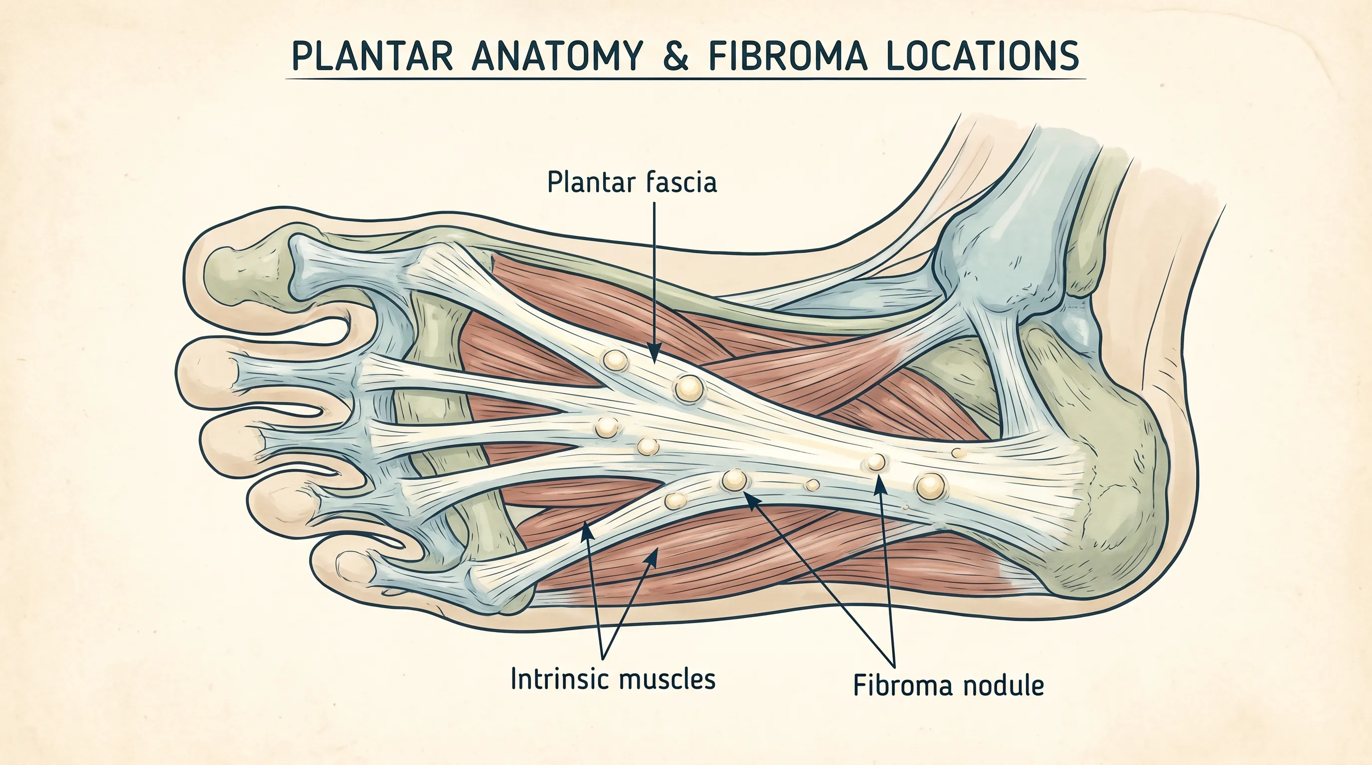
To effectively treat a knot in arch of foot, you must first understand the anatomical structures that make up the medial longitudinal arch. The arch is not a single bone but a complex suspension bridge composed of twenty-six bones, thirty-three joints, and over a hundred muscles, tendons, ligaments, and fascial bands. The primary load-bearing structure is the plantar fascia, a thick, fibrous sheet that originates at the calcaneus (heel bone) and fans out to attach to the metatarsal heads near the base of the toes. Its primary functions are shock absorption during heel strike and maintaining the structural integrity of the arch during the push-off phase of walking.
Free calculatorFoot Reflexology ChartFoot reflexology points and corresponding organsOpen the calculatorSupporting the plantar fascia are the intrinsic foot muscles, which act as dynamic stabilizers. These include the abductor hallucis along the inner border, the flexor digitorum brevis in the midline, and the quadratus plantae, which helps align the toe flexors. When these structures work in harmony, the arch acts like a spring. However, repetitive microtrauma, poor biomechanics, sudden increases in activity, or structural imbalances can disrupt this harmony. The body responds by tightening the fascia to protect vulnerable areas, forming localized inflammatory pockets, or developing fibrous nodules as a reactive tissue response. Recognizing which tissue layer is compromised allows for targeted, effective intervention rather than generic guesswork, according to anatomical guidelines from the Cleveland Clinic.
Condition 1: Plantar Fasciitis – The Tight Band That Mimics a Knot
What Is the Plantar Fascia and How Does It Inflame?
Plantar fasciitis is widely recognized as one of the most common orthopedic complaints, with approximately 2 million patients seeking treatment annually, according to the American Academy of Orthopaedic Surgeons (AAOS). Despite its name ending in "-itis" (implying pure inflammation), modern research indicates that the condition involves a degenerative process known as fasciosis, characterized by collagen breakdown, micro-tears, and disorganized healing. When the plantar fascia is subjected to excessive tensile forces, microscopic tears develop near the calcaneal insertion. The body attempts to repair these tears, but repetitive strain prevents proper healing, resulting in thickening, tightness, and a band-like sensation that many patients interpret as a knot in arch of foot.
Clinical Presentation and Diagnostic Markers
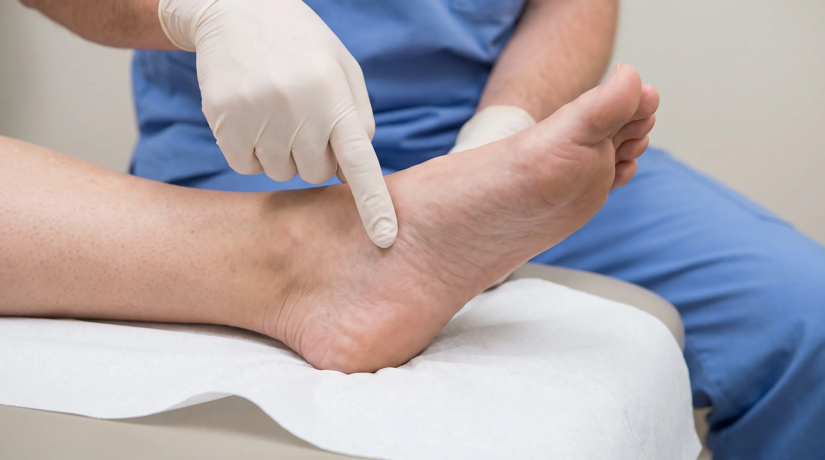
The hallmark symptom of plantar fasciitis is sharp, stabbing pain near the medial heel, typically worst during the first few steps after waking or following prolonged rest. This morning stiffness occurs because the fascia contracts and tightens overnight. As you walk, the tissue stretches and warms up, temporarily reducing pain, but it often returns after prolonged standing, descending stairs, or completing high-impact exercise. Patients frequently report a palpable tightness along the medial border of the arch, though there is usually no discrete, movable lump. Diagnosis is primarily clinical, based on history and physical examination, with point tenderness over the anterior medial calcaneal tubercle being highly indicative (Mayo Clinic).
Biomechanical and Lifestyle Risk Factors
Several factors dramatically increase susceptibility to this condition. Age is a primary variable, with peak incidence occurring between 40 and 60 years as tissue elasticity naturally declines. High-impact athletes, particularly runners and dancers, place repetitive loading cycles on the fascia. Occupations requiring prolonged standing on hard surfaces—such as nursing, manufacturing, and education—also elevate risk. Structural factors like pes planus (flat feet) or pes cavus (high arches) alter load distribution, while tight Achilles tendons or gastrocnemius-soleus complexes limit ankle dorsiflexion, transferring excessive force to the plantar fascia. Additionally, higher body mass index (BMI) increases ground reaction forces, compounding stress on the arch.
Evidence-Based Conservative Treatment Pathways
The good news is that prognosis is highly favorable with consistent conservative management. More than 90% of patients experience significant improvement within 10 months of initiating structured self-care and rehabilitation protocols. The foundation of treatment centers on load management, tissue mobility restoration, and biomechanical correction (Cleveland Clinic).
A structured approach includes:
- Relative Rest: Temporarily reducing or modifying activities that provoke sharp pain, swapping running for cycling or swimming.
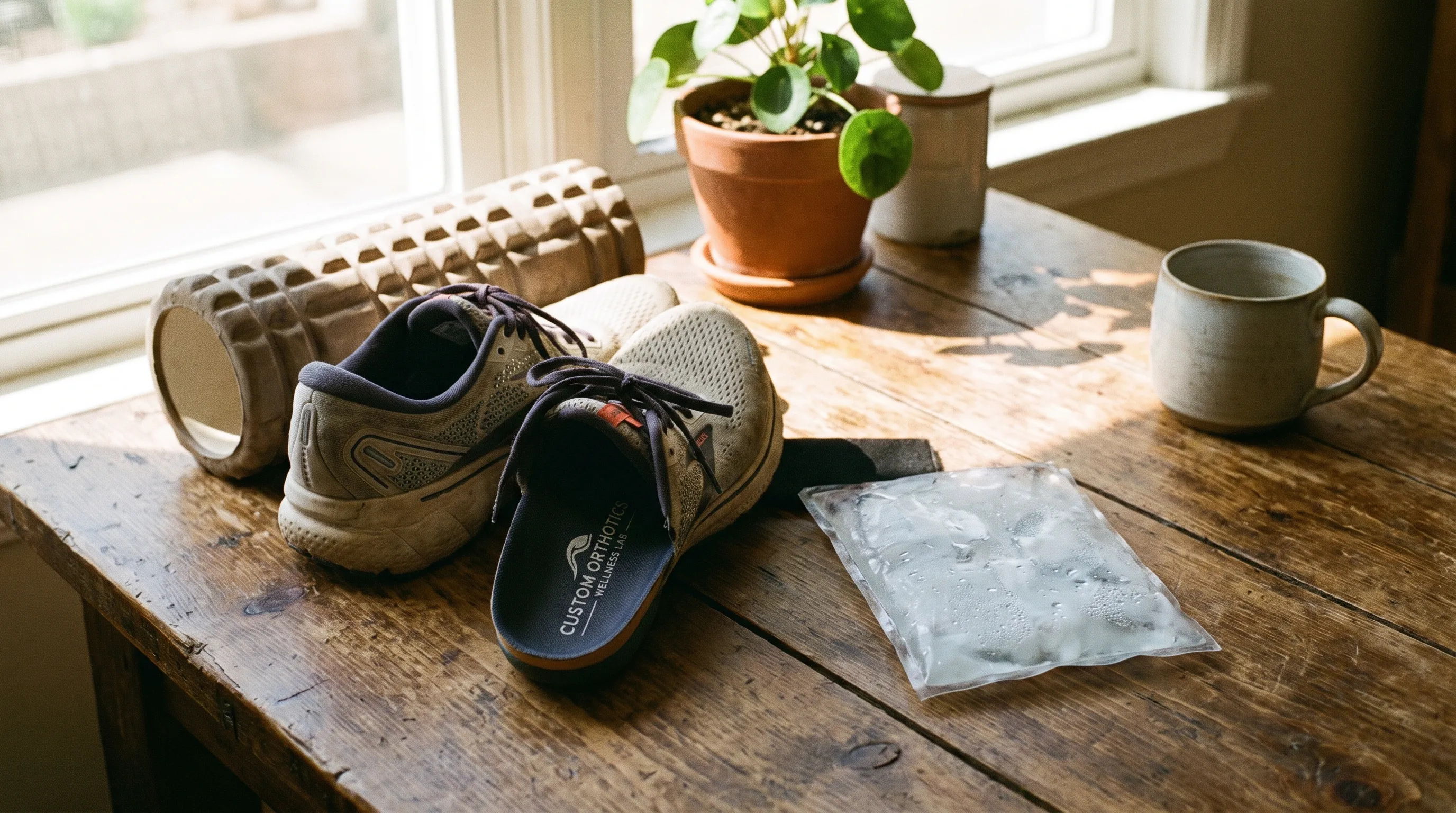
- Cryotherapy: Applying ice wrapped in a thin towel to the painful area for 15 to 20 minutes, 3 to 4 times daily, particularly after activity.
- Calf and Plantar Fascia Stretching: Gentle, sustained stretches performed multiple times daily to gradually restore tissue length.
- Supportive Footwear: Avoiding flat, unsupportive shoes and utilizing footwear with a firm midsole, adequate arch contour, and heel cushioning.
- Weight Management: Even modest weight reduction significantly decreases cumulative plantar loading forces.
Condition 2: Plantar Fibroma – A True Physical Knot
Defining the Nodule and Pathophysiology
Unlike plantar fasciitis, a plantar fibroma represents a literal, palpable mass. It is a benign, non-cancerous nodular growth that develops within the deep layer of the plantar fascia. Typically measuring less than one inch in diameter, these nodules most commonly appear in the central or medial portion of the arch. The tissue is composed of dense collagen fibers and fibroblasts, creating a firm, rubbery consistency that does not fluctuate with inflammation cycles. When multiple nodules develop, the condition progresses to plantar fibromatosis, also known as Ledderhose disease (National Institutes of Health).
Epidemiology and Genetic Associations
Research indicates a distinct demographic pattern for this condition. It predominantly affects young to middle-aged adults and shows a higher prevalence in individuals assigned male at birth. There is a well-documented genetic predisposition, particularly among those of Northern European descent. While isolated fibromas occur sporadically, multifibromatosis is frequently associated with systemic conditions such as liver disease, chronic alcohol use, diabetes mellitus, and epilepsy. Interestingly, the American Orthopaedic Foot & Ankle Society notes that direct trauma does not appear to play a primary role in solitary fibroma development, though repetitive mechanical stress may contribute to symptomatic presentation.
Symptom Progression and Clinical Monitoring
In early stages, a plantar fibroma is often asymptomatic and discovered incidentally during self-examination or a routine foot check. As the nodule slowly enlarges over months or years, it may begin to cause discomfort during weight-bearing activities, particularly when wearing minimal or poorly cushioned shoes. The mass presses against the plantar surface, creating localized pain that intensifies with direct pressure, such as walking barefoot on firm floors or stepping onto uneven terrain. Unlike a muscle knot, it does not release with massage or heat, and its firm boundaries remain distinct during palpation.
Medical Management Strategies
Treatment philosophy for plantar fibroma centers on observation and symptom control. Most clinicians recommend a watch-and-wait approach for asymptomatic or minimally symptomatic nodules, emphasizing footwear modification with custom orthotics that offload the specific pressure point. Corticosteroid injections may temporarily reduce surrounding inflammation and shrink reactive tissue, though they do not eliminate the core nodule. Physical therapy focuses on maintaining ankle and foot mobility while avoiding aggressive cross-fiber friction directly over the mass, which could theoretically stimulate fibroblast activity and encourage further growth. Surgical excision remains highly controversial due to significant risks of recurrence, wound healing complications, and potential alteration of the foot's structural integrity.
Condition 3: Myofascial Trigger Points in Intrinsic Foot Muscles
Anatomy of the Arch's Stabilizing Muscles
Beneath the plantar fascia lies a network of small intrinsic muscles responsible for fine motor control and dynamic arch support. The abductor hallucis moves the big toe, the flexor digitorum brevis curls the lesser toes, and the quadratus plantae stabilizes tendon tracking. These muscles are highly active during barefoot walking, uneven terrain navigation, and balance challenges. When fatigued, biomechanically overloaded, or deprived of proper recovery, they develop localized hyperirritable spots known as myofascial trigger points.
How Muscle Knots Form and Refer Pain
A trigger point is a discrete, hypercontractile sarcomere cluster within a muscle fiber band. It forms when repetitive micro-stress depletes local ATP stores, preventing calcium reuptake and locking actin-myosin cross-bridges in a contracted state. This creates a palpable, tender knot in arch of foot that may refer pain upward along the inner ankle or downward into the toes. Unlike fibrous nodules, trigger points exhibit a local twitch response when snapped and temporarily relieve tension when compressed and released. Factors contributing to their development include prolonged static postures, inadequate footwear support, muscle fatigue from sudden activity spikes, dehydration, and nutritional deficiencies affecting muscle recovery (Cleveland Clinic).
Targeted Myofascial Release Techniques
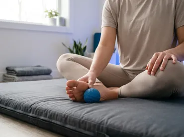
Addressing intrinsic muscle trigger points requires a different approach than treating plantar fascia degeneration. Self-myofascial release using a firm massage ball or specialized foot roller can effectively deactivate these knots. The protocol involves slowly rolling the arch while identifying localized tender spots, then holding steady pressure for 60 to 90 seconds without aggressive grinding. This ischemic compression followed by gradual release restores sarcomere length, improves local circulation, and reduces pain signaling. Combining this with toe yoga exercises, towel scrunches for strengthening, and progressive balance training ensures the intrinsic muscles regain functional endurance, reducing future knot formation.
How to Differentiate Between the Three Conditions
Accurate self-assessment can guide initial management, but a clinical diagnosis remains essential for persistent symptoms. Below is a comparative framework to help distinguish the underlying cause of your arch discomfort:
| Feature | Plantar Fasciitis | Plantar Fibroma | Myofascial Trigger Point |
|---|---|---|---|
| Primary Sensation | Tight, band-like tension; sharp morning pain | Firm, discrete, rubbery bump; pressure pain | Localized muscle knot; dull ache with tightness |
| Palpation Findings | Diffuse thickening, tender along medial band | Distinct, non-mobile nodule <1 inch | Ropy band, reproduces referred pain when pressed |
| Response to Stretch | Stiff initially, then eases | No change in nodule size; may irritate if pressed | Tension temporarily reduces with sustained pressure |
| Typical Onset | Gradual, activity-related or after rest | Slow-growing over months to years | Sudden or post-activity fatigue, poor footwear |
| Imaging Utility | Usually clinical; ultrasound shows thickening >4mm | Ultrasound/MRI shows solid hypoechoic nodule | Not visible on standard imaging; clinical diagnosis |
If you cannot confidently differentiate the cause, a podiatrist or sports medicine physician will utilize diagnostic ultrasound or MRI, perform gait analysis, and conduct palpation tests to confirm the tissue layer involved (American Academy of Orthopaedic Surgeons).
Comprehensive Self-Care & Home Treatment Protocols
Footwear Selection and Orthotic Integration
Proper footwear is the cornerstone of arch recovery. Look for shoes with a deep heel cup to stabilize the calcaneus, a semi-rigid shank to prevent excessive midfoot bending, and adequate toe box width to allow natural toe splay. Replace worn athletic shoes every 300 to 500 miles, as midsole compression reduces shock absorption. For daily wear, consider prefabricated arch supports with customizable heel wedges to correct mild pronation or supination imbalances. Avoid walking barefoot on hard floors, even at home, and use supportive slippers or recovery sandals.
Cryotherapy, Contrast Baths, and Recovery Timelines
During acute flare-ups, ice remains highly effective for modulating local inflammation and nerve conduction velocity. Apply a frozen water bottle roll under the arch for 10 to 15 minutes. For chronic, stiff tissue, contrast therapy (alternating 1 minute warm water with 2 minutes cool water for 4 to 5 cycles) can stimulate circulation and facilitate metabolic waste clearance. Consistency is critical; tissue remodeling follows the 90-day cellular turnover cycle, meaning daily adherence for at least three months yields measurable structural improvements.
Evidence-Based Stretching and Strengthening Routine
Implement a dual-phase daily routine focusing on tissue extensibility and dynamic control:
- Calf Wall Stretch: Lean against a wall with one foot forward, heel down, knee straight. Hold 45 seconds. Repeat with slight knee bend for soleus targeting.
- Plantar Fascia Towel Stretch: Sit with leg extended, loop a towel around the forefoot, gently pull until a mild arch stretch is felt. Hold 30 seconds, repeat 3 times.
- Marble Pickups & Towel Scrunches: Strengthen intrinsic stabilizers by picking up objects with toes and scrunching a towel toward the heel. Perform 2 sets of 15 daily.
- Eccentric Heel Drops: Stand on a step edge, slowly lower heels below the step line using controlled eccentric contraction. 3 sets of 10 repetitions, twice daily.
For safe, guided progression on these foundational movements, refer to evidence-based physical therapy resources and public health guidelines on musculoskeletal exercise (CDC).
Activity Modification and Inflammation Management
Cross-train with low-impact modalities like cycling, swimming, or deep-water running to maintain cardiovascular fitness without compounding arch loading. Temporarily avoid high-intensity plyometrics, hill running, and prolonged standing until morning stiffness resolves completely. Nutritionally, prioritize anti-inflammatory foods rich in omega-3 fatty acids, turmeric, and antioxidants, while ensuring adequate hydration and magnesium intake to support muscle relaxation and tissue repair.
Professional Medical Interventions & When to Escalate
Physical Therapy and Manual Techniques
When self-care plateaus, a licensed physical therapist will design an individualized program addressing hip stability, pelvic alignment, and lower extremity kinetic chain deficits. Manual therapy techniques include deep transverse massage, myofascial release, and joint mobilizations to restore talocrural and subtalar mobility. Taping methods like low-Dye taping provide immediate mechanical support and proprioceptive feedback, reducing fascial strain during rehabilitation.
Advanced Modalities and Procedural Care
For refractory cases lasting beyond 6 months, clinicians may recommend:
- Custom Orthotics: Professionally casted devices that precisely redistribute plantar pressures.
- Night Splints: Maintain the ankle at 90 degrees overnight, preventing fascial contracture and reducing morning pain.
- Corticosteroid Injections: Powerful anti-inflammatory agents for short-term relief, used sparingly due to potential fat pad atrophy and fascia weakening risks.
- Extracorporeal Shockwave Therapy (ESWT): Delivers acoustic waves to stimulate neovascularization, break down scar tissue, and reset local pain signaling. Clinical reviews indicate EWT can significantly improve functional outcomes in chronic cases (NIH/NLM).
Surgical Considerations and Realistic Expectations
Surgical intervention is reserved for severe, debilitating cases unresponsive to 12 months of comprehensive conservative care. Plantar fascia release involves partially detaching the tissue to reduce tension, while fibroma excision removes the nodule but carries a 20-50% recurrence rate and risks of arch collapse or lateral column overload. Surgery should always be a shared decision with a fellowship-trained foot and ankle surgeon, with clear rehabilitation expectations and a commitment to post-operative biomechanical correction.
Frequently Asked Questions
Can a knot in the arch of the foot go away on its own?
Yes, depending on the underlying cause. If the sensation stems from plantar fasciitis or myofascial trigger points, conservative care like rest, targeted stretching, proper footwear, and anti-inflammatory strategies typically resolve the issue within 6 to 10 months. However, a true plantar fibroma (a benign tissue nodule) rarely disappears spontaneously and requires clinical monitoring or medical intervention if it causes discomfort.
How do I know if my foot knot is a muscle knot or a fibroma?
A muscle knot or plantar fasciitis band feels like a tight, diffuse, or ropy area that changes tension when you stretch or press it. A plantar fibroma presents as a distinct, firm, pea-to-grape-sized nodule embedded directly within the plantar fascia. It usually remains stationary, feels rubbery or hard, and does not loosen with stretching. A podiatrist can differentiate them using palpation, gait analysis, and diagnostic ultrasound.
Is it safe to massage a plantar fibroma?
Light massage around the area is generally safe, but deep, aggressive pressure directly over a plantar fibroma is not recommended. Repeated trauma or intense friction may stimulate further collagen production, potentially causing the nodule to enlarge. Focus on releasing surrounding tight fascia and intrinsic foot muscles instead, and consult a foot specialist before applying deep tissue techniques.
Should I wear shoes or go barefoot if I have a knot in my arch?
During the acute pain phase, it is strongly advised to avoid walking barefoot, especially on hard surfaces. Proper supportive footwear with adequate arch support, cushioning, and a firm heel counter reduces strain on the plantar tissues. Going barefoot eliminates shock absorption and increases tensile stress on the arch, which can worsen inflammation or irritate a fibroma.
When should I consider surgery for a foot arch knot?
Surgery is considered a last resort after 12 months of consistent, supervised conservative treatments fail to provide functional relief. For plantar fasciitis, surgical options include fascia release. For plantar fibromas, partial or complete excision carries a significant risk of recurrence and potential arch instability, so it is only pursued when pain severely limits mobility or footwear wear becomes impossible.
Are there specific warning signs that require immediate medical attention?
Seek prompt medical evaluation if you experience sudden severe pain following a pop sound, signs of infection (redness, warmth, fever), numbness or tingling indicating nerve compression, open sores (especially critical for diabetic patients), or if the knot rapidly changes in size, becomes immobile, or significantly alters your walking pattern. Early intervention prevents chronic disability and ensures appropriate tissue-specific treatment.
Conclusion
Experiencing a knot in arch of foot is a common but highly manageable condition once you understand the underlying tissue involvement. Whether your discomfort stems from the degenerative tightness of plantar fasciitis, the benign but persistent growth of a plantar fibroma, or localized trigger points in the intrinsic foot muscles, the path to recovery follows a logical progression: accurate identification, load management, targeted tissue rehabilitation, and biomechanical optimization. Over 90% of individuals achieve lasting relief through consistent, evidence-based self-care, avoiding the need for invasive procedures. By prioritizing supportive footwear, implementing daily mobility routines, respecting recovery timelines, and seeking professional guidance when progress stalls, you can restore your foot's natural spring and move forward without pain. Listen to your body, treat early, and commit to the process—your foundation deserves the same care you give the rest of your body.
How we work
Researched and drafted with AI assistance, then reviewed by a human editor against the cited sources.
This article is general health information, not medical advice. Consult a qualified healthcare professional about your own situation.