Tumor Feet: Comprehensive Guide to Diagnosis, Treatment & Recovery

Discovering an unusual lump or persistent swelling on your foot can trigger immediate concern, especially when the internet quickly points toward serious possibilities. The reality is that abnormal foot masses are remarkably common, and the vast majority are benign, slow-growing, and highly manageable. However, understanding the medical landscape surrounding tumor feet requires careful differentiation between harmless cysts, benign neoplasms, and rare but aggressive malignant formations. Early recognition, precise diagnostic pathways, and evidence-based treatment strategies play a crucial role in preserving foot function, minimizing discomfort, and preventing long-term mobility restrictions. This comprehensive guide explores the clinical presentation, diagnostic standards, therapeutic interventions, and rehabilitation protocols associated with abnormal foot growths. Whether you are a patient seeking clarity, a caregiver navigating treatment options, or a health enthusiast looking to understand orthopedic pathology, you will find actionable insights grounded in peer-reviewed medical research and clinical guidelines. We will examine how specialists evaluate suspicious lesions, what imaging modalities reveal beneath the skin, how surgical techniques have evolved to prioritize limb preservation, and what daily habits support long-term foot health after treatment. Let us begin by exploring the foundational science behind foot masses and why early clinical evaluation remains the single most important step toward optimal outcomes.
Understanding Foot Masses and Growths
The human foot is a complex biomechanical structure containing twenty-six bones, thirty-three joints, over a hundred muscles, tendons, and ligaments, alongside an intricate network of blood vessels and nerves. This anatomical complexity creates multiple potential origins for localized growths. Medical professionals categorize these formations based on tissue type, cellular behavior, and growth velocity. When evaluating tumor feet, clinicians first determine whether the mass originates in the epidermis, dermis, subcutaneous fat, fibrous tissue, cartilage, bone marrow, or peripheral nerves. Each tissue type carries distinct pathological profiles, treatment algorithms, and prognostic indicators.
Free calculatorLab Values Reference ChartComplete reference chart for medical lab valuesOpen the calculatorDefining the Medical Landscape
A foot mass refers to any abnormal tissue accumulation that deviates from normal anatomical structure. These growths may remain dormant for years or exhibit progressive expansion. The terminology used in clinical settings varies significantly from colloquial descriptions. While patients often use the word tumor interchangeably with cancer, medical literature defines tumor simply as a swelling or mass. Neoplasms can be hyperplastic (excessive normal tissue growth), dysplastic (abnormal cellular development), or truly neoplastic (unregulated cellular proliferation). When discussing tumor feet, specialists rely on histological classification systems established by the World Health Organization and the American College of Foot and Ankle Surgeons. These classifications guide diagnostic imaging selection, biopsy techniques, and multidisciplinary treatment planning.
Benign formations include epidermal inclusion cysts, lipomas, plantar fibromas, ganglion cysts, and osteochondromas. These typically demonstrate slow, predictable growth patterns, well-defined margins on imaging, and lack metastatic potential. Malignant formations encompass soft tissue sarcomas like synovial sarcoma, epithelioid sarcoma, and fibrosarcoma, alongside primary bone malignancies such as osteosarcoma, chondrosarcoma, and Ewing sarcoma. Metastatic deposits from primary cancers elsewhere in the body can also manifest in the foot, though this occurs in less than three percent of all foot malignancies. Understanding this classification framework helps patients contextualize their symptoms without unnecessary alarm while recognizing when urgent evaluation is medically indicated.
Benign vs. Malignant Growths
The fundamental distinction between benign and malignant foot masses lies in cellular behavior and invasive potential. Benign tumors maintain organized tissue architecture, grow through expansion rather than infiltration, and typically cause symptoms through mechanical compression rather than tissue destruction. They rarely recur after complete excision and never metastasize to distant organs. Malignant formations demonstrate cellular atypia, loss of normal tissue boundaries, angiogenesis (new blood vessel formation to support rapid growth), and the ability to invade surrounding structures or travel through lymphatic and circulatory pathways.
Clinical differentiation relies on a combination of patient history, physical examination findings, and imaging characteristics. Benign masses often correlate with identifiable triggers such as repetitive friction, localized trauma, or degenerative joint changes. They usually present as mobile, well-circumscribed, and asymptomatic or mildly tender lumps. Malignant growths frequently lack clear triggering factors, demonstrate rapid enlargement over weeks or months, feel firm or fixed to deeper structures, and may cause unexplained nighttime pain or resting discomfort. Some malignant foot lesions initially mimic benign conditions, which underscores why clinical suspicion must remain high when warning signs emerge. Patients experiencing persistent or evolving mass characteristics should pursue professional evaluation rather than attempting self-diagnosis through online symptom checkers.
The epidemiological data reveals that approximately eighty percent of all foot masses are benign, with soft tissue origins significantly outpacing osseous formations. Plantar fibromatosis, ganglion cysts, and epidermal inclusions dominate clinical presentations across all age groups. Malignant foot tumors account for less than one percent of all cancers diagnosed globally, with soft tissue sarcomas representing the majority of cases. Despite their rarity, these malignancies require specialized sarcoma center management to optimize surgical margins, minimize functional compromise, and coordinate adjuvant therapies when indicated.
Recognizing the Early Warning Signs
Symptom awareness serves as the first line of defense against delayed diagnosis. The foot's constant weight-bearing role and complex anatomical layout mean that early-stage masses often remain asymptomatic until they reach a size that compromises footwear fit, alters gait mechanics, or compresses adjacent nerves. Recognizing subtle changes before they progress to debilitating stages significantly improves treatment options and functional preservation.
Common Symptoms of a Tumor on the Foot
Initial presentations vary widely depending on tissue origin and anatomical location. Most patients notice a visible protrusion, localized swelling, or palpable lump that gradually increases in prominence. Superficial masses often feel rubbery or fluctuant, while deeper formations may only become detectable through subtle contour changes or pressure sensitivity during weight-bearing activities. Pain typically correlates with mechanical irritation rather than malignant transformation, though this is not an absolute rule.
Nerve involvement introduces distinct symptom patterns including tingling, burning, radiating discomfort along specific dermatomal distributions, or sudden sharp sensations during foot movement. Vascular compromise may manifest as localized temperature changes, skin discoloration, delayed capillary refill, or unusual bruising without trauma. Skin surface alterations range from subtle erythema and warmth to thickened epidermis, scaling, or eventual ulceration when the mass breaches dermal layers. Shoe-related pressure marks often appear prematurely or worsen despite proper footwear adjustments, providing an indirect clue to underlying structural changes.
Patients frequently report difficulty maintaining standard shoe sizes, requiring wider toe boxes, modified lacing techniques, or custom orthotic inserts to accommodate progressive changes. Gait compensations emerge as the body unconsciously alters stride length, foot placement angles, or weight distribution patterns to avoid direct pressure on the affected area. Over time, these compensatory movements can trigger secondary conditions including plantar fasciitis, Achilles tendinopathy, knee malalignment, or lumbar discomfort. Early intervention prevents the cascade of biomechanical dysfunction that complicates both diagnosis and recovery trajectories.
Red Flags That Require Immediate Attention
Certain clinical indicators warrant expedited medical evaluation regardless of prior benign assumptions. Rapid growth exceeding half an inch within a three-month period should trigger immediate imaging referral. Masses larger than two inches in diameter carry higher statistical probability of requiring surgical intervention and histopathological analysis. Firm, fixed, or deeply adherent lesions suggest invasive characteristics that differ sharply from mobile, well-defined benign formations.
Unexplained weight loss, persistent fatigue, night sweats, or fever accompanying a foot mass raise systemic concern that may indicate paraneoplastic processes or advanced disease progression. Sudden onset of severe pain without trauma, particularly pain that intensifies during rest periods or awakens patients from sleep, requires urgent orthopedic oncology assessment. Skin changes including persistent ulceration, non-healing wounds, necrotic tissue, or spontaneous bleeding demand immediate dermatological and surgical consultation to rule out aggressive pathology.
Patients with known cancer histories, immunocompromised status, genetic predispositions to connective tissue disorders, or prior radiation exposure to the lower extremity should maintain heightened vigilance for any new foot changes. Delaying evaluation beyond four weeks when any red flag emerges significantly reduces the window for optimal treatment planning. Early referral to a multidisciplinary sarcoma center or specialized foot and ankle surgeon ensures access to advanced imaging protocols, coordinated biopsy procedures, and evidence-based therapeutic pathways tailored to your specific clinical scenario.
Diagnostic Pathways for Accurate Identification
Accurate diagnosis forms the foundation of effective treatment. Modern medicine employs a tiered diagnostic approach that progresses from non-invasive screening to tissue-level analysis, ensuring that treatment decisions rest upon definitive pathological evidence rather than clinical assumption.
Clinical Examination and Imaging
Initial evaluation begins with comprehensive physical assessment including palpation techniques to evaluate mass mobility, consistency, depth, and relationship to surrounding structures. Clinicians measure dimensions across three axes, document surface characteristics, assess overlying skin viability, and evaluate distal neurovascular function through capillary refill testing, monofilament sensitivity screening, and Doppler signal assessment. Range of motion testing identifies mechanical restrictions caused by mass impingement or joint capsule involvement.
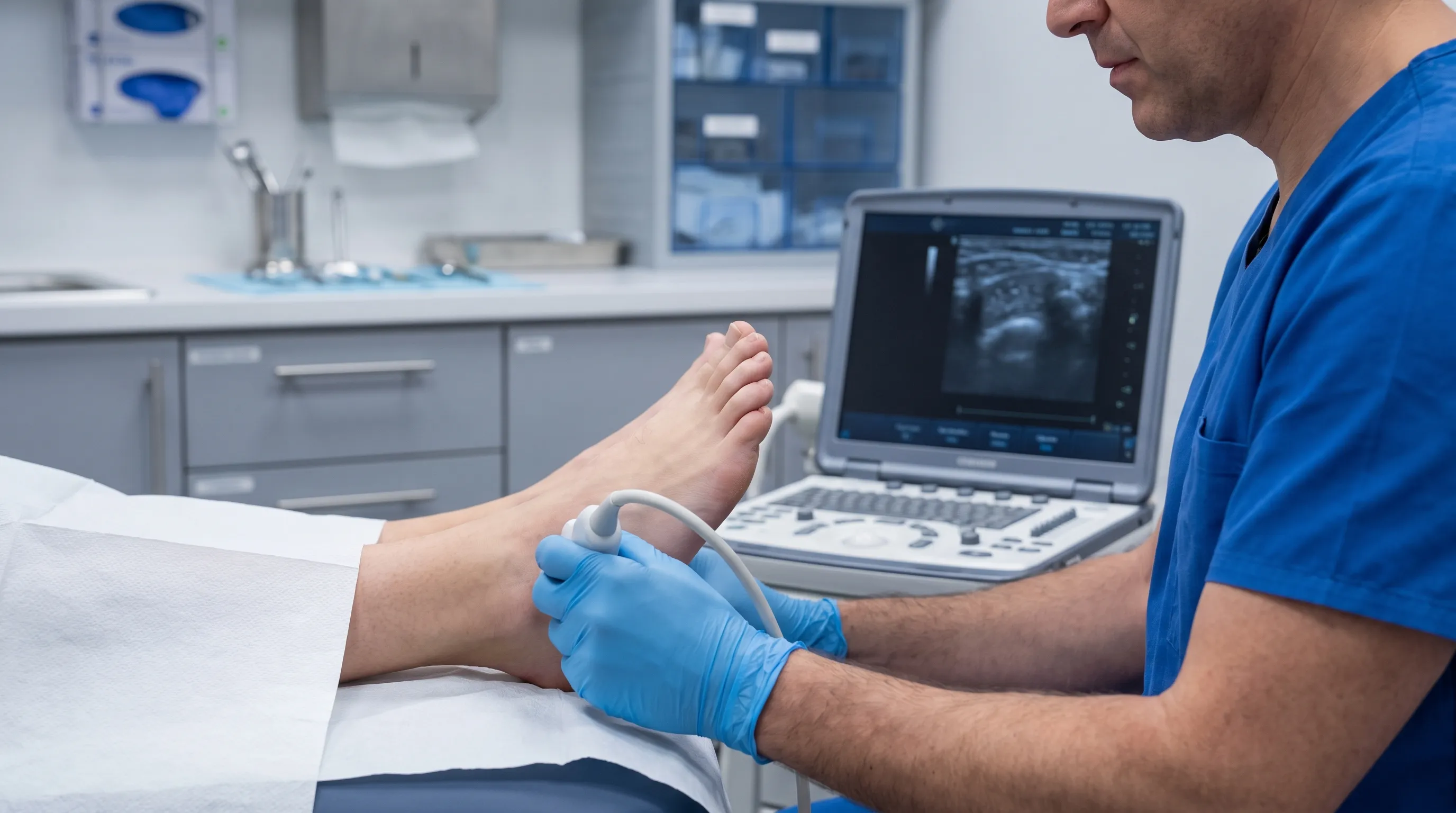
Imaging selection follows clinical suspicion patterns. Plain radiography serves as the first-line modality for osseous involvement, cortical erosion, calcification patterns, and periosteal reaction detection. Ultrasound provides dynamic, real-time evaluation of cystic versus solid components, vascularity patterns through Doppler flow mapping, and guided aspiration or injection procedures. Magnetic resonance imaging represents the gold standard for soft tissue characterization, offering superior contrast resolution for tumor boundaries, marrow infiltration, neurovascular displacement, and compartmental involvement. Contrast-enhanced protocols further differentiate viable tumor tissue from necrotic regions or inflammatory changes.
Computed tomography complements MRI when bone destruction assessment, surgical planning for complex resections, or metastatic screening is required. Positron emission tomography combined with CT scanning helps evaluate systemic disease spread, monitor treatment response, and detect early recurrence in high-risk malignancies. Each imaging modality serves distinct clinical purposes, and radiologists often correlate findings across multiple studies to construct a complete pathological profile.
| Feature | Benign Foot Mass Characteristics | Malignant Foot Mass Characteristics |
|---|---|---|
| Growth Rate | Slow, predictable over months to years | Rapid, noticeable within weeks to few months |
| Margins | Well-defined, encapsulated, mobile | Ill-defined, infiltrative, fixed to deep structures |
| Pain Pattern | Pressure-related, intermittent, activity-dependent | Rest pain, night pain, progressive intensity |
| Size Threshold | Usually under 2 inches at presentation | Frequently exceeds 2 inches before detection |
| Skin Changes | Typically normal or mild compression effects | Ulceration, discoloration, warmth, necrosis possible |
| Imaging Appearance | Homogeneous, cystic or fatty signal, no invasion | Heterogeneous, necrotic centers, cortical breach, neurovascular encasement |
| Recurrence Risk | Low after complete excision | Moderate to high without wide surgical margins |
Biopsy Procedures and Pathology
Tissue sampling remains the definitive diagnostic standard for any suspicious foot lesion. Core needle biopsy, incisional biopsy, and excisional biopsy represent the primary techniques selected based on mass size, location, depth, and clinical suspicion. Excisional biopsy typically applies to small, superficial lesions where complete removal serves both diagnostic and therapeutic purposes simultaneously. Core needle biopsy utilizes specialized coaxial needles to extract cylindrical tissue cores while preserving surrounding architecture for surgical planning. Incisional biopsy removes a representative tissue section from larger or deeper masses, ensuring pathologists receive adequate cellular material for comprehensive analysis.
Biopsy procedures execution requires meticulous planning to avoid compromising future surgical margins or contaminating adjacent compartments. Specialists follow strict protocols regarding incision placement, tract orientation, and tissue handling to prevent tumor seeding or lymphatic disruption. Histopathological evaluation includes immunohistochemical staining, molecular genetic profiling, and mitotic activity assessment to determine precise tumor classification, grade, and prognostic indicators. Modern sarcoma centers utilize multidisciplinary tumor boards where pathologists, radiologists, surgical oncologists, and medical oncologists collaboratively review biopsy results before finalizing treatment recommendations.
Patients should never undergo unsupervised or elective biopsy procedures outside specialized settings. Improper technique can obscure diagnostic accuracy, necessitate wider resection margins, or inadvertently spread malignant cells into previously unaffected tissue planes. Always seek evaluation from board-certified orthopedic surgeons, podiatric oncologists, or multidisciplinary sarcoma centers with documented experience in extremity tumor management.
Comprehensive Treatment Approaches
Treatment selection depends entirely on definitive diagnosis, tumor biology, anatomical constraints, functional goals, and patient comorbidities. Modern foot oncology emphasizes limb preservation, functional optimization, and evidence-based outcome tracking across all intervention stages.
Non-Surgical Management Strategies
Conservative management remains appropriate for confirmed benign conditions lacking mechanical compromise or malignant potential. Activity modification reduces repetitive stress on sensitive structures while inflammation resolves. Targeted physical therapy programs focus on fascial mobility restoration, proprioceptive retraining, and compensatory gait pattern correction to minimize secondary strain.
Image-guided aspiration effectively decompresses ganglion cysts and seromas, providing immediate symptomatic relief and reducing recurrence risk when combined with subsequent sclerotherapy or hyaluronidase injection protocols. Corticosteroid infiltration addresses inflammatory fibromatosis and tenosynovitis-related masses by suppressing localized cytokine activity and reducing peritumoral edema. Cryotherapy and laser ablation techniques show promise for superficial benign neoplasms and dysplastic epidermal lesions, offering minimally invasive alternatives with shorter recovery timelines.
Custom orthotic devices provide targeted pressure redistribution, arch stabilization, and gait cycle normalization that prevent compensatory overload on previously affected regions. Shockwave therapy demonstrates growing evidence for managing chronic fibromatosis and recalcitrant soft tissue proliferations by stimulating neovascularization and collagen remodeling. Conservative management requires consistent follow-up imaging and clinical reassessment to monitor growth patterns and ensure no malignant transformation occurs during the observation period.
Surgical Intervention and Reconstruction
Surgical management becomes necessary when conservative measures fail, mechanical dysfunction develops, or malignancy requires oncologic clearance. Wide-margin excision remains the standard for malignant foot tumors, ensuring complete removal with histologically negative boundaries while preserving maximal functional tissue. Marginal excision applies to benign lesions where capsule integrity can be maintained without sacrificing surrounding healthy structures.
Modern limb-sparing techniques utilize advanced reconstructive methodologies including local rotational flaps, free tissue transfers, nerve grafting, tendon realignment, and custom prosthetic integration when extensive resection compromises structural stability. Computer-assisted surgical planning and three-dimensional printing enable precise preoperative simulation, implant customization, and margin optimization that significantly reduces intraoperative uncertainty.
Adjuvant radiation therapy addresses microscopic residual disease in high-grade sarcomas, while systemic chemotherapy targets metastatic risk in aggressive histological subtypes. Targeted molecular therapies and immunotherapy protocols continue evolving, offering promising alternatives for patients with contraindications to conventional treatment regimens. Multidisciplinary coordination ensures that surgical clearance, reconstructive viability, adjuvant therapy sequencing, and rehabilitation timelines align seamlessly for optimal long-term outcomes.
Daily Care and Long-Term Management
Post-treatment success relies heavily on patient engagement with structured self-care protocols, vigilant monitoring, and lifestyle modifications that protect structural integrity while promoting sustained mobility.
Footwear Modifications and Orthotics
Appropriate footwear selection forms the cornerstone of long-term foot protection post-mass treatment. Patients require shoes with adequate toe box volume, reinforced heel counters, and shock-absorbing midsoles to minimize repetitive microtrauma during ambulation. Custom orthotic devices provide targeted pressure redistribution, arch stabilization, and gait cycle normalization that prevent compensatory overload on previously affected regions.
Moisture-wicking socks reduce friction-induced skin breakdown and maintain optimal epidermal hydration levels. Patients should inspect footwear interiors regularly for seam irregularities, structural degradation, or pressure points that could trigger new tissue irritation. Rotating between multiple shoe types throughout the week distributes mechanical load across varying contact points, preventing localized fatigue and accelerating tissue adaptation.
Regular professional pedicure services address callus formation, hyperkeratotic thickening, and nail abnormalities that commonly emerge following surgical recovery periods. Podiatric specialists can monitor scar tissue maturation, assess orthotic wear patterns, and adjust supportive devices as foot mechanics stabilize over months to years.

Rehabilitation and Physical Therapy
Structured rehabilitation programs restore functional capacity through progressive loading protocols, proprioceptive retraining, and neuromuscular re-education. Initial phases emphasize edema management, scar tissue mobilization, and protected range-of-motion exercises that prevent joint stiffness while respecting surgical healing timelines. Therapists incorporate hydrotherapy, ultrasound-assisted tissue softening, and manual fascial release techniques to accelerate recovery velocity.
Intermediate phases introduce controlled weight-bearing progression, balance training, and closed-chain strengthening that rebuilds muscular endurance without overloading healing structures. Advanced stages focus on sport-specific drills, dynamic gait optimization, and plyometric progression that safely returns patients to desired activity levels. Home exercise compliance remains the strongest predictor of functional outcomes, requiring patients to maintain consistent practice routines even when clinical appointments decrease in frequency.
Psychological support addresses body image concerns, activity anxiety, and fear of recurrence that commonly emerge during extended recovery periods. Cognitive behavioral strategies, peer support networks, and mindfulness-based stress reduction techniques improve treatment adherence and enhance overall quality of life metrics throughout the survivorship journey.
Frequently Asked Questions
What Causes Abnormal Growths to Develop on the Foot?
Abnormal foot growths develop from various triggers including genetic mutations, repetitive mechanical stress, localized trauma, chronic inflammation, and viral infections like human papillomavirus causing plantar warts. Some arise spontaneously in bone, cartilage, or soft tissues due to cellular replication errors, while others result from systemic conditions that manifest locally through tissue hyperplasia or neoplastic transformation. Environmental factors including prolonged exposure to radiation, certain chemical carcinogens, and chronic lymphatic or vascular congestion also contribute to pathological development over time.
How Can I Tell if a Foot Lump Is Cancerous?
You cannot reliably distinguish benign from malignant masses through visual inspection or self-palpation alone. Warning indicators include rapid growth exceeding half an inch within three months, firm or fixed consistency attached to deeper structures, size surpassing two inches, unexplained night pain or resting discomfort, skin ulceration, and sudden discoloration. Only advanced imaging combined with histopathological biopsy provides definitive diagnosis. Patients experiencing persistent or evolving characteristics should seek immediate evaluation from specialized foot and ankle surgeons or orthopedic oncologists rather than relying on home remedies or online symptom checkers.
What Are the Recovery Expectations After Foot Tumor Surgery?
Recovery trajectories vary significantly based on surgical extent, tissue involvement, and reconstructive requirements. Minor excisions typically require two to four weeks for initial wound closure and suture removal, followed by gradual return to standard footwear. Wide-margin resections involving tendons, nerves, or bone structures often demand three to six months of structured rehabilitation before full weight-bearing activities resume safely. Patients should anticipate initial swelling management protocols, progressive physical therapy sessions, and custom orthotic integration that supports long-term biomechanical stability. Compliance with surgeon-directed activity restrictions and scheduled follow-up imaging prevents complications and optimizes functional restoration timelines.
Can Conservative Treatments Effectively Reduce Foot Masses?
Conservative management proves highly effective for specific benign conditions including ganglion cysts, lipomas, plantar fibromatosis, and inflammatory tenosynovitis. Approaches encompass activity modification to reduce repetitive stress, targeted physical therapy addressing fascial restrictions and gait compensations, corticosteroid injections suppressing localized inflammatory cascades, and ultrasound-guided aspiration decompressing fluid-filled lesions. Specialized footwear and custom orthotics redistribute pressure away from symptomatic zones, preventing further mechanical irritation. However, conservative care does not replace surgical evaluation when red flag symptoms emerge, rapid growth occurs, or histopathological analysis indicates malignant potential. Regular monitoring ensures timely intervention if conservative management fails to control progression.
How Do I Prevent Future Foot Complications?
Preventive strategies focus on mechanical load management, early symptom recognition, and proactive foot health maintenance. Wear properly fitted shoes with adequate toe room, reinforced support structures, and shock-absorbing midsoles tailored to your activity demands. Utilize moisture-wicking socks to minimize friction-induced skin breakdown and maintain optimal epidermal hydration levels. Perform daily foot inspections using mirrors or partner assistance to detect contour changes, skin alterations, or new palpable lumps before they progress to symptomatic stages. Maintain healthy body weight to reduce excessive plantar pressure and address chronic friction points through early podiatric intervention. Follow evidence-based screening guidelines consistently if you carry genetic predispositions, prior tumor history, or immunocompromised status that elevates baseline risk.
Conclusion
Navigating the clinical pathway surrounding tumor feet demands careful observation, timely professional evaluation, and evidence-based treatment strategies tailored to your specific anatomical and pathological circumstances. While the majority of foot masses remain benign and highly manageable, early recognition of warning signs and adherence to standardized diagnostic protocols ensures that malignant possibilities are neither overlooked nor overinvestigated. Modern orthopedic oncology prioritizes limb preservation, functional optimization, and multidisciplinary coordination that addresses surgical clearance, reconstructive viability, adjuvant therapy sequencing, and comprehensive rehabilitation. Patients who actively engage with structured self-care protocols, maintain consistent follow-up imaging schedules, and collaborate closely with specialized foot and ankle teams achieve significantly better long-term outcomes than those relying on passive observation or unverified home remedies. Foot health serves as the foundation for overall mobility, independence, and quality of life. By understanding the medical landscape surrounding foot masses, recognizing early warning indicators, and committing to evidence-based management pathways, you position yourself for optimal functional restoration and sustained wellness. Prioritize professional evaluation for persistent or changing lesions, maintain realistic recovery expectations, and embrace structured rehabilitation as an active participant in your healing journey rather than a passive recipient of medical care.
How we work
Researched and drafted with AI assistance, then reviewed by a human editor against the cited sources.
This article is general health information, not medical advice. Consult a qualified healthcare professional about your own situation.