Left Eye Blurry: Causes, Diagnosis, and Evidence-Based Management

Experiencing a left eye blurry can be unsettling, especially when your right eye functions perfectly. Unilateral visual changes often prompt immediate concern, as they disrupt depth perception, reading comfort, and daily spatial awareness. Unlike generalized visual fatigue, asymmetric symptoms frequently point to localized ocular issues, neurological factors, or systemic imbalances that require targeted evaluation. Understanding why vision becomes asymmetrical is the first step toward appropriate clinical management and long-term ocular health.
Many individuals initially dismiss mild asymmetrical vision changes as simple tiredness or screen-related strain. While digital eye fatigue is incredibly common, persistent or progressive visual distortion in a single eye should never be ignored. The human visual system is highly symmetrical by design, meaning that when one eye deviates from the norm, it often signals a breakdown in tear film stability, refractive balance, retinal function, or optic nerve transmission. By examining the underlying mechanisms, recognizing clinical warning signs, and following evidence-based management strategies, patients can protect their vision and prevent irreversible damage.
This comprehensive guide explores the physiological, clinical, and lifestyle dimensions of asymmetric visual impairment. You will learn how medical professionals differentiate between benign refractive shifts and urgent pathological conditions, discover proven treatment pathways, and implement daily habits that support long-term ocular resilience. Whether you are navigating mild dryness, managing chronic systemic conditions, or simply seeking clarity on your next steps, the information provided here aligns with current ophthalmological guidelines and prioritizes patient safety and evidence-based care.
Understanding the Mechanics of Unilateral Vision Changes
The human visual pathway is a remarkably integrated system involving the cornea, lens, vitreous humor, retina, optic nerves, and occipital cortex. Light enters through the anterior segment, undergoes precise refraction, projects onto photoreceptor cells, and converts electromagnetic signals into neural impulses. When this sequence operates symmetrically, binocular vision delivers sharp depth perception and seamless visual processing. However, any disruption localized to one side of the system can produce a distinctly left eye blurry sensation that feels entirely isolated from the contralateral eye.
Free calculatorA1C CalculatorA1C to average blood glucoseOpen the calculatorThe Visual Pathway and Unilateral Focus
Each eye functions as an independent optical instrument before its neural output merges in the brain. The left eye captures information primarily from the right visual field, while the right eye processes the left visual field. When only the left eye experiences a left eye blurry sensation, the disturbance is typically localized to the anterior segment, vitreous, retina, or the initial segment of the left optic nerve. Systemic conditions can occasionally present asymmetrically due to microvascular variations, but true unilateral symptoms usually originate from structural or functional differences in one eye.
Corneal irregularities, tear film deficiencies, and lens opacifications are among the most common culprits. These structures sit at the very front of the visual pathway, meaning even minor asymmetries dramatically alter image clarity. The brain attempts to compensate by suppressing the blurrier image, which can lead to eye strain, headaches, and reduced stereopsis. Recognizing that unilateral blurriness reflects a localized optical or neural disruption helps clinicians narrow their diagnostic approach efficiently.
Why Symptoms Often Present Asymmetrically
Human biology is rarely perfectly symmetrical. Dominant eye preference, differential exposure to environmental stressors, prior microtraumas, and inherent anatomical variations all contribute to asymmetric visual function. For instance, individuals who read extensively while tilting their head may place disproportionate accommodative demand on the left eye, accelerating refractive shifts or dryness in that specific ocular surface. Additionally, sleep posture, such as consistently sleeping on the right side, can temporarily impair nocturnal tear distribution to the left eye, causing morning blurriness that gradually resolves throughout the day.
Environmental exposures also play a significant role. Occupations requiring precise lateral focus, repetitive screen positioning, or prolonged wind exposure can desiccate or irritate one eye more than the other. Contact lens wearers who do not strictly follow replacement schedules often develop unilateral dryness or mild epithelial damage that manifests exclusively as a left eye blurry complaint. These asymmetrical presentations underscore the importance of evaluating lifestyle factors alongside clinical examinations.
Common Ocular Causes of Visual Distortion in One Eye
When evaluating unilateral visual impairment, clinicians systematically assess both anterior and posterior ocular segments. The majority of left eye blurry cases stem from refractive misalignment, surface disease, or age-related structural changes that alter light transmission. Understanding these categories enables patients to recognize early warning signs and seek timely intervention before permanent vision loss occurs.
Refractive Errors and Anisometropia
Refractive errors occur when light entering the eye fails to focus precisely on the retina. While myopia, hyperopia, and astigmatism often affect both eyes, anisometropia describes a condition where each eye possesses a significantly different refractive power. This imbalance forces the visual cortex to process two mismatched images, frequently resulting in a left eye blurry experience that feels disproportionately severe.
Anisometropia can be congenital or acquired. Progressive myopic shifts in adolescence, keratoconus development, or post-surgical corneal changes all contribute to asymmetric refractive states. Patients often report difficulty with depth judgment, eyestrain during reading, and persistent unilateral blur that fluctuates with accommodation. Comprehensive refraction testing, combined with corneal topography, accurately quantifies the disparity and guides customized lens prescriptions or refractive surgery planning.
Dry Eye Syndrome and Tear Film Instability
The tear film consists of three distinct layers: mucin, aqueous, and lipid. When any layer becomes deficient or evaporates prematurely, the optical surface becomes irregular, scattering incoming light and producing transient or persistent blurriness. Dry eye disease frequently presents asymmetrically due to differential blink mechanics, meibomian gland dysfunction, or localized inflammation.
Symptoms include gritty sensations, intermittent visual fluctuation, burning, and excessive reflex tearing. Blinking temporarily restores clarity as the tear film redistributes, which helps clinicians differentiate dry eye from fixed pathological causes. Environmental factors like low humidity, prolonged screen time, and certain systemic medications accelerate tear film breakdown. Artificial tears, warm compresses, omega-3 supplementation, and prescription anti-inflammatory drops constitute first-line management strategies.
Ocular Infections and Inflammatory Conditions
Bacterial, viral, or fungal conjunctivitis can produce unilateral discharge, redness, and corneal edema that scatters light. Keratitis, an inflammation of the cornea, is particularly concerning due to its potential to cause scarring and permanent refractive distortion. Early left eye blurry symptoms often precede more pronounced pain, photophobia, and foreign body sensation.
Uveitis, affecting the middle layer of the eye, triggers cellular debris and protein leakage into the anterior chamber or vitreous, significantly degrading image quality. This condition requires prompt ophthalmological intervention, often involving corticosteroid therapy and cycloplegic agents to prevent synechiae formation and secondary glaucoma. Differentiating infectious from autoimmune etiologies requires slit-lamp examination, intraocular pressure measurement, and occasionally laboratory testing.
Age-Related Changes and Lens Opacities
The crystalline lens gradually hardens and yellows with age, leading to presbyopia and reduced accommodative range. Cataract formation typically begins asymmetrically, causing one eye to develop a milky haze that produces glare sensitivity, faded color perception, and progressive blur. A left eye blurry sensation in adults over fifty often correlates with early nuclear or cortical cataracts.
Modern phacoemulsification techniques safely replace opacified lenses with multifocal intraocular implants, restoring clarity and reducing dependence on corrective eyewear. Patients should undergo regular lens evaluations, particularly if they notice progressive difficulty with night driving or reading small print. Early detection ensures surgical timing aligns with functional impairment rather than arbitrary age thresholds.
Systemic Health Factors That Trigger Vision Changes
Ocular health does not exist in isolation. The retina contains the most concentrated microvasculature in the human body, making it highly sensitive to systemic metabolic fluctuations. Hypertension, diabetes, autoimmune disorders, and neurological conditions frequently manifest as unilateral or asymmetric visual disturbances before producing widespread systemic symptoms.
Diabetes, Blood Sugar Fluctuations, and Diabetic Retinopathy
Glucose dysregulation damages retinal capillaries, causing microaneurysms, hemorrhages, and ischemic patches. Diabetic macular edema frequently develops asymmetrically, producing a localized left eye blurry sensation that fluctuates with glycemic control. Hyperglycemia alters the osmotic balance of the crystalline lens, temporarily shifting refractive power and worsening acuity.
Tight blood sugar management, routine dilated fundus examinations, and anti-VEGF intravitreal injections for active neovascularization form the cornerstone of diabetic ocular care. Patients with newly diagnosed asymmetrical blur should request urgent retinal imaging, as early intervention significantly reduces the risk of irreversible photoreceptor loss.
Hypertension and Ocular Hypertensive Crises
Chronic elevated blood pressure stiffens retinal arterioles, leading to hypertensive retinopathy. Sudden spikes can cause retinal vein occlusion or papilledema, both of which degrade unilateral vision rapidly. A left eye blurry complaint accompanied by pulsatile headaches, transient obscurations, or visual field constriction warrants immediate blood pressure assessment.
Systemic antihypertensive therapy often stabilizes retinal perfusion within weeks, though existing ischemic damage may require prolonged rehabilitation. Collaboration between primary care physicians, cardiologists, and ophthalmologists ensures coordinated management that protects both cardiovascular and ocular endpoints.
Migraine Auras and Neurological Impacts
Ocular migraines and migraine with aura can produce temporary hemianopsia, scintillating scotomas, or unilateral blur lasting twenty to sixty minutes. Cortical spreading depression temporarily disrupts visual processing without causing permanent tissue injury. Stress, hormonal shifts, certain foods, and sleep deprivation frequently trigger these episodes.
While generally benign, atypical neurological presentations require exclusion of ischemic or demyelinating processes. Persistent unilateral blur that fails to resolve, or episodes accompanied by motor weakness or speech difficulty, necessitate urgent neuroimaging to rule out transient ischemic attack or optic neuritis. Neuro-ophthalmologists specialize in differentiating migraine variants from sight-threatening pathologies.
| Diagnostic Evaluation | Primary Indication | Typical Findings | Clinical Significance |
|---|---|---|---|
| Slit-Lamp Biomicroscopy | Anterior segment assessment | Tear film instability, corneal abrasion, cataract grading, anterior uveitis cells | Differentiates surface disease from internal pathology |
| Dilated Fundus Examination | Posterior segment evaluation | Retinal hemorrhages, macular edema, optic disc cupping, peripheral tears | Detects diabetic retinopathy, glaucoma, and retinal detachment |
| Optical Coherence Tomography (OCT) | Cross-sectional retinal imaging | Intraretinal fluid layers, retinal nerve fiber thinning, subretinal deposits | Quantifies macular degeneration progression and monitors treatment response |
| Visual Field Testing | Functional optic pathway assessment | Scotomas, arcuate defects, generalized depression | Identifies glaucomatous damage and neurological visual pathway disruption |
| Intraocular Pressure Measurement | Glaucoma screening | Elevated tonometry readings, asymmetric fluctuations | Early detection of optic nerve compression risk |
Clinical Diagnosis and Professional Evaluation Protocols
Accurate diagnosis of asymmetric visual impairment requires systematic evaluation. Clinicians integrate subjective symptom reporting with objective measurement tools to construct a comprehensive clinical picture. Patients should expect a structured assessment that examines both anatomical integrity and functional performance.
Comprehensive Ophthalmic Assessment
Initial evaluation begins with a detailed history, documenting onset, duration, progression, associated symptoms, and systemic health factors. Visual acuity testing establishes baseline clarity, while refraction quantifies refractive error. Cover testing evaluates ocular alignment and binocular coordination, revealing subtle muscle imbalances that contribute to unilateral strain.
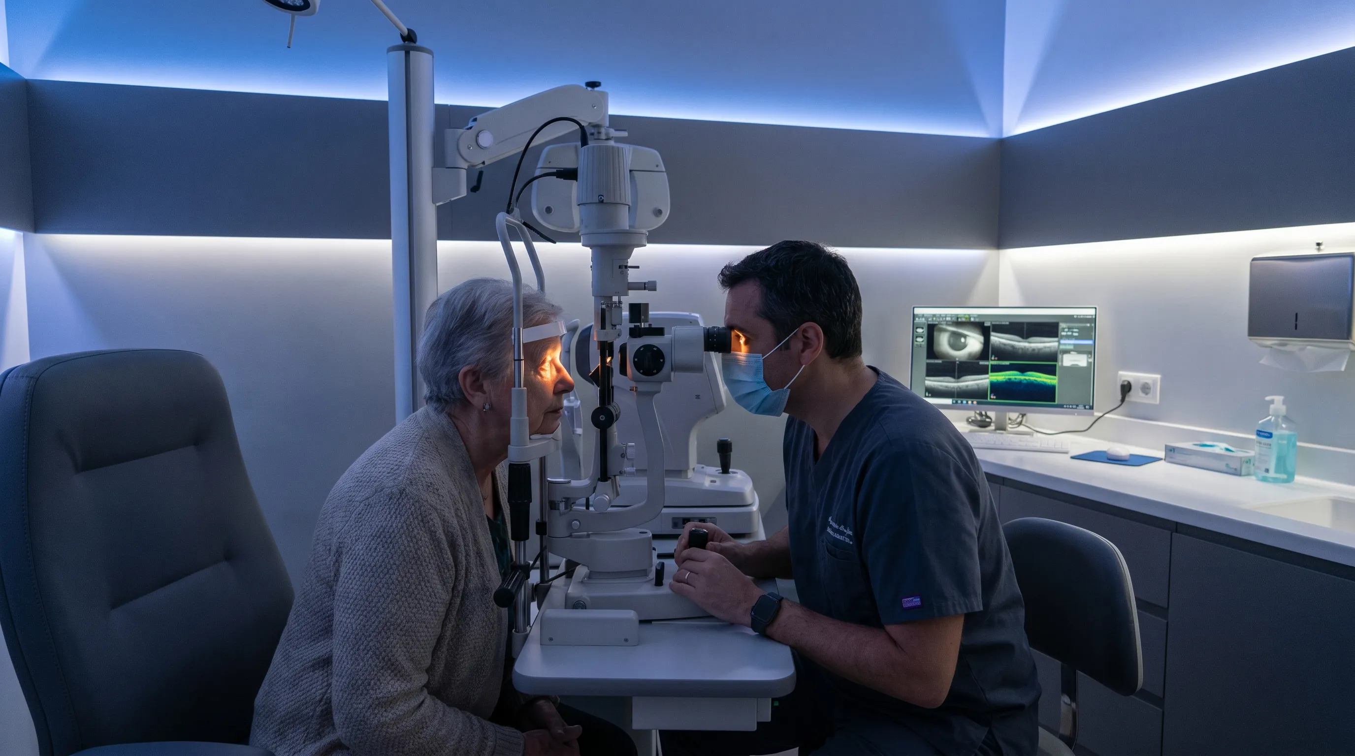
Slit-lamp examination provides magnified visualization of the eyelids, conjunctiva, cornea, anterior chamber, and iris. Pupil reactivity assessment detects afferent or efferent pathway abnormalities. Dilating drops temporarily paralyze accommodation, allowing thorough lens and retinal inspection. Patients should arrange transportation for dilated exams, as temporary photophobia and near-vision blur are expected.
Advanced Imaging and Functional Testing
When standard examination reveals structural concerns, clinicians utilize specialized imaging modalities. Optical coherence tomography generates micrometer-resolution cross-sections of the retina, identifying edema, drusen, or nerve fiber thinning before they manifest as severe vision loss. Fluorescein angiography highlights vascular leakage and ischemic zones, guiding targeted anti-VEGF or laser interventions.
Corneal topography maps surface curvature irregularities, essential for diagnosing keratoconus or post-surgical astigmatism. Visual field analyzers measure peripheral sensitivity, detecting early glaucomatous damage or neurological deficits. Integration of these technologies ensures precise disease staging and individualized treatment planning.
Differentiating Benign from Urgent Pathologies
Not every left eye blurry warrants emergency intervention, but certain red flags demand immediate attention. Sudden vision loss without pain suggests vascular occlusion or retinal detachment. Gradual blur with halos and severe eye pain indicates acute angle-closure glaucoma. Associated neurological deficits raise concern for stroke or demyelinating disease.
Clinicians utilize symptom onset timelines, pain profiles, pupillary findings, and intraocular pressure measurements to triage urgency. Patients experiencing progressive symptoms should never wait for routine appointments to expire. Prompt referral to emergency ophthalmology or neurology preserves visual potential and prevents irreversible neural damage.
Evidence-Based Treatment and Management Strategies
Once the underlying etiology is established, targeted interventions restore clarity and prevent progression. Treatment pathways vary significantly depending on whether symptoms originate from refractive imbalance, surface disease, retinal pathology, or neurological dysfunction. Patients benefit from understanding the therapeutic options available and the expected timelines for improvement.
Pharmacological Interventions and Eye Drops
Medications form the first line of defense for many inflammatory and infectious ocular conditions. Preservative-free artificial tears lubricate compromised tear films, reducing optical scatter and improving surface regularity. Short-term topical corticosteroids suppress acute uveitis or post-surgical inflammation, while cyclosporine or lifitegrast modulate chronic dry eye inflammation over months.
Antibiotic, antiviral, or antifungal drops eradicate microbial keratitis, though adherence to dosing schedules prevents resistance and recurrence. Glaucoma management utilizes prostaglandin analogs, beta-blockers, alpha-agonists, and carbonic anhydrase inhibitors to lower intraocular pressure and protect the optic nerve. Patients must understand that ophthalmic medications require precise administration techniques and scheduled follow-ups to monitor efficacy and adverse effects.
Optical Corrections and Vision Rehabilitation
Corrective eyewear remains the most accessible and reversible intervention for refractive asymmetry. Custom lenses compensate for anisometropia, astigmatism, and presbyopic shifts, immediately improving binocular fusion. Progressive or bifocal designs accommodate varying focal distances without requiring frequent lens changes.
Contact lenses provide wider fields of view and eliminate peripheral distortion from frames. Orthokeratology lenses temporarily reshape the cornea overnight, offering daytime clarity without surgical intervention. Vision therapy exercises strengthen accommodative flexibility and convergence, particularly beneficial for patients experiencing strain-induced unilateral blur. Consistent follow-up ensures prescriptions adapt to changing ocular physiology.
Surgical and Procedural Options
When conservative measures fail to restore functional vision, procedural interventions offer definitive solutions. Laser refractive surgery reshapes the corneal curvature, reducing dependence on corrective eyewear. Candidates require stable refraction for at least one year, adequate corneal thickness, and absence of progressive retinal disease.
Cataract extraction remains the most frequently performed ophthalmic procedure worldwide. Phacoemulsification fragments opacified lens material, which is suctioned through a micro-incision before implanting an advanced intraocular lens. Retinal specialists utilize intravitreal injections, laser photocoagulation, and vitrectomy to address hemorrhages, edema, and tractional detachments. Surgical success relies heavily on patient selection, postoperative compliance, and multidisciplinary coordination.
Daily Habits, Prevention, and When to Seek Emergency Care
Long-term ocular health depends on consistent protective behaviors, environmental modifications, and nutritional support. Preventing a left eye blurry from recurring or progressing requires integrating clinical guidance into daily routines. Simple adjustments yield substantial cumulative benefits for surface integrity, neural resilience, and overall visual comfort.
Digital Hygiene and the 20-20-20 Rule
Prolonged screen exposure reduces blink frequency by up to sixty percent, accelerating tear film evaporation and triggering accommodative spasm. Implementing the 20-20-20 rule significantly mitigates digital strain: every twenty minutes, shift focus to an object twenty feet away for at least twenty seconds. Adjusting screen brightness to match ambient lighting reduces glare and minimizes pupillary fluctuation.
Ergonomic positioning ensures the monitor sits slightly below eye level, promoting complete eyelid closure during blinking. Using humidity chambers or desktop humidifiers counteracts environmental dryness from heating or air conditioning systems. Patients who experience persistent left eye blurry despite lifestyle modifications should request a comprehensive dry eye workup and discuss prescription tear film stabilizers with their clinician.
Nutritional Support for Ocular Resilience
Dietary patterns directly influence retinal microcirculation, tear film composition, and oxidative defense mechanisms. Lutein and zeaxanthin accumulate in the macula, filtering harmful blue light and neutralizing free radicals. Leafy greens, yellow vegetables, and egg yolks provide bioavailable carotenoids that accumulate gradually over months of consistent intake.
Omega-3 fatty acids, particularly EPA and DHA, improve meibomian gland secretion quality, stabilizing the lipid layer of the tear film. Fatty fish, walnuts, and flaxseeds support both ocular surface health and systemic cardiovascular function. Vitamin C and zinc enhance collagen synthesis and immune response, while hydration maintains aqueous humor production. Patients should view nutrition as foundational therapy rather than standalone treatment, integrating supplements under medical supervision.

Red Flags That Require Immediate Medical Attention
While many unilateral visual changes respond well to conservative management, certain symptoms demand urgent evaluation. Sudden painless vision loss suggests vascular compromise or retinal detachment. Intense eye pain with nausea and halos indicates acute glaucoma. New-onset double vision, facial asymmetry, or slurred speech alongside blur raises neurological concern.
Traumatic injuries causing corneal laceration or hyphema require immediate shielding and emergency transport. Chemical exposures demand immediate irrigation for at least fifteen minutes before seeking care. Patients should never self-prescribe steroid drops without diagnosis, as they can accelerate corneal melting or fungal proliferation. Trusting clinical expertise ensures accurate triage and prevents irreversible damage.
Frequently Asked Questions
Is a sudden left eye blurry an emergency?
A rapid onset of left eye blurry without prior warning often signals an acute pathological process requiring immediate attention. Retinal detachment, central retinal vein occlusion, optic neuritis, and ischemic events can all manifest as unilateral visual disruption within minutes or hours. When accompanied by severe pain, sudden floaters, a dark curtain obscuring peripheral vision, or neurological deficits, patients should bypass routine clinics and proceed directly to emergency ophthalmology or a hospital emergency department. Prompt intervention dramatically improves the prognosis and preserves functional vision.
Can stress or fatigue cause blurry vision in only one eye?
Chronic stress and systemic fatigue typically produce bilateral visual strain, but they frequently exacerbate underlying asymmetrical conditions. Individuals with preexisting anisometropia, unilateral dry eye, or subtle binocular vision dysfunction may experience pronounced left eye blurry sensations during periods of heightened cortisol, sleep deprivation, or prolonged screen exposure. Fatigue reduces blink frequency and impairs tear film regeneration, making the less dominant eye disproportionately uncomfortable. Addressing sleep hygiene, implementing regular visual breaks, and managing stress through mindfulness or physical activity often alleviates these symptoms without requiring medical intervention.
How long should I wait before seeing a doctor for a left eye blurry?
Mild, transient blurriness that resolves with blinking or rest typically indicates surface dryness or temporary accommodative fatigue, which can be monitored for twenty-four to forty-eight hours. However, if the left eye blurry persists beyond two days, progressively worsens, or associates with redness, discharge, light sensitivity, or headache, scheduling an appointment with an optometrist or ophthalmologist becomes essential. Patients with diabetes, autoimmune disorders, or glaucoma risk factors should seek evaluation immediately upon noticing unilateral changes, as delayed assessment increases the likelihood of irreversible tissue damage and complicates long-term management strategies.
Will wearing glasses or contacts fix a left eye blurry permanently?
Corrective lenses effectively compensate for refractive errors by redirecting incoming light to focus precisely on the retina. They provide immediate clarity while worn and significantly improve binocular coordination, reducing strain and enhancing quality of life. However, lenses do not halt disease progression or reverse structural pathology. Cataracts, macular degeneration, diabetic retinopathy, and corneal scarring require targeted medical or surgical interventions for permanent resolution. Regular prescription updates ensure optical devices align with current ocular anatomy, preventing compensatory strain that can exacerbate underlying left eye blurry sensations over time.
What vitamins or supplements support unilateral vision recovery?
Nutritional supplementation plays a supportive role in maintaining retinal integrity, optimizing tear film stability, and reducing oxidative stress. Lutein, zeaxanthin, omega-3 fatty acids, vitamin C, vitamin E, and zinc demonstrate clinical efficacy in preserving macular health and improving dry eye symptoms. The Age-Related Eye Disease Study formulations remain the most extensively researched protocols for slowing retinal deterioration progression. However, supplements cannot replace comprehensive medical evaluation or targeted pharmaceutical therapy. Patients should discuss dosage, potential drug interactions, and quality verification with their healthcare provider before initiating any regimen, particularly when managing chronic systemic conditions or preparing for ocular surgery.
Conclusion
Navigating a left eye blurry requires a balanced approach that integrates clinical evaluation, lifestyle modification, and informed decision-making. While many unilateral visual changes stem from manageable refractive shifts or temporary surface dryness, others signal underlying systemic imbalances or progressive ocular pathology that demand timely intervention. By recognizing early warning signs, understanding diagnostic pathways, and adhering to evidence-based treatment protocols, patients can preserve visual acuity, prevent irreversible damage, and maintain functional independence.
Ocular health is fundamentally intertwined with systemic wellness, neurological function, and daily environmental exposures. Consistent preventive habits, regular comprehensive examinations, and proactive communication with eye care professionals create a robust framework for long-term visual resilience. Whether addressing mild dryness, managing chronic metabolic conditions, or exploring surgical options, the journey toward clear vision relies on patience, precision, and professional guidance. Prioritizing timely assessment and embracing holistic ocular care ensures that a left eye blurry remains a manageable symptom rather than a permanent limitation.
Learn more about comprehensive eye exams and refractive evaluation from the American Academy of Ophthalmology and explore clinical management protocols through Mayo Clinic's visual symptom guidelines. For detailed information on retinal health and age-related changes, visit the National Eye Institute's resource library.
How we work
Researched and drafted with AI assistance, then reviewed by a human editor against the cited sources.
This article is general health information, not medical advice. Consult a qualified healthcare professional about your own situation.