Eye Blurry in One Eye: Causes, Diagnosis, and Treatment Guide
Waking up to an experience of eye blurry in one eye can be unsettling, especially when you expect your vision to be symmetrical and reliable. Our visual system relies heavily on binocular coordination, meaning that when one eye suddenly or gradually loses clarity, the brain struggles to merge the two inputs seamlessly. This disruption can manifest as mild fuzziness, significant distortion, or even temporary blindness on the affected side. While many causes are benign and easily correctable, unilateral visual impairment can occasionally signal serious ocular or neurological conditions that demand prompt medical intervention. Understanding the underlying mechanisms, recognizing red flag symptoms, and implementing evidence-based self-care strategies are essential steps in preserving your long-term ocular health. In this comprehensive guide, we will explore the physiological triggers, diagnostic pathways, clinical treatments, and preventative measures that address this common yet potentially complex symptom. By the end, you will have a clear, actionable roadmap for when to monitor, when to intervene at home, and when to seek professional care to ensure optimal visual outcomes.
Understanding the Nature of Unilateral Visual Disturbance
Visual acuity depends on a complex interplay between optical structures, neural pathways, and vascular supply. When eye blurry in one eye occurs, it indicates a localized disruption rather than a systemic refractive error. The human visual system processes light through the cornea, passes it through the aqueous humor and pupil, focuses it via the crystalline lens, and projects it onto the retina. The retina then converts light into electrochemical signals transmitted through the optic nerve to the visual cortex. Any interruption along this precise pathway can compromise clarity in a single eye while leaving the contralateral eye completely unaffected.
Gradual vs. Sudden Onset
The temporal progression of symptoms provides critical diagnostic clues. Gradual blurring that develops over months or years typically points to structural degeneration or refractive shifts. Age-related conditions such as early cataracts, progressive astigmatism, or early-stage age-related macular degeneration often present with slow, unilateral deterioration. These changes allow the brain to gradually compensate, which is why patients sometimes do not notice the deficit until the affected eye is closed. Conversely, sudden onset symptoms that appear within seconds or minutes suggest acute events like retinal vascular occlusion, optic neuritis, acute angle-closure glaucoma, or retinal detachment. Sudden visual loss requires immediate triage because neural tissue damage becomes irreversible after a short ischemic window. Differentiating between these timelines during your initial clinical consultation will significantly accelerate the diagnostic process.
Temporary vs. Persistent Symptoms
Temporary episodes lasting minutes to hours often correlate with vascular spasms, migraines with aura, dry eye exacerbations, or corneal edema following overnight contact lens wear. These transient disturbances frequently resolve spontaneously once the underlying trigger subsides. Persistent symptoms that remain unchanged over days or progressively worsen indicate structural pathology requiring targeted intervention. Conditions like keratoconus, chronic uveitis, untreated glaucoma, or macular pathology will not resolve without medical management. Tracking symptom duration, frequency, and associated factors such as lighting conditions, hydration levels, or physical exertion will help your eye care provider narrow the differential diagnosis efficiently.
Primary Medical Causes of Vision Changes
When patients report eye blurry in one eye, clinicians systematically evaluate multiple anatomical regions to identify the source of optical degradation. The most common etiologies span refractive, lens-related, corneal, and retinal domains. Each category follows distinct pathophysiological pathways, necessitating tailored diagnostic and therapeutic approaches.
Refractive Errors and Astigmatism
Refractive anomalies represent the most frequent cause of unilateral visual blurriness, particularly when patients experience gradual symptom onset. Myopia, hyperopia, and astigmatism can develop asymmetrically, meaning one eye may require a significantly different prescription than the other. Anisometropia, the clinical term for unequal refractive power between eyes, forces the visual cortex to suppress input from the weaker eye, leading to perceived blurriness. Corneal irregularities such as mild keratoconus or post-surgical refractive changes can also create asymmetric focusing errors. Comprehensive autorefraction and subjective refraction testing during a routine eye exam will quantify the discrepancy, allowing precise lens correction through glasses, toric contact lenses, or refractive surgery. Early intervention prevents amblyopia in children and reduces visual fatigue in adults.
Cataracts and Age-Related Changes
Cataract formation involves the progressive opacification of the crystalline lens, often beginning unilaterally before eventually affecting both eyes. Protein denaturation and water content imbalances scatter incoming light, reducing contrast sensitivity and creating a foggy visual experience. Patients frequently describe difficulty reading, increased glare sensitivity at night, and yellowish color distortion. While cataracts develop slowly, they remain the leading cause of treatable unilateral blurriness worldwide. Advanced diagnostic phacoemulsification combined with premium intraocular lens implantation offers highly predictable visual rehabilitation. Regular monitoring through slit-lamp biomicroscopy and lens grading scales ensures optimal surgical timing before the condition significantly impacts quality of life.
Ocular Surface and Retinal Conditions
Beyond the anterior segment and lens, the tear film and posterior retinal structures play pivotal roles in maintaining optical clarity. Disruptions in these systems frequently manifest as asymmetric visual degradation.
Dry Eye Syndrome and Meibomian Gland Dysfunction
Ocular surface disease remains an underdiagnosed contributor to intermittent unilateral blurriness. When the tear film destabilizes due to aqueous deficiency or evaporative dysfunction from meibomian gland blockage, the corneal surface becomes microscopically irregular. This optical turbulence scatters light, creating temporary blur that often fluctuates throughout the day. Patients typically notice improvement after blinking vigorously or applying lubricating drops, as these actions temporarily restore tear film integrity. Diagnosis involves tear break-up time measurement, Schirmer testing, and meibography imaging. Management encompasses warm compress therapy, lid hygiene, prescription anti-inflammatory drops, omega-3 supplementation, and procedural treatments like LipiFlow or intense pulsed light therapy. Addressing ocular surface health frequently eliminates unexplained blurriness without requiring complex interventions.
Macular Degeneration and Retinal Vascular Disorders
Retinal pathology demands particular attention because delayed treatment can result in irreversible central vision loss. Dry age-related macular degeneration involves gradual photoreceptor atrophy beneath the fovea, while the wet form features abnormal choroidal neovascularization leaking fluid and blood. Both conditions initially present with unilateral distortion, metamorphopsia, or central scotoma. Diabetic retinopathy, retinal vein occlusion, and retinal artery occlusion similarly cause asymmetric vision changes through microvascular compromise or ischemic injury. Diagnostic confirmation relies heavily on optical coherence tomography, fluorescein angiography, and comprehensive dilated fundus examination. Treatment protocols range from anti-vascular endothelial growth factor intravitreal injections and laser photocoagulation to strict glycemic and blood pressure management. Early detection remains the single most critical factor in preserving functional vision.
Neurological and Systemic Triggers
Unilateral visual impairment does not always originate within the eye itself. The optic nerve and visual pathways can be affected by systemic conditions, inflammatory processes, and neurological events.
Ocular Migraine and Transient Ischemic Attack
Migraine with visual aura frequently produces temporary unilateral blurriness, zigzag lines, or scintillating scotomas that precede headache onset. These phenomena result from cortical spreading depression across the occipital lobe, temporarily disrupting visual processing rather than causing structural damage. Episodes typically resolve within 60 minutes. However, distinguishing migraine aura from transient ischemic attacks requires careful evaluation. A transient retinal ischemic event, often termed amaurosis fugax, manifests as a sudden graying or black curtain descending over one eye, caused by microemboli lodging in the central retinal artery. This represents a vascular emergency requiring immediate cardiovascular workup, carotid imaging, and antithrombotic therapy to prevent permanent stroke or vision loss. Proper differentiation prevents inappropriate management and ensures appropriate preventive care.
Optic Neuritis and Multiple Sclerosis Links
Inflammation of the optic nerve, clinically recognized as optic neuritis, produces painful unilateral vision loss exacerbated by eye movement. Demyelination disrupts signal transmission along the afferent visual pathway, resulting in reduced visual acuity, impaired color perception, and central scotoma. While optic neuritis can occur idiopathically, it frequently serves as an early clinical indicator of multiple sclerosis or neuromyelitis optica spectrum disorders. Diagnosis involves visual evoked potentials, magnetic resonance imaging of the brain and orbits, and comprehensive neurological assessment. High-dose corticosteroid therapy accelerates recovery, although most patients regain baseline vision within weeks. Long-term neurological monitoring ensures appropriate disease-modifying therapy initiation if systemic autoimmune conditions are confirmed.
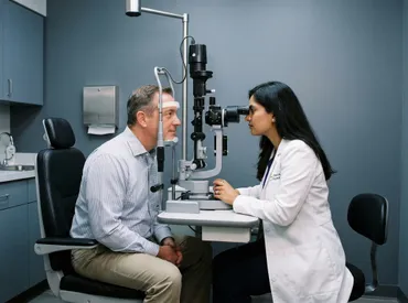
Diagnostic Approach and Clinical Evaluation
Accurate identification of the underlying etiology requires a systematic, stepwise clinical workflow. Eye care professionals utilize standardized examination protocols combined with advanced imaging to isolate the precise anatomical location of dysfunction.
Comprehensive Eye Exam Components
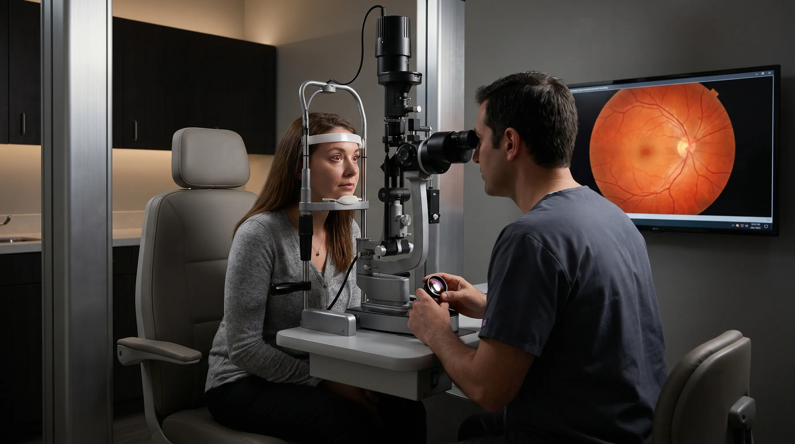
The foundational evaluation begins with uncorrected and best-corrected visual acuity testing, followed by pinhole assessment to differentiate refractive from non-refractive causes. Slit-lamp biomicroscopy examines the cornea, anterior chamber, lens, and eyelids for structural abnormalities, inflammation, or surface irregularities. Intraocular pressure measurement screens for glaucomatous damage, while pupillary response testing evaluates afferent and efferent neural pathway integrity. The swinging flashlight test specifically identifies relative afferent pupillary defects, which strongly suggest optic nerve pathology. Dilated fundus examination allows direct visualization of the retina, optic disc, and macula, revealing hemorrhages, exudates, neovascularization, or detachment margins. These core assessments establish the initial diagnostic framework before proceeding to specialized imaging.
Imaging Modalities and Functional Testing
Modern ophthalmology relies heavily on non-invasive imaging technologies to characterize subtle structural changes invisible during conventional examination. Optical coherence tomography provides cross-sectional retinal scans measuring macular thickness, identifying fluid accumulation, and detecting early neurodegenerative thinning. Fundus fluorescein angiography evaluates vascular perfusion and identifies leakage sites in conditions like diabetic retinopathy or macular edema. Visual field testing maps peripheral and central sensitivity, revealing characteristic patterns associated with glaucoma, optic neuropathy, or cerebral lesions. Ultrasound biomicroscopy becomes essential when dense media opacity prevents direct visualization. Combining these modalities creates a comprehensive diagnostic profile that guides targeted therapeutic interventions with high precision.
| Condition Category | Typical Onset | Associated Symptoms | Primary Diagnostic Tools | Standard Treatment Approach |
|---|---|---|---|---|
| Refractive Error / Astigmatism | Gradual | Eye strain, headaches, difficulty focusing | Autorefractor, subjective refraction | Corrective lenses, refractive surgery |
| Cataract | Gradual | Glare, color fading, progressive fogging | Slit-lamp exam, glare testing | Phacoemulsification with IOL implantation |
| Dry Eye Syndrome | Intermittent | Burning, fluctuating blur, foreign body sensation | TBUT, Schirmer test, meibography | Artificial tears, anti-inflammatory drops, thermal pulse |
| Macular Degeneration | Gradual (Dry) / Rapid (Wet) | Central distortion, straight line waviness | OCT, fundus angiography | Anti-VEGF injections, AREDS2 supplements |
| Retinal Vascular Occlusion | Sudden | Painless vision loss, visual field defect | Fundus exam, OCT, carotid ultrasound | Urgent ophthalmology referral, systemic management |
| Optic Neuritis | Acute to Subacute | Pain on eye movement, color desaturation | MRI, visual evoked potentials, RAPD test | High-dose IV corticosteroids, neurology referral |
Evidence-Based Treatment Approaches
Therapeutic strategies depend entirely on accurate etiological classification. Interventions range from conservative management to advanced surgical procedures, each supported by rigorous clinical evidence.
Pharmacological and Topical Therapies
Medication-based management targets inflammation, vascular abnormalities, and ocular surface dysfunction. Artificial tear formulations and lipid-based lubricants restore tear film stability, eliminating intermittent blur caused by surface irregularity. Prescription cyclosporine or lifitegrast eye drops modulate localized immune responses, providing long-term relief for autoimmune dry eye conditions. Anti-vascular endothelial growth factor agents represent the gold standard for wet macular degeneration and diabetic macular edema, delivered via intravitreal injection at regular intervals to prevent abnormal vessel proliferation and reduce fluid leakage. Corticosteroid regimens address optic neuritis, uveitis, and post-surgical inflammation through controlled immune suppression. Antimicrobial and antiviral therapies resolve infectious corneal ulcers or herpetic keratitis that distort optical clarity. Strict adherence to prescribed dosing schedules ensures therapeutic efficacy while minimizing ocular toxicity.
Surgical and Laser Interventions
When conservative management fails or structural damage progresses, procedural intervention becomes necessary. Cataract extraction using ultrasonic phacoemulsification combined with premium multifocal or toric intraocular lens implantation restores refractive precision and eliminates lens opacity. Laser photocoagulation seals leaking retinal vessels in diabetic retinopathy, preventing macular edema progression. Photodynamic therapy selectively destroys choroidal neovascularization while preserving surrounding retinal architecture. Glaucoma filtration procedures and minimally invasive surgical stents reduce intraocular pressure, protecting optic nerve fibers from progressive degradation. Retinal detachment repair utilizes pars plana vitrectomy, pneumatic retinopexy, or scleral buckling to reposition detached tissue and prevent permanent photoreceptor death. Corneal cross-linking halts keratoconus progression by strengthening stromal collagen bonds. Each procedure requires careful patient selection, risk-benefit analysis, and structured postoperative rehabilitation.
Practical Self-Care and Prevention Strategies
While clinical intervention addresses established pathology, daily habits and environmental modifications play a crucial role in preventing symptom onset and supporting long-term visual resilience. Implementing proactive self-care measures significantly reduces the likelihood of experiencing eye blurry in one eye due to preventable factors.
Digital Eye Strain Management
Prolonged screen exposure reduces blink rate by up to 60 percent, accelerating tear film evaporation and inducing corneal surface irregularity. Implementing structured visual breaks using the 20-20-20 rule effectively resets accommodative fatigue and restores tear distribution. Adjusting monitor brightness to match ambient lighting, utilizing blue light filtering applications during evening hours, and maintaining a viewing distance of 20 to 30 inches minimizes optical stress. Ergonomic workstation positioning ensures proper gaze alignment, preventing sustained downward viewing that increases tear evaporation rates. Regularly cleaning spectacle lenses with microfiber cloths removes particulate matter that scatters light and contributes to perceived blurriness. These simple behavioral modifications consistently improve comfort and visual stability for digital professionals.
Nutritional Optimization and Lifestyle Modifications
Systemic health profoundly influences ocular tissue integrity and microvascular perfusion. Consuming a diet rich in lutein, zeaxanthin, vitamins C and E, zinc, and omega-3 fatty acids supports macular pigment density and reduces oxidative retinal damage. Leafy greens, fatty fish, eggs, and citrus fruits provide essential micronutrients clinically proven to slow age-related degenerative processes. Maintaining strict glycemic control prevents diabetic microvascular complications that frequently manifest as unilateral visual changes. Regular cardiovascular exercise improves systemic circulation, enhancing choroidal blood flow and retinal oxygenation. Adequate hydration maintains tear production volume, while avoiding tobacco smoke eliminates exposure to toxic compounds that accelerate cataract formation and macular degeneration. Scheduling comprehensive dilated eye examinations annually ensures early detection of asymptomatic pathology before functional impairment occurs.

Frequently Asked Questions
Why is my vision suddenly blurry in one eye?
Sudden blurriness typically indicates an acute event such as retinal vascular occlusion, optic nerve inflammation, ocular migraine, or retinal detachment. Because these conditions involve time-sensitive neural or vascular compromise, immediate evaluation by an ophthalmologist or emergency department is strongly recommended. Do not wait to see if symptoms improve independently.
Can eye strain cause vision blurriness in just one eye?
Yes, asymmetric refractive errors or prolonged unilateral focusing during digital tasks can strain the ciliary muscles unevenly, leading to temporary monocular blur. Rest, targeted visual exercises, and updated prescriptions typically resolve these symptoms. If the blurriness persists beyond a few days despite rest, professional assessment is warranted.
When should I go to the emergency room for blurry vision?
Seek immediate emergency care if you experience abrupt vision loss, a dark curtain descending across your visual field, severe ocular pain, halos around lights, double vision, or accompanying neurological symptoms like facial drooping or slurred speech. These signs indicate potential retinal detachment, acute glaucoma, or cerebrovascular events requiring urgent intervention.
Is blurry vision in one eye a sign of diabetes?
Uncontrolled diabetes frequently manifests through unilateral or asymmetric visual changes caused by fluctuating blood glucose affecting lens hydration, diabetic macular edema, or retinal ischemia. Regular annual dilated examinations and strict metabolic management significantly reduce the risk of diabetic visual complications.
How long does it take for blurry vision to resolve naturally?
Transient causes like dry eye, fatigue, or migraine aura typically resolve within minutes to several hours with appropriate rest and hydration. Structural, vascular, or degenerative causes will not resolve spontaneously and require medical treatment. Persistent symptoms lasting longer than 48 hours mandate clinical evaluation to prevent irreversible damage.
Conclusion
Experiencing eye blurry in one eye should never be dismissed as a minor inconvenience or normal aging process. While many underlying triggers remain easily manageable through corrective lenses, lubricating therapies, or lifestyle adjustments, others require urgent clinical intervention to preserve long-term visual function. The distinction between benign and pathological causes hinges on symptom onset, duration, associated clinical features, and comprehensive diagnostic evaluation. By understanding the anatomical pathways involved, recognizing red flag indicators, and prioritizing regular ophthalmic assessments, you can maintain optimal ocular health throughout your lifetime. Implementing preventive strategies such as digital hygiene, nutritional optimization, and systemic condition management significantly reduces the likelihood of unilateral visual disruption. If you notice persistent, worsening, or sudden vision changes, consult a licensed eye care professional promptly to receive accurate diagnosis and evidence-based treatment tailored to your specific clinical profile.
For authoritative guidance on eye health and symptom management, visit the American Academy of Ophthalmology and the National Eye Institute. Your vision is irreplaceable, proactive care remains the foundation of lasting visual clarity.

About the author
David Chen, DO, is a board-certified neurologist specializing in neuro-oncology and stroke recovery. He is the director of the Comprehensive Stroke Center at a New Jersey medical center and has published numerous articles on brain tumor treatment.