Prism Glasses for Double Vision: Complete Guide, Benefits & Adaptation Tips

Waking up to the unsettling experience of seeing two overlapping images can instantly disrupt your daily routine, compromise mobility, and trigger persistent headaches. This condition, clinically known as diplopia, affects millions of individuals worldwide according to global vision health data from the WHO and stems from a breakdown in the intricate coordination between the eyes and the brain. When the visual axes fail to align properly, the brain receives conflicting signals that it cannot seamlessly merge, resulting in doubled vision, visual fatigue, and impaired spatial judgment. Fortunately, modern optical science has developed highly effective interventions, and prism glasses for double vision have emerged as one of the most reliable, non-invasive solutions available today. By precisely redirecting light before it reaches the retina, these specialized lenses restore comfortable binocular function without requiring surgical intervention. This comprehensive guide explores the clinical mechanisms, prescription protocols, adaptation strategies, and evidence-based best practices surrounding this transformative visual therapy, ensuring patients and caregivers have the accurate information needed to navigate treatment safely and effectively.
Understanding Double Vision and Binocular Misalignment
To fully appreciate how prism glasses for double vision restore visual harmony, it is essential to first understand the underlying pathophysiology of diplopia. Normal vision relies on the precise coordination of six extraocular muscles in each eye, all governed by cranial nerves III, IV, and VI. These muscles work in tandem to ensure both eyes track the same target simultaneously. When this neuromuscular synchronization is disrupted, the images projected onto each retina no longer align with the foveal center. The brain, which is hardwired to interpret a single coherent image, struggles to reconcile the disparity, leading to the perception of two distinct pictures.
Free calculatorBMI CalculatorCalculate Body Mass IndexOpen the calculatorMonocular vs. Binocular Diplopia
Diplopia is broadly categorized into two distinct types, each requiring fundamentally different management strategies. Monocular diplopia persists even when the unaffected eye is closed, indicating that the problem originates within a single eye. Common culprits include corneal irregularities like keratoconus, early cataract formation, uncorrected astigmatism, or severe dry eye syndrome. Since the issue is optical rather than neuromuscular, prism correction offers no therapeutic benefit. In contrast, binocular diplopia disappears as soon as either eye is covered, confirming that the misalignment stems from extraocular muscle dysfunction, neurological impairment, or restrictive orbital conditions. This is the precise clinical scenario where prism glasses for double vision provide measurable, life-changing relief.
How Misalignment Affects Your Brain
When the eyes are chronically misaligned, the brain employs several compensatory mechanisms that often lead to secondary symptoms. Some individuals develop suppression, a neurological process where the brain actively ignores input from one eye to eliminate confusion. Others may adopt abnormal head postures or tilt their faces to align the visual axes, which frequently triggers chronic neck pain, shoulder tension, and postural fatigue. Prolonged effort to fuse disparate images overloads the fusional vergence system, resulting in debilitating eye strain, migraines, difficulty reading, and reduced depth perception. In older adults, untreated binocular misalignment significantly increases the risk of falls, stair navigation errors, and mobility limitations, making timely optical intervention a critical public health consideration.
The Science Behind Prism Glasses for Double Vision
At their core, prism lenses operate on fundamental principles of geometric optics. Rather than focusing light like traditional spherical or cylindrical lenses, prismatic lenses are wedge-shaped optical elements designed to deviate light rays toward the thicker base of the lens. By precisely calculating the angle and direction of deviation, eye care professionals can shift the incoming image onto the functional retinal area of the misaligned eye. This optical realignment allows the visual cortex to receive two congruent images that it can effortlessly fuse into a single, three-dimensional perception.
How Light Bending Corrects Visual Axes
The prismatic effect works by exploiting the refractive properties of glass or polycarbonate materials. When light passes through the angled wedge, it bends toward the base. For example, if the left eye is turned inward (esotropia), the image falls on the temporal retina rather than the fovea. A base-out prism placed in front of the affected eye bends the light nasally, effectively relocating the image back to the central foveal zone. This eliminates the need for the extraocular muscles to exert excessive corrective effort, immediately reducing strain and allowing the fusional centers in the brainstem to operate within their physiological range. Research from the National Eye Institute confirms that prism correction is a safe, reversible intervention that restores comfortable binocular vision without altering the underlying anatomical structure.
Measuring Prism Strength in Diopters
Prism power is quantified using prism diopters (Δ), a standardized unit of measurement that represents one centimeter of image displacement at a distance of one meter. Clinical prescriptions typically range from 0.5Δ to 30Δ or higher, depending on the severity and axis of misalignment. Lower powers are often sufficient for decompensated phorias or mild convergence insufficiency, while larger deviations require higher diopter values. It is important to note that prism strength is not linear; higher diopters increase lens thickness, weight, and potential peripheral distortion. Clinicians frequently employ the Prentice formula and cover-uncover testing to determine the exact diopter value that neutralizes the deviation across primary gaze, downgaze, and near fixation. The American Academy of Ophthalmology emphasizes that prism glasses for double vision are most effective when the deviation remains stable and falls within the tolerable range for optical compensation.
Types of Prism Lenses: Fresnel vs. Ground-In
Not all prismatic corrections are created equal. The choice between temporary and permanent prism technology depends entirely on the patient’s clinical stability, lifestyle demands, and adaptation timeline. Understanding the structural and optical differences ensures patients make informed decisions alongside their eye care provider.
When to Use Temporary Fresnel Prisms
Fresnel prisms consist of thin, flexible vinyl sheets containing microscopic parallel grooves that replicate the refractive properties of traditional prisms while adding minimal thickness. Because they are applied directly to the back surface of existing corrective lenses using static-adhesive backing, they can be easily removed, repositioned, or replaced as prescriptions change. This adaptability makes them the gold standard for trial periods, post-stroke rehabilitation, recovering cranial nerve palsies, and unstable strabismus. Their lightweight construction reduces frame weight, though the grooved surface can slightly reduce contrast sensitivity and attract cleaning difficulties. Many neuro-ophthalmologists prescribe Fresnel strips initially to assess fusional tolerance before committing to permanent lens fabrication.
Benefits of Permanent Ground-In Lenses
Once the ocular deviation stabilizes for at least three to six months, patients typically transition to ground-in prisms. These lenses are optically manufactured by grinding the prismatic angle directly into the prescription material during the surfacing process. The result is a seamless, highly transparent lens that delivers superior optical clarity, scratch resistance, and cosmetic appearance. Ground-in prisms eliminate the visual artifacts associated with Fresnel grooves and integrate effortlessly into standard eyeglass designs. However, they are significantly more expensive, require highly accurate final measurements, and cannot be adjusted if the underlying strabismus progresses. They represent a long-term commitment best suited for stable, chronic diplopia management.
Comparison Table
| Feature | Fresnel Prisms | Ground-In Prisms |
|---|---|---|
| Material | Flexible vinyl adhesive sheet | Polycarbonate, Trivex, or high-index glass |
| Thickness/Weight | Extremely thin, lightweight | Increases with diopter strength, heavier frames |
| Optical Clarity | Slight reduction due to surface grooves | Exceptional, seamless visual quality |
| Adjustment Period | Easily modified or removed during adaptation | Fixed prescription; requires new lenses to change |
| Ideal Use Case | Unstable deviations, trial periods, post-surgical recovery | Chronic, stable diplopia requiring long-term wear |
| Cost | Low to moderate | Moderate to high |

Clinical Indications and Who Benefits Most
The therapeutic application of prism glasses for double vision extends far beyond simple eye misalignment. Numerous systemic, neurological, and ocular conditions create fusional disruptions that respond remarkably well to prismatic correction. Recognizing these indications helps patients seek appropriate referrals and avoid delayed interventions.
Strabismus and Cranial Nerve Palsies
Adult-onset strabismus remains one of the most common indications for prism therapy. Esotropia, exotropia, and hypertropia that cannot be immediately addressed with strabismus surgery or Botox injections are routinely managed with optical correction. Similarly, acquired cranial nerve palsies, often resulting from microvascular ischemia in diabetic or hypertensive patients, frequently resolve spontaneously within three to six months. During this recovery window, prism glasses for double vision provide immediate symptomatic relief while the nerve regenerates. The Cleveland Clinic notes that prism therapy serves as a first-line, non-invasive intervention for stable binocular diplopia, often used alongside active vision therapy or as a bridge to surgical correction.
Neurological and Autoimmune Conditions
Systemic diseases that affect neuromuscular transmission or orbital tissue frequently disrupt binocular alignment. Thyroid eye disease (Graves’ ophthalmopathy) causes restrictive strabismus due to inflammation and fibrosis of extraocular muscles, making prisms highly effective during the quiescent phase. Patients with multiple sclerosis may experience internuclear ophthalmoplegia or cranial nerve demyelination that triggers intermittent diplopia. Myasthenia gravis causes fluctuating ocular weakness that worsens with fatigue, requiring adaptive prism management. In these complex cases, regular neuro-ophthalmology follow-ups ensure prescriptions align with disease activity.
Post-Surgical and Trauma Recovery
Ocular trauma, orbital fractures, and post-surgical misalignment frequently leave patients struggling with intractable double vision. Following retinal surgery, cataract extraction, or strabismus procedures, the extraocular muscles may temporarily lose coordination or become restricted by scar tissue. Prism therapy offers a non-surgical pathway to restore spatial awareness during rehabilitation. Additionally, traumatic brain injury survivors often benefit from low-to-moderate prism diopters that reduce visual overload, improve reading stamina, and decrease motion-induced nausea. The StatPearls clinical database (NIH/NCBI) confirms that prism glasses are indicated for small-to-moderate angle deviations, particularly in patients who are poor surgical candidates or wish to explore conservative management first.
The Prescription and Fitting Process
Successfully integrating prism glasses for double vision into daily life requires meticulous clinical assessment, precise optical fabrication, and personalized patient education. The process extends far beyond a standard refraction, demanding specialized binocular vision expertise.
Comprehensive Binocular Vision Assessment
Before any lens is manufactured, clinicians perform a series of diagnostic evaluations to quantify the deviation. The cover-uncover test and alternate cover test determine the magnitude and direction of misalignment across multiple gaze positions. Prism bar measurements and Maddox rod testing allow practitioners to isolate vertical and horizontal components separately. Patients are evaluated at both distance and near fixation to account for convergence demands. If the deviation varies significantly depending on gaze direction (incomitant strabismus), the clinician may limit prism power or recommend alternative interventions. Accurate base-in, base-out, base-up, or base-down orientation ensures the light shift precisely compensates for the anatomical misalignment.
Base Direction and Orientation
Vertical prism prescriptions are generally less physiologically tolerated than horizontal ones. Even small vertical misalignments can cause severe symptoms, but the brain struggles to adapt to vertical prismatic shifts exceeding two to three diopters without inducing spatial distortion. Horizontal prisms, conversely, can accommodate higher powers with minimal disruption. Clinicians carefully balance prescription strength against visual comfort, often splitting prism power between both lenses to reduce thickness and weight. Proper centration and pupillary distance measurements are critical, as decentration can induce unwanted prismatic effects that exacerbate symptoms rather than alleviate them.
Professional Guidelines from Leading Institutions
Both the Mayo Clinic and the American Academy of Ophthalmology stress the importance of ruling out acute neurological or vascular emergencies before initiating prism therapy. Sudden-onset diplopia accompanied by headache, ptosis, pupillary abnormalities, or focal neurological deficits requires immediate emergency department evaluation. Once life-threatening causes are excluded, conservative management with prism correction becomes the standard of care. The National Eye Institute emphasizes that while prisms compensate for misalignment, they do not cure the underlying pathology. Therefore, they should always be paired with appropriate disease management, whether that involves thyroid hormone regulation, glycemic control, or targeted vision therapy programs.
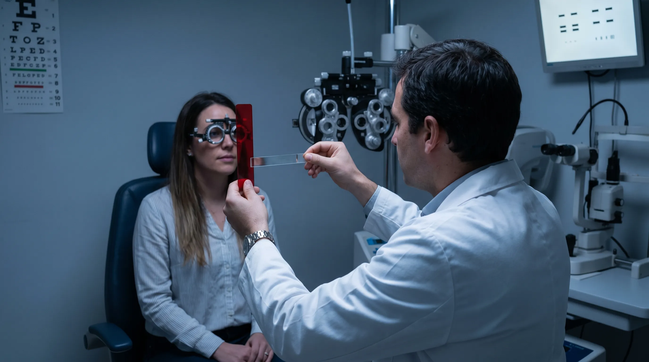
Adapting to Your New Prescription: What to Expect
Neuro-adaptation to prism lenses is a highly individualized process that requires patience, gradual exposure, and proactive symptom management. The brain must recalibrate its spatial mapping algorithms to accommodate the newly aligned visual input.
The First 2 Weeks: Dizziness and Spatial Awareness
During the initial adaptation window, patients commonly report mild nausea, floor curvature illusions, peripheral blur, or a sensation that objects appear closer or farther than expected. These symptoms reflect the visual cortex recalibrating proprioceptive and vestibular signals to match the corrected retinal input. A landmark study published in the Journal of AAPOS found that 78% of patients prescribed Fresnel prisms reported significant symptom improvement within two weeks, with adaptation rates improving dramatically when patients followed structured wear schedules. Pushing through discomfort too aggressively can prolong neuroplastic adjustment and increase rejection risk.
Tips for a Smoother Transition
Successful adaptation hinges on gradual exposure and environmental optimization. Patients should begin wearing their new glasses for short intervals indoors, starting with stationary tasks like reading or watching television before progressing to walking and dynamic movement. Maintaining consistent lighting reduces pupil fluctuation, which can temporarily alter perceived prism strength. Staying hydrated and avoiding excessive screen time during the first week minimizes visual fatigue. If dizziness persists beyond three weeks, or if headaches intensify rather than improve, a follow-up measurement is essential. Sometimes minor diopter adjustments or axis rotations resolve lingering discomfort without requiring a full prescription overhaul.
Driving and Reading with Prism Correction
Reading often becomes dramatically more comfortable once prism glasses for double vision are properly integrated, as the binocular convergence demand decreases significantly. However, driving requires fully stabilized depth perception and reliable peripheral awareness. High-powered prisms can induce chromatic aberration and image jump when looking through the periphery, making quick head turns or lane changes potentially disorienting. Most specialists recommend waiting until adaptation is complete and performing supervised test drives in familiar, low-traffic environments before returning to highway navigation. Polarized lenses and anti-reflective coatings are frequently recommended to reduce glare and enhance contrast during mobility.
Safety Precautions and Long-Term Management
While prism therapy is remarkably safe when properly prescribed, certain clinical scenarios demand caution. Misapplication of prismatic correction can mask progressive pathology, delay necessary surgical intervention, or induce unsustainable compensatory strain.
Contraindications and Medical Warnings
Prisms are strictly contraindicated for monocular diplopia, as bending light will not resolve intraocular optical irregularities. Rapidly progressing strabismus, such as that seen in untreated myasthenia gravis or expanding intracranial masses, requires definitive treatment of the underlying cause rather than optical masking. Incomitant deviations that fluctuate dramatically across gaze positions may only be partially corrected with prisms, potentially inducing secondary torsional strain. Additionally, vertical prism powers exceeding 6Δ frequently cause intolerable spatial distortion, making alternative interventions like strabismus surgery or botulinum toxin injection more appropriate. Patients must disclose all systemic diagnoses to ensure the prescription aligns with their overall medical trajectory.
Follow-Up Protocols and Monitoring
Long-term success depends on structured clinical follow-ups. Initial re-evaluation typically occurs within two to four weeks to assess neuro-adaptation, verify lens positioning, and screen for residual fusional stress. Once the prescription stabilizes, patients should schedule annual comprehensive exams to monitor underlying disease progression and detect age-related refractive changes. The transition from temporary Fresnel to permanent ground-in lenses usually occurs after three to six months of stable alignment, though complex neurological recoveries may require longer observation periods. Systematic reviews published in Ophthalmology conclude that prism glasses provide excellent symptomatic relief for stable deviations but require close monitoring when applied to fluctuating or progressive conditions.
Frequently Asked Questions
How long does it take to adjust to prism glasses for double vision?
Most patients require between three days and three weeks for full neuro-adaptation. During the initial phase, the brain recalibrates spatial processing to accommodate the newly aligned visual axes. Mild symptoms like transient dizziness, slight floor curvature illusions, or peripheral distortion are normal and typically resolve as cortical plasticity adjusts. Consistent daily wear and gradual exposure to varied environments accelerate this process.
Can prism lenses permanently cure strabismus or diplopia?
No. Prism glasses for double vision function as an optical compensation mechanism, not a curative treatment. They redirect incoming light so that both retinas receive aligned images, allowing the brain to fuse them comfortably. The underlying neuromuscular imbalance, cranial nerve dysfunction, or restrictive orbital condition remains unchanged. In some pediatric cases, early optical correction may prevent amblyopia development, but adults rely on prisms for symptom management rather than anatomical correction.
Are Fresnel prisms better than ground-in prisms for beginners?
Fresnel prisms are widely considered the optimal starting point for new patients or those with unstable ocular alignment. Their adhesive vinyl construction allows clinicians to rapidly modify diopter strength and base orientation without manufacturing an entirely new pair of lenses. They are lightweight, cost-effective, and ideal for trial periods during post-stroke recovery, thyroid eye disease management, or evolving cranial nerve palsies. Once the deviation remains consistent for several months, patients typically upgrade to ground-in prisms for superior optical clarity.
Is it safe to drive with prism glasses?
Driving is generally safe once the adaptation period concludes and spatial awareness stabilizes. However, initial wearers should avoid operating vehicles until peripheral distortion and depth perception normalize. High prism powers can cause image displacement at the lens edges, making quick head turns or checking blind spots temporarily disorienting. Most clinicians recommend practicing in familiar, low-traffic areas first and upgrading to high-quality anti-reflective coatings to minimize glare-induced visual stress.
Do insurance plans typically cover prism lenses?
Coverage varies significantly depending on the provider, geographic location, and documented diagnosis. When prism glasses for double vision are prescribed for medically recognized conditions such as cranial nerve palsy, stroke recovery, traumatic brain injury, or congenital strabismus, many vision and medical insurance plans classify them as medically necessary and provide partial coverage. Cosmetic adaptations or elective prism adjustments may require out-of-pocket payment. Patients should request a pre-authorization letter from their optometrist detailing the ICD-10 diagnostic codes and clinical necessity documentation.
Conclusion
Managing chronic visual misalignment requires a thoughtful, evidence-based approach that prioritizes both optical precision and neurological adaptation. Prism glasses for double vision represent a cornerstone of modern conservative ophthalmology, offering immediate, non-invasive relief for patients who would otherwise endure persistent eye strain, impaired mobility, and compromised quality of life. By understanding the distinction between monocular and binocular diplopia, recognizing the clinical applications of Fresnel versus ground-in lenses, and committing to structured adaptation protocols, individuals can safely integrate optical therapy into their daily routines. Leading medical organizations consistently emphasize that successful outcomes depend on accurate diagnosis, specialized fitting, and proactive follow-up care. Whether recovering from a neurological event, managing autoimmune eye disease, or addressing adult-onset strabismus, patients who partner with experienced optometrists and neuro-ophthalmologists achieve the highest rates of symptom resolution and functional visual recovery. With ongoing advancements in optical fabrication and binocular vision research, prism therapy will continue to serve as a vital, patient-centered intervention in the broader landscape of visual health and neurological rehabilitation.
How we work
Researched and drafted with AI assistance, then reviewed by a human editor against the cited sources.
This article is general health information, not medical advice. Consult a qualified healthcare professional about your own situation.