Understanding the Eyeball Socket: Anatomy, Health Conditions & Expert Care
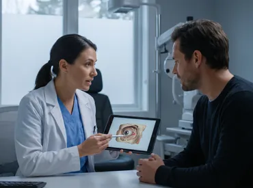
The human visual system is a marvel of biological engineering, and at its very foundation lies a protective bony enclosure known as the eyeball socket. Often overlooked until injury or disease strikes, this intricate cavity plays a vital role in safeguarding one of our most critical sensory organs. Understanding its structure, function, and vulnerabilities is essential for maintaining long-term ocular health and preventing irreversible damage. Whether you are an athlete, a healthcare professional, or simply someone looking to protect your vision, knowing how to support and maintain orbital health can make a significant difference in your overall wellness journey.
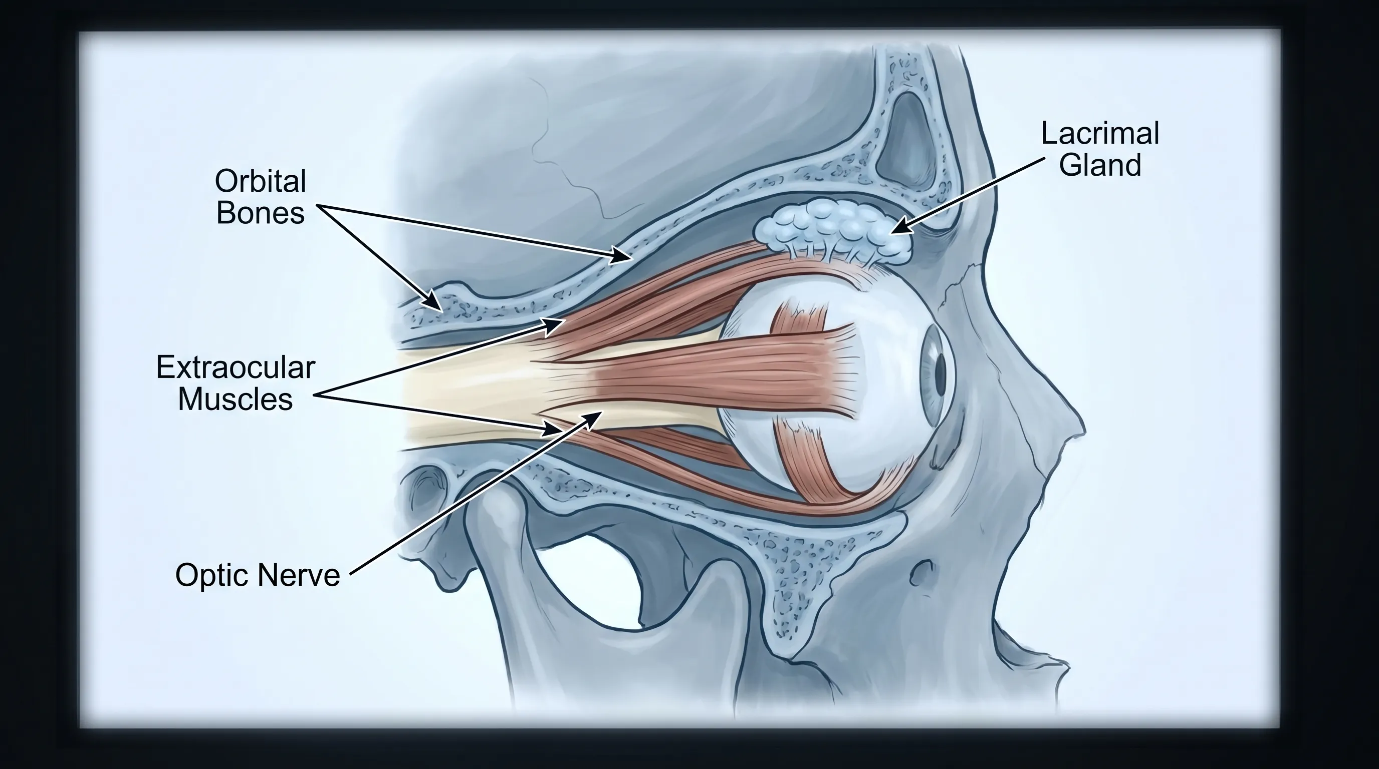
The eyeball socket, clinically referred to as the orbit, is far more than a simple anatomical void (learn more about orbital anatomy). It is a meticulously designed, pyramid-shaped chamber located on either side of the facial skeleton. Each cavity houses not only the globe of the eye but also a complex network of extraocular muscles, cranial nerves, vascular supply, the lacrimal drainage system, and protective adipose tissue. These components work in seamless harmony to facilitate precise eye movements, maintain intraocular pressure, and shield delicate neural pathways from mechanical stress. Because the orbital environment operates in such close proximity to the brain, sinuses, and nasal passages, systemic inflammation or localized trauma can quickly escalate into serious medical conditions. This article provides a comprehensive, evidence-based exploration of orbital anatomy, common pathologies, diagnostic methodologies, and actionable strategies for preserving your visual and structural eye health.
Anatomy of the Eyeball Socket
A thorough grasp of orbital anatomy is the cornerstone of understanding how injuries occur, why certain conditions manifest, and how medical professionals approach treatment. The human orbit is not merely an empty space waiting to hold the eyeball; it is a dynamic, multi-layered system designed for both protection and functional mobility. Each structural element serves a specific physiological purpose, and disruptions to any of these layers can compromise vision and comfort.
The Bony Orbital Walls
The skeletal framework of the eyeball socket consists of seven craniofacial bones that interlock to form a remarkably strong yet delicate enclosure. The roof is primarily composed of the orbital plate of the frontal bone and a small portion of the lesser wing of the sphenoid bone. This roof separates the orbit from the anterior cranial fossa and the frontal sinuses, providing critical protection to the brain above. The medial wall is the thinnest and most fragile part, formed largely by the ethmoid bone's lamina papyracea, alongside contributions from the lacrimal, sphenoid, and maxillary bones. Because of its thinness, this wall is highly susceptible to fractures during blunt trauma and can easily transmit sinus infections into the orbital space.
The lateral wall, constructed from the zygomatic bone and greater wing of the sphenoid, is the strongest portion of the orbit. It provides robust structural support against direct impacts and serves as an attachment point for several crucial muscles. The floor, or orbital floor, consists mainly of the maxillary bone, with minor contributions from the zygomatic and palatine bones. This region separates the orbit from the maxillary sinus and is the most common site for blowout fractures. Understanding this bony architecture is crucial because the structural integrity of each wall directly influences how forces are distributed during trauma, how surgical implants are positioned, and how sinus drainage pathways interact with orbital health.
Muscles and Nerves
Movement and precise visual alignment depend on the coordinated action of six extraocular muscles: the superior, inferior, medial, and lateral rectus muscles, along with the superior and inferior oblique muscles. These muscles originate from the orbital apex or annulus of Zinn and insert into the sclera, enabling smooth, rapid, and highly coordinated eye movements. The intricate innervation of these muscles involves three primary cranial nerves. The oculomotor nerve (CN III) controls the medial, superior, and inferior rectus muscles, as well as the inferior oblique and levator palpebrae superioris. The trochlear nerve (CN IV) uniquely innervates the superior oblique muscle, while the abducens nerve (CN VI) governs the lateral rectus.
Damage to these neuromuscular pathways can result in diplopia (double vision), strabismus (misaligned eyes), or ptosis (drooping eyelid). Furthermore, the optic nerve (CN II) passes through the optic canal at the posterior apex of the eyeball socket, transmitting visual information from the retina to the occipital cortex. The optic canal also houses the ophthalmic artery, making it a critical anatomical bottleneck where swelling or compression can rapidly lead to ischemic optic neuropathy and permanent blindness if not addressed promptly.
Soft Tissue and Fat Pads
Beyond bone, muscle, and nerve, the eyeball socket contains a highly specialized arrangement of soft tissues. Orbital fat is not merely passive filler; it acts as a shock-absorbing cushion that allows the globe to glide smoothly during ocular movements while maintaining proper intraorbital pressure. These fat compartments are separated by fibrous septa that help localize infections and limit inflammatory spread. The fibrous tissue also includes Tenon's capsule, a transparent membrane that envelops the globe and separates it from the extraocular muscles, reducing friction during contraction.
The lacrimal system, responsible for tear production and drainage, resides primarily in the superotemporal quadrant of the orbit. The lacrimal gland secretes tears that flow across the ocular surface before draining through the puncta, canaliculi, lacrimal sac, and nasolacrimal duct into the nasal cavity. Dysfunction at any point along this pathway can lead to dry eye syndrome, chronic epiphora (excessive tearing), or dacryocystitis. The orbital venous plexus also features valveless veins that allow blood to flow in either direction, which explains why sinus infections or facial trauma can sometimes trigger retrograde orbital infections. Recognizing these soft tissue dynamics is essential for clinicians managing inflammatory, infectious, or neoplastic orbital conditions.
Common Conditions Affecting the Eyeball Socket
Pathologies involving the orbit range from acute, life-altering emergencies to chronic autoimmune disorders that progressively alter facial aesthetics and visual function. Early recognition and appropriate intervention are crucial for preventing permanent damage. Understanding the clinical presentation, underlying pathophysiology, and standard treatment pathways empowers patients to seek timely care and adhere to medical recommendations.
Thyroid Eye Disease and Exophthalmos
Thyroid eye disease (TED), also known as Graves' ophthalmopathy, is one of the most prevalent causes of progressive orbital changes (read more about TED from the National Eye Institute). It is an autoimmune condition characterized by inflammation, glycosaminoglycan accumulation, and subsequent enlargement of the extraocular muscles and orbital fat. This expansion occurs within the rigid confines of the eyeball socket, leading to increased intraorbital pressure and forward displacement of the globe, clinically termed exophthalmos or proptosis. Patients often experience eyelid retraction, periorbital edema, conjunctival injection, and a gritty sensation.
In severe cases, the enlarged muscles can compress the optic nerve at the orbital apex, resulting in compressive optic neuropathy. This medical emergency presents with declining visual acuity, color vision defects, and relative afferent pupillary defects. The active inflammatory phase typically lasts 6 to 24 months, followed by a fibrotic phase where structural changes become permanent. Management strategies include smoking cessation (a major exacerbating factor), selenium supplementation for mild cases, intravenous corticosteroids, targeted biologic therapies like teprotumumab, and orbital radiotherapy. Refractory or sight-threatening cases often require surgical orbital decompression to create additional space within the cavity.
Orbital Cellulitis and Preseptal Inflammation
Infectious processes involving the orbit are categorized by their anatomical location relative to the orbital septum, a fibrous layer that extends from the orbital rim to the levator and inferior rectus muscles. Preseptal cellulitis involves the superficial eyelid tissues and generally responds well to oral antibiotics. Orbital cellulitis, however, penetrates the septum and involves the deeper structures of the eyeball socket, often originating from ethmoid or maxillary sinusitis, dental infections, or trauma (Cleveland Clinic guidelines on orbital cellulitis).
Clinical hallmarks include painful eye movement, proptosis, restricted ocular motility, fever, and potential vision loss. Unlike preseptal cellulitis, orbital cellulitis can rapidly progress to cavernous sinus thrombosis, meningitis, or intracranial abscess formation. Diagnostic confirmation typically requires computed tomography (CT) imaging to visualize subperiosteal abscesses and bony erosion. Immediate hospitalization and intravenous broad-spectrum antibiotics are standard. Surgical drainage may be necessary if there is no clinical improvement within 24 to 48 hours or if optic nerve compromise is evident.
| Feature | Preseptal Cellulitis | Orbital Cellulitis |
|---|---|---|
| Anatomical Boundary | Anterior to orbital septum | Posterior to orbital septum, within the eyeball socket |
| Eye Movement Pain | Typically absent | Severe, limits ocular motility |
| Proptosis | None | Common |
| Vision Changes | Unaffected | Potential optic nerve compression |
| Primary Etiology | Local skin trauma, insect bites, hordeolum | Sinusitis, dental infection, penetrating trauma |
| Management | Oral antibiotics, outpatient | IV antibiotics, possible surgical drainage, inpatient |
Traumatic Injuries and Fractures
Blunt and penetrating trauma to the midface frequently compromises the structural integrity of the orbit. The most common traumatic injury is the orbital blowout fracture, typically caused by an object larger than the orbital rim striking the globe and transmitting force to the orbital floor or medial wall. The sudden increase in intraorbital pressure causes the thin bones to buckle inward. Classic symptoms include enophthalmos (sunken eye), diplopia (especially on upward gaze due to inferior rectus or inferior oblique entrapment), infraorbital nerve hypoesthesia, and ecchymosis.
Surgical repair is indicated when fracture size exceeds 50% of the orbital floor, when there is significant enophthalmos greater than 2 mm, when muscle entrapment restricts eye movement, or when the "white-eyed blowout fracture" occurs in children (which carries high risk of ischemic muscle necrosis). Surgeons typically utilize titanium mesh, porous polyethylene, or absorbable plates to reconstruct the bony defect. Recovery involves avoiding nose blowing, sneezing with an open mouth, and using decongestants to prevent surgical emphysema or pressure spikes that could disrupt the healing orbit.
Diagnostic Procedures for Orbital Health
Accurate diagnosis of orbital disorders requires a systematic approach that combines clinical examination, specialized testing, and advanced imaging modalities. Because the eyeball socket houses critical neural and vascular structures, misdiagnosis can lead to delayed treatment and irreversible vision loss. Ophthalmologists and oculoplastic surgeons employ a combination of functional assessments and radiographic visualization to pinpoint pathology.
Clinical Examination Techniques
The initial assessment begins with a comprehensive history and physical examination. Clinicians evaluate visual acuity, pupillary reflexes, extraocular motility, and stereopsis. Hertel exophthalmometry is a standardized tool used to measure anterior-posterior globe position, providing objective data to track progression or improvement. Slit-lamp examination reveals anterior segment involvement, conjunctival congestion, or corneal exposure keratopathy secondary to proptosis. Gonioscopy and funduscopy help rule out glaucoma or papilledema.
Neurological evaluation focuses on cranial nerve integrity. The cover-uncover test and prism diopter measurements quantify strabismus, while forced duction testing differentiates restrictive myopathy from paralytic nerve palsies. In cases of suspected inflammatory disease, laboratory workups may include thyroid function panels, complete blood counts, inflammatory markers (ESR, CRP), and autoimmune antibodies. These baseline assessments guide further imaging and determine whether the condition requires medical, surgical, or multidisciplinary management.
Advanced Radiographic Imaging
Imaging is indispensable for visualizing the deep anatomy of the eyeball socket and identifying subtle structural changes. High-resolution computed tomography (CT) of the orbits is the gold standard for evaluating bony fractures, detecting foreign bodies, and assessing sinus-orbital relationships. Thin-slice axial and coronal reconstructions with bone and soft tissue windows allow surgeons to plan precise reconstructive approaches. Magnetic resonance imaging (MRI) excels in visualizing soft tissue pathology, including optic nerve lesions, inflammatory pseudotumors, vascular malformations, and intracranial extensions. Contrast-enhanced MRI differentiates between abscesses, cysts, and neoplastic tissue with superior tissue characterization.
Ultrasound biomicroscopy and orbital ultrasonography provide rapid, non-invasive bedside assessments, particularly useful for evaluating extraocular muscle thickness, retrobulbar hematomas, or optic nerve sheath diameter in emergency settings. Doppler ultrasound assesses vascular flow within the ophthalmic artery and central retinal artery, detecting ischemic events early. Selecting the appropriate imaging modality depends on clinical suspicion, radiation exposure considerations, and the need for surgical precision. Integrating these diagnostic tools ensures that orbital pathologies are accurately staged and managed according to established clinical guidelines.
Evidence-Based Treatment Strategies
Management of orbital conditions requires a tailored approach that balances symptom relief, disease modification, and structural preservation. Treatment protocols are continuously refined through clinical trials and multidisciplinary consensus guidelines. Understanding the spectrum of therapeutic options helps patients make informed decisions and set realistic expectations for recovery.
Pharmacological Management
Medical therapy forms the first line of intervention for inflammatory and infectious orbital diseases. Corticosteroids, administered orally or intravenously, remain highly effective for rapidly suppressing acute inflammation in conditions like TED, orbital myositis, and nonspecific orbital inflammation. Dosing regimens are typically tapered over weeks to months to minimize adverse effects such as osteoporosis, hyperglycemia, and immunosuppression. For patients with moderate-to-severe Graves' ophthalmopathy, the insulin-like growth factor-1 receptor inhibitor teprotumumab has revolutionized treatment outcomes by directly targeting the inflammatory cascade responsible for muscle and fat expansion.
Antibiotic selection for orbital infections follows pathogen-specific guidelines. Staphylococcus aureus, Streptococcus pneumoniae, and anaerobic sinus flora are common culprits. Empiric therapy often combines vancomycin with third-generation cephalosporins or metronidazole until culture and sensitivity results refine the regimen. Antiviral agents are indicated for herpes zoster ophthalmicus involving the orbit, while antifungals address mucormycosis in immunocompromised patients. Adjunctive treatments include lubricating eye drops to prevent corneal exposure, antihistamines for allergic orbital conjunctivitis, and immunomodulators like methotrexate or mycophenolate mofetil for chronic refractory cases. Medication management requires careful monitoring of liver function, renal clearance, and ocular surface health to optimize therapeutic efficacy.
Surgical Interventions and Rehabilitation
When medical therapy fails to arrest disease progression or when structural compromise threatens vision, surgical intervention becomes necessary. Orbital decompression surgery involves removing segments of the orbital walls (typically the medial wall, floor, or lateral roof) or debulking orbital fat to create additional space for the globe. This procedure significantly reduces proptosis, relieves optic nerve compression, and improves cosmetic appearance. Endoscopic techniques now allow surgeons to access the medial and inferior walls transnasally, minimizing external incisions and accelerating recovery.
Strabismus surgery corrects misalignment caused by muscle fibrosis or nerve palsy through adjustable suture techniques that fine-tune ocular positioning. Eyelid retraction repair restores normal palpebral fissure width, improves blink mechanics, and protects the cornea. Traumatic reconstruction utilizes biocompatible implants to restore orbital volume and prevent late-stage enophthalmos. Postoperative rehabilitation includes head elevation, cold compresses, nasal saline irrigation, and strict avoidance of activities that increase intraorbital pressure. Physical therapy focusing on vestibular adaptation helps patients adjust to post-surgical diplopia, while psychological support addresses body image concerns and anxiety related to orbital surgery. A coordinated care team ensures comprehensive recovery and long-term functional restoration.
Prevention, Self-Care, and Daily Maintenance
Proactive measures significantly reduce the risk of orbital trauma, inflammatory flare-ups, and degenerative changes. While not all conditions are preventable, adopting evidence-based lifestyle modifications, utilizing protective strategies, and maintaining nutritional balance can profoundly impact orbital longevity and visual function. Prevention is always more effective than reactive treatment.
Protective Gear and Safety Protocols
Traumatic injuries remain a leading cause of structural damage to the eyeball socket, particularly in sports, industrial environments, and outdoor activities (CDC eye safety recommendations). Polycarbonate safety glasses or full-face shields should be worn during high-velocity sports, woodworking, laboratory work, and metal grinding. Unlike standard glass lenses, polycarbonate is virtually shatterproof and provides superior impact resistance, absorbing and dispersing kinetic energy before it reaches the orbital rim. Prescription safety eyewear with ANSI Z87.1 certification meets stringent occupational safety standards and should not be substituted with regular sunglasses.
For contact sport athletes, helmets with integrated face guards reduce blunt force transmission to the midface. Proper technique training, situational awareness, and adherence to safety regulations further mitigate injury risk. In automotive and cycling environments, properly fitted eyewear prevents debris intrusion and reduces wind-induced dry eye, which can compromise the ocular surface. Educating children on playground safety and supervising tool usage at home are additional critical layers of prevention. Consistent protective habits form the foundation of lifelong orbital preservation.
Nutritional Support for Ocular Health
The tissues comprising the eyeball socket require sustained nutritional input to maintain structural resilience, immune competence, and microvascular integrity. Omega-3 fatty acids, particularly EPA and DHA, reduce intraorbital inflammation and support tear film stability (NIH guidelines on eye-healthy nutrition). Fatty fish like salmon, sardines, and mackerel, along with flaxseeds and walnuts, provide optimal dietary sources. Lutein and zeaxanthin accumulate in the macula but also exhibit antioxidant properties that protect retinal and orbital tissues from oxidative stress.
Vitamin D and calcium are essential for bone density maintenance within the orbital walls. Deficiency increases susceptibility to stress fractures and compromises postoperative healing. Citrus fruits, leafy greens, and bell peppers supply vitamin C, crucial for collagen synthesis and capillary strength. Zinc and vitamin A support mucosal immunity and epithelial regeneration in the conjunctiva and lacrimal glands. Hydration remains equally vital; adequate water intake ensures optimal viscosity of lacrimal secretions and prevents dry eye exacerbations that can indirectly strain orbital mechanics. Limiting refined sugars and trans fats reduces systemic inflammatory load, creating a favorable internal environment for orbital tissue health.
When to See a Specialist
Recognizing the threshold between self-manageable discomfort and urgent medical necessity is critical for preserving vision and orbital function. Minor eyelid styes, mild seasonal allergies, and transient dryness often resolve with conservative care. However, specific symptom clusters demand immediate ophthalmological or emergency evaluation. Persistent pain with eye movement, sudden double vision, progressive forward globe displacement, or visual field defects suggest deep orbital involvement.
Fever accompanied by erythema, warmth, and swelling extending beyond the eyelid margin raises suspicion for infectious spread. Trauma resulting in numbness of the cheek, upper lip, or teeth indicates infraorbital or maxillary nerve compromise. Unexplained bruising behind the ear or raccoon eyes may signal basilar skull fracture extension into the orbital roof. Patients experiencing these red flags should seek immediate care at an emergency department or specialized eye trauma center (Mayo Clinic guidelines for urgent eye care). Early intervention minimizes tissue destruction, reduces surgical complexity, and dramatically improves functional outcomes.
Frequently Asked Questions
What exactly is the eyeball socket and what bones form it?
The eyeball socket, medically termed the orbit, is a pyramidal-shaped cavity in the skull that houses and protects the eyeball, optic nerve, muscles, blood vessels, and lacrimal apparatus. It is composed of seven bones: the frontal, maxillary, zygomatic, sphenoid, ethmoid, palatine, and lacrimal bones. Together, they form a highly structured bony enclosure that shields delicate ocular tissues while allowing precise muscular control for vision and eye movement.
What causes swelling or bulging around the eyeball socket?
Swelling or bulging, often referred to as proptosis or exophthalmos, typically results from thyroid eye disease (Graves' disease), where autoimmune inflammation expands the orbital fat and extraocular muscles. Other causes include orbital cellulitis (bacterial infection), traumatic hemorrhage, vascular malformations, or benign and malignant tumors. Prompt medical evaluation is essential to determine the underlying cause and prevent vision-threatening complications.
How long does it take for a fractured eyeball socket to heal?
Recovery from an orbital fracture depends on severity and whether surgical intervention is required. Minor fractures without muscle entrapment or vision impairment typically heal within two to three weeks with conservative management. Complex fractures involving the orbital floor, roof, or walls often require surgical reconstruction, followed by a rehabilitation period of six to eight weeks. Full structural stabilization may take several months, and follow-up imaging ensures proper alignment and bone fusion.
Can nutritional deficiencies affect the health of the eyeball socket?
Yes. While bones and soft tissues in the orbit are largely resilient, chronic nutritional deficiencies can compromise overall ocular and orbital health. Inadequate vitamin D and calcium weaken the bony orbital walls. Omega-3 fatty acid and antioxidant deficiencies increase susceptibility to dry eye and inflammatory conditions. A balanced diet rich in leafy greens, lean proteins, and essential micronutrients supports tissue integrity and immune defense.
What are the warning signs that require immediate medical attention?
Seek immediate care if you experience sudden vision loss, double vision, severe pain with eye movement, noticeable forward displacement of the eyeball, difficulty moving the eye, fever with eyelid swelling, or trauma resulting in facial numbness or breathing changes. These symptoms may indicate orbital compartment syndrome, cellulitis, nerve compression, or severe fracture, all of which require urgent ophthalmological or emergency intervention.
Conclusion
The eyeball socket is a highly specialized anatomical chamber that demands careful attention and informed management. From its intricate bony architecture and neuromuscular coordination to its complex soft tissue networks, every component plays a vital role in preserving vision, enabling precise ocular motility, and shielding critical neural pathways. Conditions affecting this region range from manageable inflammatory disorders to sight-threatening emergencies, underscoring the importance of early recognition and professional intervention. By integrating protective habits, maintaining nutritional balance, and adhering to clinical guidance, individuals can significantly reduce orbital risk factors and support long-term visual wellness.
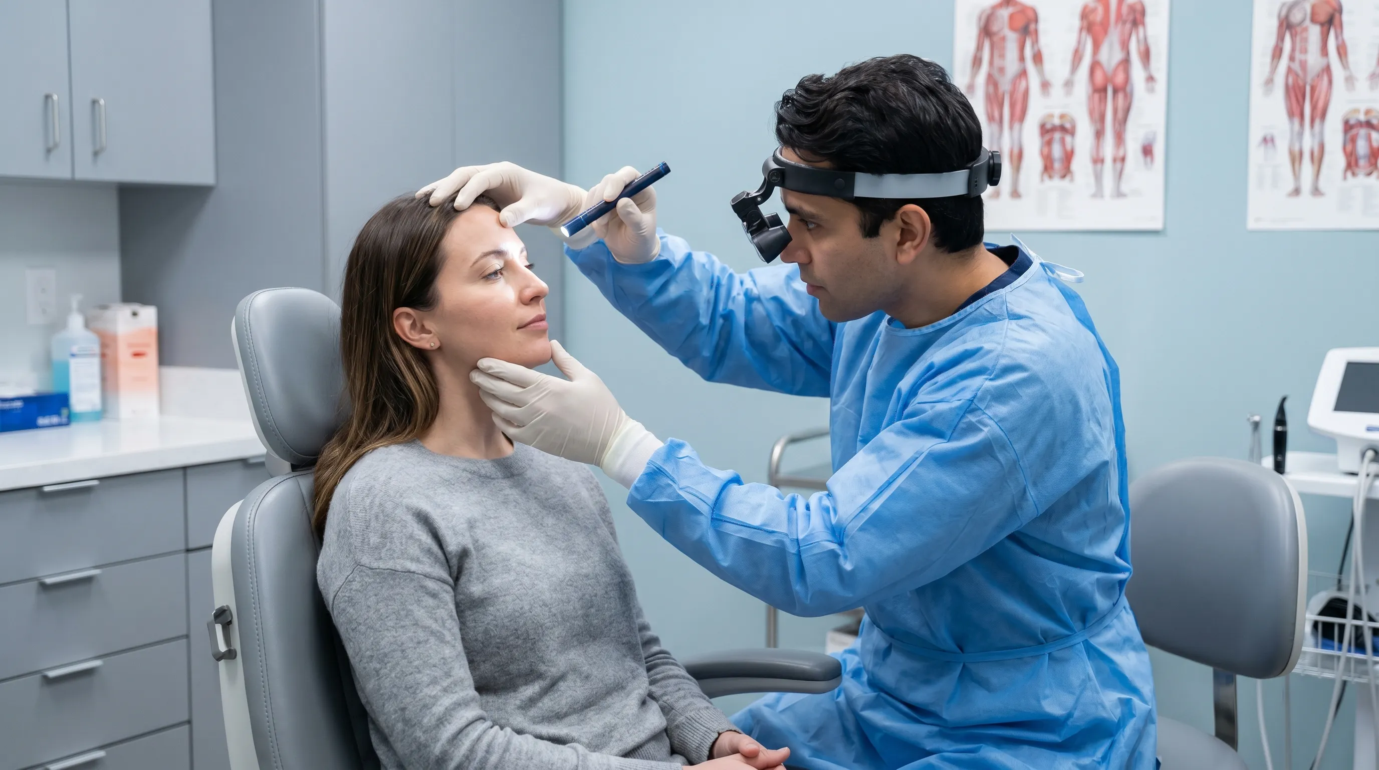
Modern diagnostic imaging, advanced pharmacological therapies, and minimally invasive surgical techniques have transformed orbital care, offering patients unprecedented outcomes and recovery trajectories. However, proactive prevention remains the most powerful tool in maintaining orbital health. Regular eye examinations, consistent use of certified protective eyewear, and prompt evaluation of unusual symptoms create a comprehensive defense against trauma and disease. Whether managing a chronic autoimmune condition or recovering from acute injury, collaboration with qualified ophthalmologists and oculoplastic specialists ensures personalized, evidence-based care. Prioritizing the health of your eyeball socket is not merely an aesthetic or functional investment; it is a foundational commitment to lifelong clarity, comfort, and quality of life.

About the author
Benjamin Carter, MD, is a board-certified otolaryngologist specializing in head and neck surgery, with an expertise in treating throat cancer. He is an associate professor and the residency program director at a medical school in North Carolina.