Sudden Blurry Vision in One Eye: Causes, Emergencies, and Expert Care

Imagine sitting at your desk, preparing for a routine meeting, when without warning, the world on one side of your field of view dissolves into a hazy, unfocused blur. You blink repeatedly, rub your eyes, and wonder if it is merely fatigue or a temporary screen-related issue. Yet, when the distortion persists, a wave of concern washes over you. Sudden blurry vision in one eye is a symptom that demands immediate attention, not dismissal. While it can occasionally stem from benign triggers like prolonged digital screen use or transient blood pressure fluctuations, it frequently signals an underlying ocular or systemic condition that requires prompt medical intervention. The human visual system is remarkably complex, integrating intricate networks of nerves, blood vessels, and delicate tissue layers that can be compromised in an instant. Understanding the mechanisms behind unilateral visual disturbances is the first step toward protecting your long-term sight and navigating the healthcare system effectively.
This comprehensive guide explores the clinical causes, diagnostic pathways, treatment options, and preventive strategies associated with abrupt monocular vision changes. You will learn how to distinguish between harmless episodes and sight-threatening emergencies, what to expect during an ophthalmic evaluation, and which lifestyle modifications significantly reduce your risk. By arming yourself with evidence-based knowledge, you can respond quickly, communicate clearly with medical professionals, and take proactive steps to safeguard your visual health.
Understanding Sudden Visual Changes and Ocular Physiology
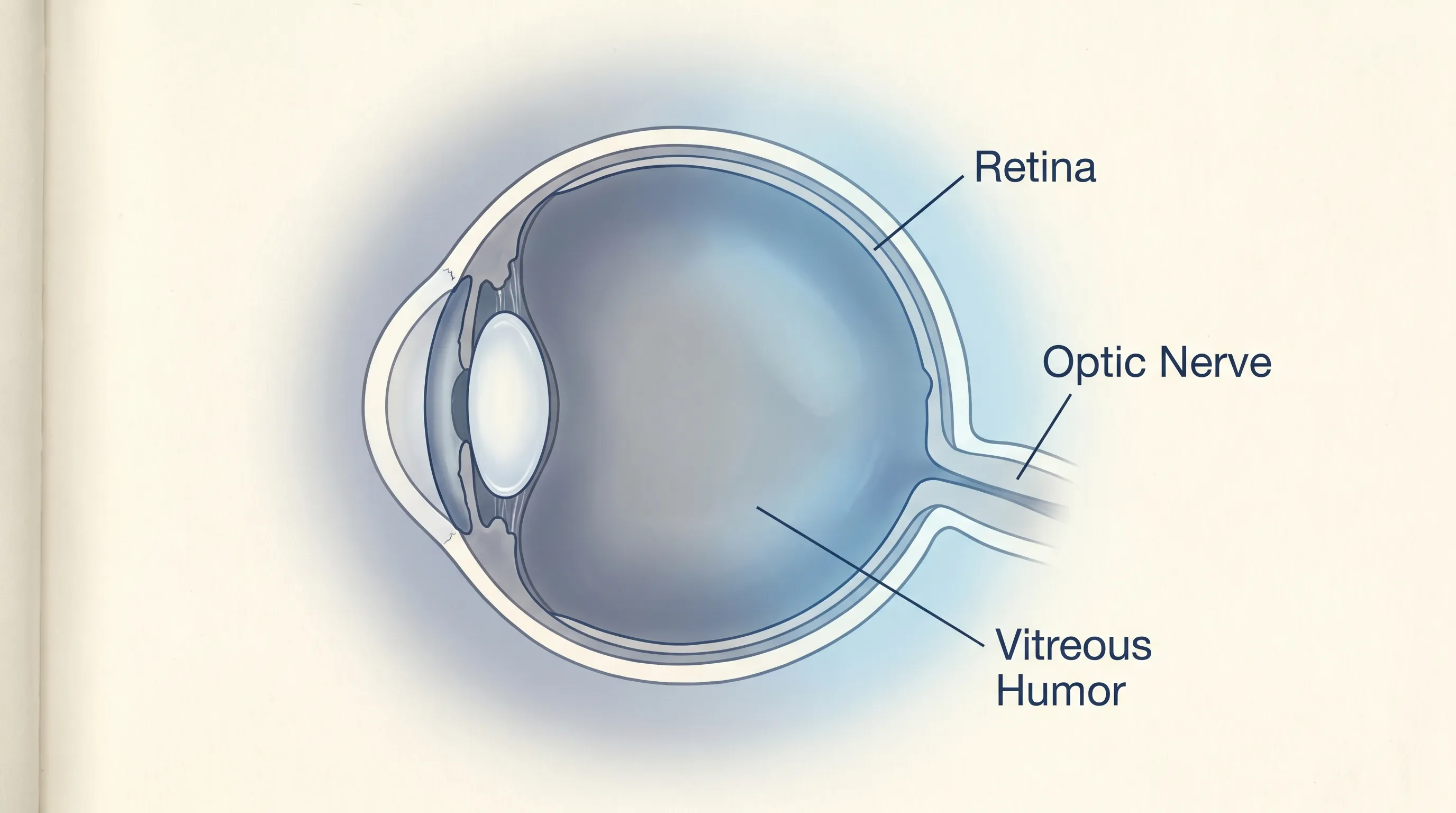
The eye functions as a sophisticated optical instrument, converting light waves into electrical signals that the brain interprets as coherent images. Light enters through the transparent cornea, passes through the pupil regulated by the iris, and is precisely focused by the crystalline lens. It then travels through the vitreous chamber before striking the retina, a thin layer of photoreceptor cells located at the back of the eye. These cells initiate neural impulses that travel along the optic nerve to the visual cortex. Any disruption along this pathway can manifest as sudden blurry vision in one eye, but the specific nature of the blurring often reveals which structure has been affected. For example, corneal irregularities cause surface-level distortion, while retinal or optic nerve issues create central or peripheral vision deficits.
Free calculatorMAP CalculatorMean Arterial Pressure calculatorOpen the calculatorHow the Eye Processes Light and Focus
Focusing light accurately requires seamless coordination between multiple anatomical components. The ciliary muscles adjust the lens curvature to shift focus between near and distant objects, a process known as accommodation. The tear film maintains a smooth corneal surface essential for light refraction. When these systems malfunction due to inflammation, vascular blockage, or mechanical detachment, light scatters improperly on the retina. Patients experiencing sudden blurry vision in one eye often report a cloudy film, a darkened shadow, or a complete loss of sharpness that does not improve with blinking. Understanding this optical cascade helps patients recognize that unilateral blurring is rarely a standalone phenomenon and usually indicates structural or vascular compromise.
Differentiating Monocular vs. Binocular Vision Loss
A critical clinical distinction lies in whether visual impairment affects one eye or both simultaneously. Binocular blurring typically points to neurological issues, systemic medication side effects, or bilateral metabolic fluctuations. Monocular blurring, however, isolates the problem to the specific eye, its anterior structures, the retina, or the optic nerve before it joins the chiasm. Clinicians rely on a simple covering test during initial evaluations to confirm unilaterality. If covering the unaffected eye eliminates the patient's complaint, but covering the symptomatic eye reveals normal vision, the issue is strictly localized. This differentiation dramatically narrows the diagnostic search and accelerates appropriate treatment protocols.
The Significance of Unilateral Symptoms
Unilateral symptoms carry substantial diagnostic weight because they bypass systemic causes and focus attention on localized pathology. Sudden blurry vision in one eye frequently correlates with vascular events like retinal artery occlusion, inflammatory conditions like optic neuritis, or mechanical complications like retinal tears. Unlike gradual vision decline associated with aging, abrupt unilateral changes rarely improve spontaneously. Medical guidelines universally recommend same-day evaluation for any unexplained monocular vision alteration, particularly when accompanied by pain, photophobia, or visual field restrictions. Recognizing this clinical urgency can mean the difference between temporary visual disturbance and permanent impairment.
Common Medical Causes of Sudden Monocular Blurring
Unilateral vision changes stem from a diverse spectrum of pathologies, ranging from acute vascular occlusions to chronic inflammatory diseases that suddenly exacerbate. Identifying the precise etiology requires correlating the onset timeline, accompanying symptoms, and patient medical history with clinical examination findings. The following conditions represent the most frequently diagnosed sources of abrupt unilateral visual impairment.
Retinal Detachment and Tears
The retina adheres tightly to the underlying retinal pigment epithelium and choroid. When this attachment weakens or experiences mechanical stress, a tear can develop, allowing vitreous fluid to seep underneath and peel the retina away from its nourishing blood supply. Patients typically report a sudden cascade of floaters, intermittent photopsia (flashes of light), and a progressive curtain-like shadow that correlates with sudden blurry vision in one eye. Risk factors include advanced myopia, prior ocular trauma, previous cataract surgery, and familial predisposition. Without prompt laser photocoagulation or pneumatic retinopexy, complete retinal detachment leads to irreversible photoreceptor death. For detailed clinical guidelines, refer to the National Eye Institute's comprehensive overview of retinal disorders.
Central Retinal Artery Occlusion (CRAO)
Often described as an eye stroke, CRAO occurs when an embolus or thrombus blocks the central retinal artery, instantly cutting off oxygenated blood flow to the inner retinal layers. Vision loss is characteristically profound, painless, and instantaneous, though some patients experience milder, transient blurring if the occlusion is partial. The affected eye displays a classic pale retina with a cherry-red spot at the fovea during fundoscopy. Immediate intervention focuses on lowering intraocular pressure and attempting embolus displacement, though therapeutic windows are notoriously narrow. Managing underlying cardiovascular risk factors like hypertension, hyperlipidemia, and carotid artery disease is essential for prevention. Review the Mayo Clinic's clinical guidance on retinal artery occlusion for emergency management protocols.
Optic Neuritis and Neurological Links
Inflammation of the optic nerve, most commonly triggered by autoimmune responses, disrupts signal transmission from the retina to the brain. Patients frequently experience subacute blurring over hours to days, accompanied by pain during eye movement and impaired color perception. Multiple sclerosis and neuromyelitis optica spectrum disorders are strongly associated with optic neuritis, making it a potential early indicator of broader neurological pathology. Diagnosis relies on MRI imaging revealing optic nerve enhancement and abnormal visual evoked potentials. High-dose corticosteroids accelerate recovery, while long-term management targets underlying autoimmune activity to prevent recurrence. The National Institutes of Health provides comprehensive resources on optic nerve inflammation.
Vitreous Hemorrhage and Floaters
Bleeding into the vitreous cavity obscures light transmission to the retina, causing sudden blurring, dark spots, or a reddish hue in the visual field. This condition frequently stems from proliferative diabetic retinopathy, posterior vitreous detachment with torn retinal vessels, or ocular trauma. While mild hemorrhages may clear spontaneously as macrophages absorb the blood, dense bleeds require surgical vitrectomy. Controlling blood glucose levels and blood pressure remains the cornerstone of preventing recurrent vitreous bleeding.
Acute Glaucoma Attacks
Primary angle-closure glaucoma manifests when the iris physically blocks aqueous humor drainage, causing intraocular pressure to spike rapidly. Patients report severe ocular pain, headache, nausea, halos around lights, and pronounced sudden blurry vision in one eye. The cornea becomes hazy due to epithelial edema, and the pupil often appears fixed and mid-dilated. This is a true ophthalmic emergency requiring immediate pressure-lowering medications like acetazolamide, topical beta-blockers, and ultimately laser peripheral iridotomy to restore normal aqueous outflow and prevent optic nerve damage. Consult the Cleveland Clinic's detailed overview of acute glaucoma management for further information.
Macular Degeneration Complications
While age-related macular degeneration typically progresses gradually, the wet form involves rapid choroidal neovascularization that leaks fluid or blood beneath the macula. This abrupt exudation distorts central vision, causing straight lines to appear wavy and reading to become difficult. Prompt anti-VEGF intravitreal injections can stabilize leakage and preserve functional acuity, highlighting the necessity of regular retinal screening for individuals over sixty. Learn more about macular degeneration treatment pathways from the NEI.
When Symptoms Indicate an Immediate Medical Emergency
Time is retinal tissue. The longer a structural or vascular compromise remains untreated, the higher the likelihood of permanent neural loss. Recognizing red flag combinations allows patients to bypass home monitoring and proceed directly to emergency ophthalmic care.
Red Flags That Demand Immediate Care
Seek emergency services or proceed directly to an urgent care ophthalmology clinic if sudden blurry vision in one eye is accompanied by any of the following: severe unilateral eye pain, sudden onset of numerous floaters with photopsia, a distinct shadow or curtain obscuring part of the visual field, profound vision loss that does not improve with blinking, or systemic symptoms like facial drooping, limb weakness, or speech difficulties indicating a possible cerebrovascular event. These clinical presentations require rapid imaging, pressure assessment, and specialist intervention within hours rather than days. For standardized symptom evaluation guidelines, refer to the Mayo Clinic's vision emergency protocols.
The Critical First 90 Minutes
For ischemic events like CRAO or acute glaucoma, the first ninety minutes represent a crucial therapeutic window. Retinal cells begin irreversible apoptosis within approximately ninety minutes of complete arterial occlusion. Rapid administration of ocular massage, anterior chamber paracentesis, or intraocular pressure-lowering agents can sometimes restore perfusion and salvage central acuity. Delayed presentation significantly diminishes prognosis, making public awareness of time-sensitive eye emergencies vital for community health.
Differentiating Transient vs. Persistent Blurring
Transient monocular blurring, often termed amaurosis fugax, typically resolves completely within minutes. It commonly results from temporary retinal ischemia due to emboli breaking off from carotid atherosclerotic plaques or transient hypotension. Persistent blurring lasting hours or days suggests structural damage, active inflammation, or hemorrhage. Patients should never assume transient episodes are benign, as they frequently serve as warning signs for impending permanent vision loss or cerebral stroke. Comprehensive vascular workup is mandatory following any episode of transient monocular blindness.
Diagnostic Procedures and Clinical Evaluation
Accurate diagnosis hinges on a systematic evaluation combining patient history, clinical examination, and advanced imaging modalities. Ophthalmologists employ a structured approach to isolate the anatomical site of dysfunction and determine the underlying pathophysiology.
Comprehensive Eye Examination
Initial assessment begins with visual acuity testing, pupillary response evaluation, and extraocular motility assessment. Slit-lamp biomicroscopy examines the cornea, anterior chamber, iris, and lens for inflammation, trauma, or opacification. Tonometry measures intraocular pressure to rule out glaucomatous crises. Dilated fundus examination allows direct visualization of the retina, macula, optic disc, and retinal vasculature, providing immediate clues regarding hemorrhage, detachment, or ischemia. Clinicians also assess contrast sensitivity, color vision, and visual field boundaries using Goldmann or Humphrey perimetry.
Imaging and Advanced Diagnostic Tools
Optical coherence tomography (OCT) generates high-resolution cross-sectional images of retinal layers, detecting subtle macular edema, epiretinal membranes, or early detachment with micron-level precision. Fluorescein angiography involves intravenous dye injection to map retinal blood flow, highlighting leaks, blockages, or abnormal neovascularization. B-scan ultrasonography evaluates the posterior segment when media opacities like dense hemorrhage or cataract prevent direct fundus visualization. These technologies transform subjective symptoms into objective, quantifiable data that guides precise therapeutic decisions. Learn more about modern ophthalmic imaging techniques from the National Institutes of Health.
Neurological and Vascular Assessments
When optic nerve involvement or ischemic etiology is suspected, systemic evaluation becomes imperative. MRI of the brain and orbits with gadolinium contrast identifies optic nerve inflammation, demyelination, or compressive lesions. Carotid duplex ultrasonography and echocardiography screen for embolic sources like atherosclerotic plaques or valvular thrombi. Blood panels assess inflammatory markers, autoimmune antibodies, coagulation profiles, and metabolic parameters. Multidisciplinary collaboration between ophthalmologists, neurologists, and cardiologists ensures comprehensive management of complex ocular presentations.
| Symptom Cluster | Likely Pathology | Primary Diagnostic Test | Immediate Intervention |
|---|---|---|---|
| Painless vision loss, cherry-red spot | Central Retinal Artery Occlusion | Fundoscopy, Carotid Ultrasound | Ocular massage, IOP lowering, ER referral |
| Painful blurring, halos, fixed pupil | Acute Angle-Closure Glaucoma | Tonometry, Gonioscopy | Acetazolamide, topical drops, laser iridotomy |
| Curtain shadow, flashes, floaters | Retinal Detachment | Dilated exam, OCT | Laser photocoagulation, pneumatic retinopexy |
| Pain with eye movement, color desaturation | Optic Neuritis | MRI orbits, Visual Evoked Potentials | IV corticosteroids, neurological workup |
| Sudden floaters, red-tinted haze | Vitreous Hemorrhage | B-scan ultrasound, Fundoscopy | Head elevation, observation, possible vitrectomy |
Treatment Pathways and Medical Interventions
Once the underlying etiology is confirmed, treatment shifts from diagnostic urgency to targeted therapeutic intervention. Management strategies vary significantly based on the anatomical structure affected, disease severity, and patient comorbidities. Evidence-based protocols emphasize rapid stabilization, anatomical restoration, and long-term prevention.
Emergency Procedures for Vascular Events
Ischemic ocular conditions require immediate hemodynamic optimization. Ocular massage temporarily lowers intraocular pressure, potentially dislodging emboli into peripheral retinal branches. Intravenous hyperosmotic agents like mannitol reduce vitreous volume, while anterior chamber paracentesis rapidly decompresses the globe. Hyperbaric oxygen therapy may be considered in select cases to temporarily oxygenate ischemic retina via choroidal circulation. Concurrent cardiovascular management includes antiplatelet therapy, statins, and blood pressure optimization to prevent recurrent embolization.
Surgical Interventions for Retinal Conditions
Retinal tears and detachments demand precise surgical repair. Laser photocoagulation or cryopexy creates localized scars that seal retinal breaks, preventing fluid ingress. Pneumatic retinopexy involves injecting a gas bubble to tamponade the tear while positioning the patient to facilitate retinal reattachment. Pars plana vitrectomy removes scarred or hemorrhagic vitreous, flattens the retina, and often combines with silicone oil or gas tamponade. Postoperative positioning, strict activity modification, and avoidance of nitrous oxide anesthesia (if gas tamponade is used) are critical for successful anatomical outcomes.
Pharmacological Treatments and Corticosteroids
Inflammatory and autoimmune conditions respond to targeted pharmacological regimens. Intravenous methylprednisolone followed by oral prednisone taper remains the standard for optic neuritis, accelerating functional recovery. Anti-VEGF agents like ranibizumab and aflibercept are administered via intravitreal injection to inhibit abnormal blood vessel growth and reduce macular leakage. Topical corticosteroids and cycloplegic drops manage anterior uveitis and ciliary spasm. Patients must adhere strictly to dosing schedules and attend regular intraocular pressure monitoring to prevent steroid-induced glaucoma.
Managing Underlying Systemic Diseases
Ocular pathology rarely exists in isolation. Diabetes mellitus, hypertension, hyperlipidemia, and autoimmune disorders frequently drive recurrent or bilateral eye complications. Tight glycemic control, evidenced by HbA1c targets below 7.0%, dramatically reduces microvascular damage progression. Blood pressure management below 130/80 mmHg protects retinal autoregulation. Statin therapy stabilizes atherosclerotic plaques, reducing embolic risk. Multidisciplinary coordination ensures that systemic disease modification runs parallel to ocular treatment, maximizing visual preservation and overall health outcomes. For detailed guidance on hypertensive retinopathy and systemic eye care, visit the Cleveland Clinic.
Lifestyle Modifications and Preventive Strategies
Prevention remains the most effective defense against sudden ocular events. While not all vision changes are avoidable, consistent lifestyle modifications significantly reduce risk factors and enhance ocular resilience. Integrating these practices into daily routines creates a sustainable foundation for long-term visual health.
Daily Eye Care and Protection Routines
Establishing consistent ocular hygiene minimizes environmental stressors. Wear wraparound sunglasses with 100% UVA/UVB protection during outdoor activities to prevent cumulative photochemical damage. Use safety-rated protective eyewear during sports, home improvement projects, or occupational tasks involving flying debris or chemical exposure. Implement the 20-20-20 rule during prolonged near-work: every twenty minutes, focus on an object twenty feet away for twenty seconds to reduce accommodative spasm and surface dryness. Proper contact lens hygiene, including strict adherence to replacement schedules and avoiding tap water exposure, prevents microbial keratitis.
Nutritional Support for Ocular Health
The retina possesses some of the highest metabolic rates in the body, demanding consistent antioxidant and micronutrient supply. A diet rich in dark leafy greens, colorful vegetables, fatty fish, and nuts delivers essential compounds like lutein, zeaxanthin, omega-3 fatty acids, and vitamins C and E. Clinical studies demonstrate that AREDS2-formulated supplements significantly slow macular degeneration progression in intermediate stages. Maintaining adequate hydration supports tear film stability and vitreous composition. Limiting processed sugars and saturated fats reduces systemic inflammation and microvascular dysfunction.
Managing Screen Time and Visual Fatigue
Prolonged digital device use disrupts normal blink patterns, reduces tear film stability, and induces accommodative strain. Adjust screen brightness to match ambient lighting, increase text size to reduce squinting, and maintain a viewing distance of at least twenty inches. Consider installing blue light filtering lenses or software adjustments, though clinical evidence for blue light blocking remains mixed compared to behavioral modifications. Ensure proper ergonomic setup with screens slightly below eye level to minimize lid aperture exposure and evaporative tear loss.

Regular Screening and Monitoring Protocols
Preventive ophthalmic care follows evidence-based screening intervals aligned with age, risk factors, and baseline health status. Adults with no risk factors require comprehensive dilated exams every two to four years between ages twenty and forty, with increased frequency as aging progresses. Individuals with diabetes require annual screening regardless of age or symptom presence. Hypertensive patients, those with high myopia, family history of glaucoma, or prior ocular trauma should undergo evaluation every one to two years. Documenting baseline visual acuity, intraocular pressure, and retinal appearance creates a longitudinal reference that accelerates detection of subtle pathological changes.
Frequently Asked Questions
Is sudden blurry vision in one eye always an emergency?
While not every episode results in permanent damage, it must be treated as a potential medical emergency. The symptom can indicate retinal detachment, vascular occlusion, or acute glaucoma, all of which have narrow treatment windows. Delaying evaluation risks irreversible neural loss. Always seek same-day ophthalmic assessment for abrupt unilateral vision changes.
How do I differentiate between an eye strain and a serious condition?
Eye strain typically presents with bilateral discomfort, mild dryness, and resolves after rest or screen breaks. Serious conditions usually affect one eye, persist beyond several hours, worsen over time, or accompany pain, flashes, floaters, or visual field defects. If symptoms are strictly unilateral and unexplained, professional evaluation is mandatory.
Can high blood pressure cause sudden monocular blurring?
Yes. Hypertensive retinopathy can cause retinal artery spasms, vascular leakage, or even retinal vein occlusion, leading to abrupt unilateral visual impairment. Chronically elevated blood pressure damages the delicate microvasculature of the retina. Managing systemic hypertension through medication, diet, and stress reduction is essential for ocular protection.
What should I do while waiting to see a doctor?
Remain calm, avoid rubbing or applying pressure to the affected eye, and keep your head elevated if you suspect bleeding or increased intraocular pressure. Do not drive yourself to the appointment, as impaired vision poses safety risks. Arrange immediate transportation to an urgent care ophthalmology clinic or emergency department. Bring a list of current medications and relevant medical history.
Are there any supplements that help restore sudden vision loss?
Supplements cannot reverse acute structural damage or vascular occlusion. While formulations like AREDS2 support macular health and slow chronic degeneration, they are not emergency treatments. Sudden vision loss requires immediate medical intervention. Always consult an ophthalmologist before initiating any supplement regimen, as certain vitamins interact with blood thinners or alter intraocular pressure.
Conclusion
Sudden blurry vision in one eye is a symptom that should never be ignored or dismissed as temporary fatigue. The intricate architecture of the eye, combined with its reliance on precise vascular supply and neural signaling, means that abrupt changes frequently indicate underlying pathology requiring expert evaluation. From retinal detachment and ocular strokes to inflammatory optic neuropathies and acute glaucoma, the spectrum of potential causes demands rapid clinical assessment and targeted intervention. Understanding the warning signs, recognizing the urgency of the first ninety minutes, and knowing what diagnostic procedures to expect empowers patients to advocate effectively for their own care.
Long-term visual preservation extends beyond emergency response. It encompasses consistent preventive screenings, rigorous management of systemic conditions like diabetes and hypertension, nutritional optimization, and mindful daily habits that protect against environmental and digital stressors. By integrating these evidence-based strategies into your healthcare routine, you significantly reduce the likelihood of sudden ocular events and maintain functional vision throughout your lifetime. If you experience abrupt unilateral blurring, act swiftly, seek professional care immediately, and trust in modern ophthalmic medicine's ability to diagnose, treat, and protect your most vital sensory asset.
How we work
Researched and drafted with AI assistance, then reviewed by a human editor against the cited sources.
This article is general health information, not medical advice. Consult a qualified healthcare professional about your own situation.