Chest Pain After Workout: Causes, Red Flags, and Evidence-Based Management

Pushing your physical limits in the gym or on the track is a rewarding endeavor, but encountering unexpected discomfort can quickly turn triumph into anxiety. Experiencing chest pain after workout is a surprisingly common complaint among fitness enthusiasts, yet it rarely comes with a straightforward explanation. For some, it is a fleeting reminder of intense pectoral engagement or heavy compound movements. For others, it may signal a deeper physiological imbalance that requires immediate medical attention. Navigating this symptom requires a balanced, evidence-based approach that respects both the body’s adaptive capacity and its warning mechanisms. Understanding the precise origin of the discomfort—whether it stems from skeletal muscle microtrauma, joint inflammation, gastrointestinal reflux, or cardiovascular strain—is essential for making safe, informed decisions about recovery and future training. This comprehensive guide will walk you through the underlying physiology, clinical red flags, diagnostic pathways, and practical management strategies to help you safely return to peak performance while safeguarding your long-term heart health.
Understanding the Physiology of Exercise-Induced Chest Discomfort
The human chest is a complex anatomical region housing the heart, lungs, major blood vessels, the sternum, multiple rib articulations, and overlapping muscle layers including the pectoralis major and minor, serratus anterior, and intercostal groups. When you engage in structured exercise, blood flow is redirected toward working muscles, metabolic byproducts accumulate, and the autonomic nervous system shifts into a heightened sympathetic state. These physiological shifts are normal and necessary for adaptation, but they also create a cascade of responses that can manifest as discomfort in the thoracic region. Recognizing the difference between adaptive physiological stress and pathological strain begins with understanding how tissue mechanics and cardiovascular demand interact during and immediately following physical exertion.
Free calculatorAnion Gap CalculatorAcid-base status assessmentOpen the calculator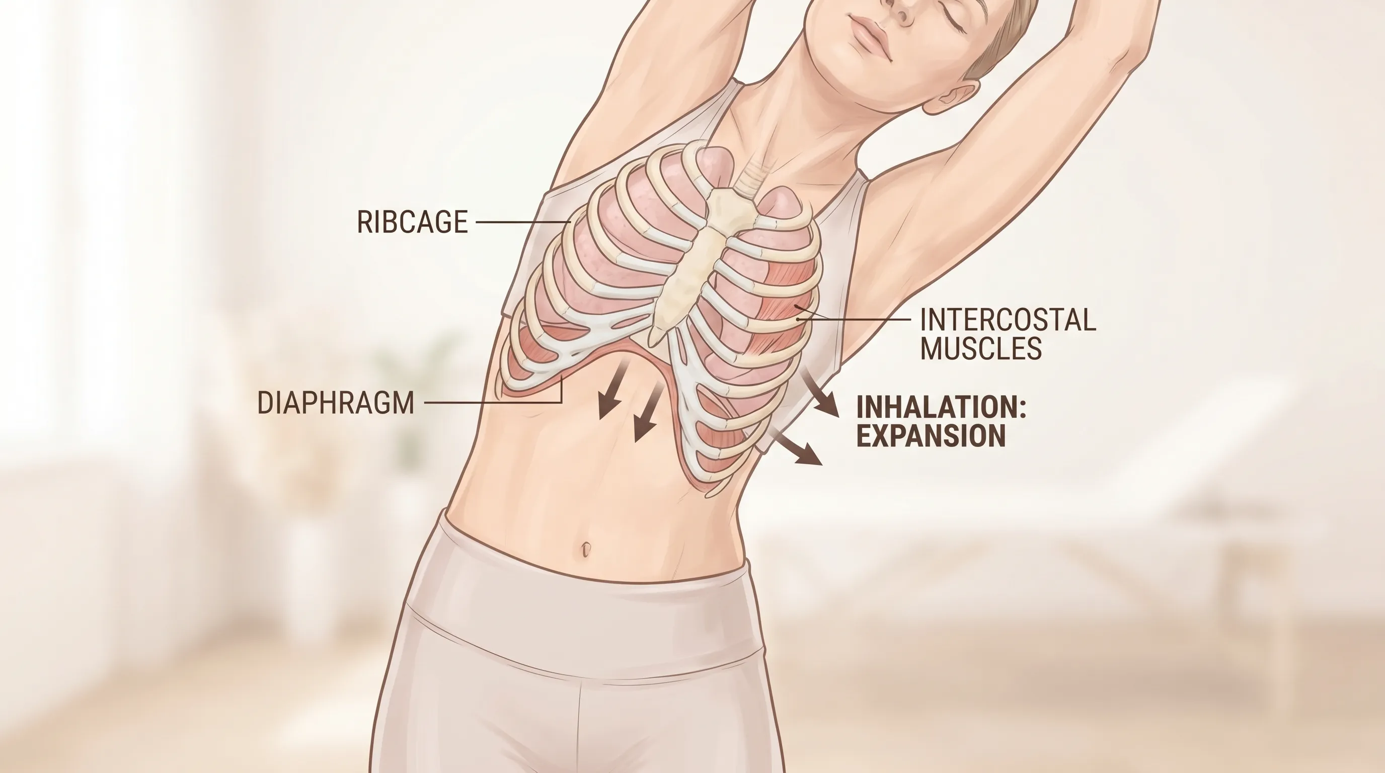
The Role of Cardiovascular Adaptation
During moderate to vigorous activity, cardiac output can increase three to fivefold to meet the oxygen and nutrient demands of active tissue. This surge requires the coronary arteries to dilate and deliver adequate perfusion to the myocardium. In healthy individuals, endothelial function supports this demand seamlessly. However, if underlying atherosclerosis is present, or if blood pressure spikes dramatically due to improper breathing or extreme exertion, the myocardial oxygen supply-to-demand ratio can become compromised. This mismatch, known as relative ischemia, often manifests as a dull ache, tightness, or pressure rather than a sharp sting. Learn more about exercise-induced myocardial strain and angina. Additionally, post-exercise vasodilation and sudden drops in venous return (especially if you stop abruptly) can trigger transient hypotension and compensatory tachycardia, which some interpret as chest discomfort. Learning to monitor heart rate variability, implement active cool-downs, and respect intensity thresholds significantly reduces the likelihood of these hemodynamic fluctuations.
Musculoskeletal vs. Cardiac Pain Mechanisms
Differentiating pain origins relies on understanding nerve pathway divergence. Somatic pain, originating from muscles, tendons, fascia, or cartilage, travels through well-localized spinal nerves and typically presents as sharp, stabbing, or aching sensations that worsen with movement, palpation, or specific postures. Visceral pain, originating from internal organs like the heart or esophagus, travels through autonomic nerve fibers and tends to be diffuse, poorly localized, and often described as pressure, burning, or heaviness. When you experience chest pain after workout caused by a strained intercostal muscle or inflamed costosternal joint, the discomfort usually reproduces when you twist, cough, or press on the exact spot. Conversely, cardiac-related discomfort rarely changes with touch but may intensify with emotional stress, cold exposure, or sustained aerobic demand. Understanding this neurophysiological distinction prevents both unnecessary panic and dangerous complacency, allowing for more accurate self-assessment and appropriate clinical referral. Mayo Clinic guide to chest pain evaluation.
Differentiating Benign Causes from Serious Red Flags
Not every instance of thoracic discomfort following physical activity warrants an emergency department visit, yet dismissing warning signs can have severe consequences. Clinical experience and epidemiological data consistently show that the vast majority of post-exercise chest complaints stem from musculoskeletal strain, exercise-induced bronchospasm, or gastroesophageal reflux. However, because cardiovascular events remain the leading cause of exercise-related sudden morbidity, establishing clear boundaries between benign and high-risk presentations is non-negotiable. A structured approach to symptom tracking, combined with awareness of systemic red flags, empowers individuals to make safe decisions without resorting to guesswork.
| Symptom Characteristic | Likely Musculoskeletal/Gastrointestinal Origin | Potential Cardiac or Vascular Origin |
|---|---|---|
| Pain Quality | Sharp, stabbing, localized, reproducible | Pressure, squeezing, dull heaviness |
| Radiation | Stays localized to chest wall or ribs | Spreads to jaw, neck, left arm, back |
| Trigger/Aggravation | Movement, deep breathing, palpation, meals | Sustained exertion, stress, cold air |
| Associated Symptoms | Mild soreness, stiffness, belching, heartburn | Diaphoresis, nausea, dizziness, dyspnea |
| Response to Rest | Gradual relief over hours/days | Persistent or worsening despite rest |
Common Non-Cardiac Triggers
Pectoral and intercostal muscle overuse is the most prevalent non-cardiac cause, particularly following push-ups, bench presses, overhead throws, or sudden changes in training volume. The microtrauma that occurs during eccentric loading triggers localized inflammation and delayed onset muscle soreness (DOMS), which peaks 24 to 48 hours post-activity. Costochondritis, an inflammation of the cartilage connecting ribs to the sternum, frequently mimics cardiac pain but responds well to targeted stretching, anti-inflammatory protocols, and load modification. Exercise-induced bronchoconstriction narrows the airways during intense aerobic efforts, producing a burning chest sensation alongside wheezing and coughing. CDC information on managing exercise-induced airway restriction. Finally, heavy lifting increases intra-abdominal pressure, which can force gastric contents upward, triggering acid reflux that feels indistinguishable from cardiac discomfort without clinical correlation. NIH resources on gastroesophageal reflux disease (GERD). Identifying these triggers through movement analysis and symptom mapping is the first step toward targeted intervention.
When to Recognize Cardiac Warning Signs
Cardiovascular compromise should always be treated as the primary rule-out condition, especially in individuals over 40, those with a family history of premature coronary disease, smokers, diabetics, or sedentary individuals who recently initiated high-intensity training. Angina equivalents may present atypically in women and older adults, sometimes manifesting as profound fatigue, indigestion-like discomfort, or isolated shortness of breath rather than classic crushing chest pain. If discomfort persists beyond twenty minutes of rest, occurs at baseline without recent exertion, or escalates rapidly in intensity, it requires immediate emergency evaluation. Recognizing heart attack warning signs and symptoms can be life-saving. Diagnostic markers such as elevated high-sensitivity troponin, ischemic electrocardiogram changes, or abnormal wall motion on echocardiography are definitive indicators that cannot be ignored. Delaying care under the assumption that symptoms are muscle-related significantly increases morbidity risk.
Gastrointestinal and Respiratory Contributors
The thoracic cavity shares innervation and anatomical proximity with both the digestive tract and pulmonary system. Heavy compound lifts, inverted positions, or high-impact running can disrupt lower esophageal sphincter function, allowing hydrochloric acid to irritate sensitive mucosal tissue. This creates a retrosternal burning sensation that often worsens when lying down and improves with upright positioning or antacids. Simultaneously, exercise-induced asthma or vocal cord dysfunction restricts airflow, forcing accessory breathing muscles to work overtime. This compensatory overuse leads to anterior chest wall fatigue that many misinterpret as cardiac strain. Recognizing meal timing, hydration status, and breathing patterns as contributing variables allows for simple, highly effective modifications like avoiding large meals within two hours of training, incorporating extended warm-ups, and prioritizing nasal-dominant breathing during moderate sessions.
Evidence-Based Assessment and Diagnostic Pathways
When self-assessment proves inconclusive, clinical evaluation provides definitive answers through standardized protocols validated by major cardiology and sports medicine organizations. The diagnostic pathway for thoracic discomfort begins with a thorough history, physical examination, and baseline vitals, followed by targeted investigations based on pre-test probability. Understanding what to expect during a clinical workup reduces anxiety and encourages proactive health management. Physicians utilize risk stratification algorithms to determine whether symptoms warrant outpatient monitoring, immediate imaging, or urgent intervention. Familiarity with these pathways ensures that you can advocate for appropriate testing without requesting unnecessary procedures.
Clinical Evaluation Protocols
Standard clinical assessment follows the OPQRST mnemonic (Onset, Provocation, Quality, Radiation, Severity, Time) combined with a focused cardiovascular and musculoskeletal exam. Blood pressure, oxygen saturation, and resting heart rate establish baseline hemodynamic status. Auscultation detects murmurs, rubs, or crackles that may indicate valvular pathology, pericarditis, or pulmonary involvement. If musculoskeletal origin is suspected, physicians will perform palpation and range-of-motion testing to reproduce symptoms. Cardiac probability dictates subsequent steps: low-risk patients may receive reassurance and conservative management, while intermediate-to-high risk individuals proceed to objective testing. Cleveland Clinic guidelines on comprehensive chest pain evaluation emphasize that atypical presentations in active adults still require baseline electrocardiography and lipid profiling to establish long-term cardiovascular risk baselines.
Self-Assessment Checklists
Before stepping into a clinic, tracking symptom patterns provides invaluable data for healthcare providers. Maintain a training log that records exercise type, duration, intensity, nutrition timing, and exact symptom onset. Note whether pain correlates with specific movements, breathing depths, or emotional states. Rate severity on a 0-10 scale and document response to rest, positioning, and over-the-counter remedies. This structured tracking eliminates recall bias and accelerates diagnostic accuracy. If symptoms consistently follow heavy upper-body sessions, improve with stretching, and lack systemic features, they are highly likely musculoskeletal. Conversely, if discomfort appears unpredictably, escalates with minimal effort, or clusters around high-stress periods, it warrants expedited medical review.
Imaging and Stress Testing Explained
Objective diagnostic tools eliminate uncertainty when clinical history remains ambiguous. Electrocardiography captures electrical conduction abnormalities and prior infarction markers. Stress testing, either treadmill or pharmacologic, evaluates coronary flow reserve under controlled exertion. Echocardiography visualizes chamber size, valve function, and wall motion in real time. Computed tomography coronary angiography (CTCA) provides non-invasive plaque burden assessment with high sensitivity. While imaging is not routinely indicated for isolated, clearly mechanical discomfort, it becomes essential when red flags exist, risk factors cluster, or symptoms recur despite conservative management. Understanding the purpose and limitations of each modality prevents overreliance on single tests and supports a comprehensive, longitudinal approach to cardiac and musculoskeletal health.
Immediate Response and Acute Management Strategies
How you respond in the first fifteen to thirty minutes of symptom onset dictates both short-term safety and long-term tissue recovery. Panic elevates heart rate and muscular tension, potentially exacerbating both benign and serious presentations. Implementing a calm, structured response protocol minimizes secondary stress and allows accurate symptom monitoring. Whether the origin is muscular, respiratory, or cardiovascular, immediate management follows a hierarchy of stabilization, assessment, and escalation based on clinical evolution.
First Steps When Pain Strikes
Upon noticing chest pain after workout, immediately cease all physical exertion and assume a stable, supported position—typically seated with back support or standing leaning slightly forward. Avoid lying completely flat if respiratory or reflux components are suspected. Initiate slow, rhythmic diaphragmatic breathing: inhale through the nose for four seconds, hold for two, exhale through pursed lips for six. This technique stimulates vagal tone, lowers sympathetic drive, and reduces intercostal muscle guarding. Monitor pulse rate and oxygen saturation if a wearable is available. Record exact symptom quality, location, and any accompanying features. If discomfort remains isolated, sharp, and unchanged after ten minutes of quiet rest, gentle thoracic mobility drills may be cautiously introduced. If symptoms intensify, radiate, or trigger autonomic responses, activate emergency services immediately.
Rest, Recovery, and Active Rehabilitation
Once acute discomfort stabilizes, a phased recovery strategy prevents re-injury and restores functional capacity. The initial 48 hours prioritize inflammation control through relative rest, meaning you avoid aggravating movements while maintaining gentle lower-body circulation and light walking. After pain-free baseline returns, introduce dynamic stretching targeting the pectorals, lats, thoracic spine, and serratus anterior. Foam rolling along the lateral ribcage and anterior shoulder complex reduces myofascial adhesions that perpetuate tension patterns. Progressive reloading begins with low-intensity, high-rep movements that emphasize mind-muscle connection over absolute force. Only when full pain-free range of motion, symmetric strength, and normalized breathing mechanics return should you advance to moderate loading. Rushing this timeline is the most common cause of recurrent strain.
Over-the-Counter Interventions and Precautions
Pharmacological support can accelerate recovery when used appropriately, but improper application masks warning signs and delays diagnosis. Nonsteroidal anti-inflammatory drugs (NSAIDs) like ibuprofen or naproxen effectively reduce intercostal inflammation and costochondritis pain when taken with food for short durations. Topical diclofenate or menthol-based gels provide localized relief without systemic gastrointestinal impact. Antacids or H2-receptor antagonists address reflux-triggered burning that mimics cardiac pain. However, these interventions must never be used to push through persistent, unexplained, or escalating discomfort. Masking symptoms during heavy training increases tissue damage risk and delays recognition of underlying pathology. Always consult a physician before initiating regular NSAID use, particularly if you have renal, gastrointestinal, or cardiovascular comorbidities.
Long-Term Prevention and Training Optimization
Sustainable fitness progression relies on intelligent programming that respects tissue adaptation timelines, biomechanical limits, and autonomic recovery capacity. Chest pain after workout frequently emerges from training errors rather than structural pathology. Addressing these foundational variables through evidence-based periodization, technical refinement, and physiological monitoring transforms vulnerability into resilience. Prevention is not about avoiding intensity; it is about applying intensity strategically.

Progressive Overload and Warm-Up Science
Progressive overload requires gradual increases in volume, intensity, or frequency to stimulate adaptation without overwhelming connective tissue tolerance. Sudden jumps in training load bypass collagen remodeling timelines, increasing microtrauma risk exponentially. Implementing the RAMP protocol (Raise, Activate, Mobilize, Potentiate) establishes a 10-15 minute neuromuscular preparation phase that elevates core temperature, lubricates joint surfaces, and primes motor pathways. Dynamic movements like arm circles, band pull-aparts, and scapular wall slides specifically target anterior chest wall mobility. Research consistently demonstrates that structured warm-ups reduce musculoskeletal injury incidence by over 30 percent. Explore evidence-based exercise safety and warm-up guidelines. Pairing warm-up consistency with logarithmic load progression (e.g., 5-10 percent weekly increases) creates predictable adaptation curves that minimize unexpected symptom flare-ups.
Breathing Mechanics and Core Engagement
Breathing is the hidden foundation of thoracic stability and force transfer. The Valsalva maneuver, commonly used during maximal lifts, dramatically increases intrathoracic pressure and can compress coronary vessels or strain costochondral junctions when misapplied. Mastering diaphragmatic breathing ensures optimal ribcage expansion, maintains intra-abdominal pressure balance, and reduces accessory muscle fatigue. Practice coordinated breath timing: exhale during concentric (lifting) phases, inhale during eccentric (lowering) phases, and maintain continuous airflow during sustained holds. Incorporate dedicated respiratory training using incentive spirometry or paced breathing drills 3-4 times weekly. Cleveland Clinic guidance on proper lifting mechanics and breath control highlights how strengthening the deep core stabilizers (transversus abdominis, multifidus, pelvic floor) creates a rigid midsection that distributes load away from the anterior chest, significantly reducing mechanical strain during compound movements.
Nutritional Support and Electrolyte Balance
Metabolic fatigue and electrolyte imbalances directly influence muscular excitability and pain perception. Magnesium deficiency, highly prevalent among endurance athletes and frequent gym-goers, impairs calcium clearance from muscle cells, prolonging contraction cycles and increasing cramping likelihood. Potassium and sodium losses through heavy sweating disrupt action potential thresholds, causing irregular twitching and localized soreness. NIH Office of Dietary Supplements on magnesium intake recommends prioritizing whole-food sources like leafy greens, almonds, bananas, and coconut water, supplemented strategically based on sweat rate testing and dietary gaps. Timing carbohydrates and electrolytes within the peri-workout window (60 minutes before and 30 minutes after) stabilizes blood glucose, reduces cortisol spikes, and accelerates glycogen replenishment. Chronic underfueling or aggressive caloric deficits compound recovery delays, making minor strain feel disproportionately painful. Aligning macronutrient intake with training demands transforms recovery from an afterthought into a predictable physiological process.
Lifestyle Modifications for Sustainable Cardiovascular Health
Exercise does not exist in a vacuum. The body’s ability to tolerate training stress depends on cumulative lifestyle factors including sleep architecture, chronic stress levels, nutritional quality, and environmental exposures. Optimizing these variables reduces baseline inflammation, improves autonomic balance, and creates a resilient physiological foundation that minimizes exercise-related symptom flare-ups. Long-term prevention extends far beyond the gym walls.
Stress Management and Autonomic Balance
Chronic psychological stress maintains elevated cortisol and catecholamine levels, which impair endothelial function, increase resting heart rate, and lower pain thresholds. The autonomic nervous system requires deliberate parasympathetic activation to restore homeostasis after sympathetic training stress. Implement daily practices that stimulate the vagus nerve: extended exhalation breathing, mindfulness meditation, cold facial immersion, or gentle yoga flows. Limit digital overstimulation and high-intensity screen exposure within two hours of bedtime. Regularly assess your stress-to-recovery ratio using heart rate variability (HRV) tracking. Consistently suppressed HRV indicates systemic fatigue that demands training deload regardless of motivation levels. Honoring recovery signals prevents chronic sympathetic dominance that frequently manifests as unexplained chest tightness and exercise intolerance.
Sleep Architecture and Recovery Metrics
Sleep is the single most potent anabolic and restorative intervention available. Deep non-REM sleep triggers growth hormone release, facilitates cellular repair, and clears metabolic waste products from muscle tissue and neural pathways. Fragmented sleep or chronic sleep debt increases inflammatory cytokine production, slows tissue remodeling, and amplifies pain perception through central sensitization mechanisms. Maintain consistent sleep-wake cycles, optimize bedroom temperature between 60-67 degrees Fahrenheit, and limit caffeine intake after 2:00 PM. Track sleep duration and quality alongside training load. CDC sleep recommendations for optimal recovery emphasize that when recovery metrics consistently trail performance demands, proactively reducing volume or intensity until balance restores is critical. Sustainable progress requires viewing rest not as the absence of training, but as the active foundation that enables it.
Frequently Asked Questions
Is it normal to feel sharp chest pain after workout?
Sharp, localized chest pain that worsens with movement, twisting, or deep inhalation is typically musculoskeletal in nature, often resulting from intercostal muscle strain, pectoral microtrauma, or costochondral inflammation. These tissues bear significant mechanical load during upper-body training and require adequate recovery time. However, any persistent, unexplained, or pressure-like discomfort should be clinically evaluated to exclude cardiac or pulmonary involvement. Tracking symptom onset, reproducibility, and associated features helps differentiate adaptive soreness from pathological warning signs.
How do I know if my chest pain after workout is muscular or heart-related?
Muscular pain is usually well-localized, reproducible through palpation or specific movements, and lacks systemic symptoms. It typically follows a predictable timeline linked to recent training volume or new exercise introduction. Cardiac discomfort tends to present as diffuse pressure, heaviness, or burning, often radiating to the jaw, neck, left arm, or upper back. It is rarely affected by touch but may worsen with sustained exertion, emotional stress, or cold exposure. Accompanying features like cold sweats, nausea, lightheadedness, or disproportionate shortness of breath strongly favor cardiovascular evaluation over musculoskeletal assumptions.
Should I stop exercising completely after experiencing chest discomfort?
Complete cessation is unnecessary for clearly mechanical, mild soreness that resolves with rest and mobility work. Temporary modification of training variables—reducing load, avoiding aggravating planes of motion, and extending warm-ups—is sufficient for musculoskeletal recovery. However, if pain is severe, unpredictable, occurs at rest, or clusters with systemic warning signs, halt all exertion immediately and seek medical clearance. Training through unexplained symptoms increases re-injury risk and delays diagnosis of underlying conditions. Use objective recovery markers rather than motivation alone to guide return-to-activity decisions.
Can pre-workout supplements cause chest pain after workout?
Yes, many popular pre-workout formulations contain high doses of stimulants like caffeine, synephrine, yohimbine, and beta-alanine, which significantly elevate sympathetic nervous system activity. This increases heart rate, blood pressure, and myocardial oxygen demand, potentially triggering palpitations, arrhythmias, or ischemic-type discomfort in susceptible individuals. Combining multiple stimulant sources or consuming supplements without adequate food or hydration amplifies cardiovascular strain. Always start with half the recommended dose, cycle off stimulants periodically, and prioritize whole-food energy sources when experiencing recurrent post-exercise discomfort.
When should I go to the emergency room for post-exercise chest pain?
Activate emergency services immediately if discomfort lasts longer than fifteen to twenty minutes, occurs at rest without recent exertion, feels like crushing pressure or squeezing, radiates to the jaw or left arm, or is accompanied by diaphoresis, nausea, dizziness, syncope, or severe shortness of breath. Do not drive yourself to the hospital. Do not wait to see if symptoms improve. Time-critical cardiac interventions yield exponentially better outcomes when administered within the first golden hour. Err on the side of caution; false alarms are vastly preferable to delayed treatment.
Conclusion
Navigating chest pain after workout demands a balanced integration of physiological knowledge, clinical awareness, and disciplined self-monitoring. While musculoskeletal strain and benign training errors account for the majority of cases, the stakes of misidentifying cardiovascular compromise require unwavering respect for red flag symptoms and prompt medical evaluation when indicated. Implementing structured warm-ups, mastering breathing mechanics, aligning nutrition with training demands, and prioritizing sleep architecture creates a resilient foundation that minimizes future flare-ups and optimizes long-term athletic longevity. Track your symptoms objectively, modify training intelligently, and never sacrifice safety for short-term gains. Sustainable fitness progression relies on listening to your body’s feedback loops, applying evidence-based recovery protocols, and partnering with healthcare professionals when warning signs emerge. By honoring both intensity and recovery, you transform uncertainty into confidence, ensuring that every training session builds strength without compromising health.
How we work
Researched and drafted with AI assistance, then reviewed by a human editor against the cited sources.
This article is general health information, not medical advice. Consult a qualified healthcare professional about your own situation.