Narrow Angle Eye: Comprehensive Guide to Anatomy, Symptoms, and Evidence-Based Treatment
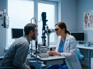
Vision is one of our most vital senses, and preserving its health requires understanding the complex structures that make up the human eye. When patients hear the term narrow angle eye, it often triggers concern, and rightfully so. This condition refers to an anatomical variation where the drainage angle of the eye is physically constricted, potentially leading to dangerous pressure buildup and irreversible damage if left unmanaged. Unlike common refractive errors, a structurally restricted drainage pathway operates silently in many individuals, making routine screening and patient awareness absolutely critical. By exploring the underlying mechanisms, recognizing early warning signs, and understanding modern treatment protocols, you can take proactive steps toward safeguarding your ocular health. This comprehensive guide breaks down everything you need to know about angle configuration, diagnostic procedures, therapeutic interventions, and daily habits that support long-term vision preservation. Whether you have received a preliminary diagnosis or simply want to stay informed, the following information is designed to empower you with evidence-based insights and actionable strategies for optimal eye care.
Understanding the Anatomy and Mechanics of a Narrow Angle Eye
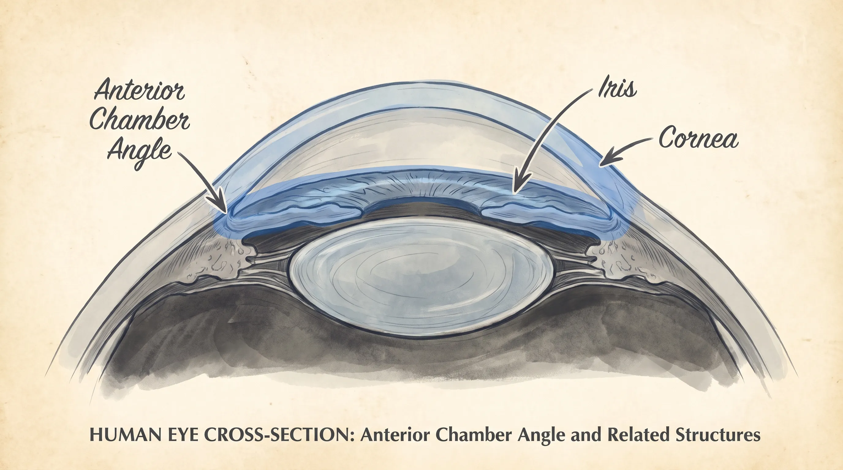
The human eye maintains a delicate fluid balance through a continuous cycle of aqueous humor production and drainage. Inside the front portion of the eye, the iris and the cornea meet to form a specialized structure known as the drainage angle. In a typical healthy eye, this space remains sufficiently wide, allowing fluid to flow freely through the trabecular meshwork and into Schlemm’s canal before exiting via the venous system. When the space narrows, however, the iris sits dangerously close to the cornea, physically obstructing this vital outflow pathway. This anatomical configuration is commonly referred to as a narrow angle eye. It is important to recognize that having a narrow angle does not automatically mean a patient will develop glaucoma, but it does significantly elevate the statistical risk for acute or chronic pressure-related complications. Genetic predisposition, age-related lens thickening, and certain ethnic backgrounds play substantial roles in determining angle width. Research from the National Eye Institute consistently highlights that individuals of East Asian, Native American, and Inuit descent face a higher prevalence of restricted anterior chambers, making demographic awareness a crucial component of preventive screenings. The physiological implications of this anatomical variation extend beyond simple fluid dynamics, influencing corneal clarity, iris mobility, and overall anterior segment stability.
Free calculatorBMI CalculatorCalculate Body Mass IndexOpen the calculatorWhat Is the Anterior Chamber Angle?
The anterior chamber angle is a three-dimensional corridor where multiple critical tissues converge. It houses the trabecular meshwork, a sieve-like network of collagenous beams that acts as the primary filtration site for intraocular fluid. Beneath this meshwork lies the scleral spur, which provides structural support, while the ciliary band and iris processes complete the complex architecture. Understanding this region is essential because fluid resistance at this junction determines intraocular pressure regulation. In eyes with ample space, the iris maintains a concave configuration, keeping peripheral tissue away from the meshwork. Conversely, a restricted angle forces the iris into a convex, forward-bowing position that physically blocks filtration sites. This mechanical obstruction initiates a cascade of physiological stress that can compromise optic nerve perfusion if unaddressed. Clinical grading systems, such as the Shaffer classification, quantify this space based on angular width, providing ophthalmologists with standardized metrics for risk stratification and treatment planning.
How a Narrow Angle Eye Develops Over Time
The development of this anatomical variant is typically multifactorial, involving both hereditary traits and age-related physiological changes. During early adulthood, the crystalline lens remains relatively thin and positioned posteriorly, maintaining a comfortable clearance between the iris and cornea. As we age, the lens naturally continues to grow in thickness, gradually shifting forward and pushing the iris ahead. This forward displacement progressively reduces the available drainage space, which is why the incidence of angle restriction peaks in individuals over the age of fifty. Additionally, hyperopia, or farsightedness, is strongly correlated with shorter axial lengths of the eye, which inherently creates a shallower anterior chamber. Environmental factors rarely cause the initial narrowing, but they can trigger sudden pressure spikes in those already predisposed. Understanding this progressive nature is essential for early intervention. When a narrow angle eye is identified during routine examinations, ophthalmologists can implement monitoring protocols that prevent sudden deterioration and preserve functional vision long-term. Regular documentation of anterior chamber depth allows clinicians to track subtle shifts and intervene before irreversible damage occurs.
Signs and Symptoms to Watch For
Clinical presentations vary dramatically depending on whether the condition manifests chronically or acutely. Chronic narrowing typically develops gradually, allowing the trabecular meshwork to adapt temporarily while subtle pressure elevations cause silent optic nerve stress. Patients in this category often report mild brow aching, occasional blurry vision after reading in dim light, or seeing faint halos around streetlights during evening hours. Because these symptoms are intermittent and easily dismissed as fatigue, chronic progression frequently goes unnoticed until standardized testing reveals compromised drainage. Conversely, acute closure represents a sudden, complete blockage of the outflow pathway. When the iris bows forward and completely seals the trabecular meshwork, intraocular pressure can skyrocket within minutes, creating a true ocular emergency. Classic indicators include severe ocular pain, nausea, vomiting, profound redness, fixed mid-dilated pupils, and sudden vision loss. Both scenarios stem from the same underlying narrow angle eye anatomy, but the clinical urgency and treatment pathways differ significantly. Recognizing this distinction helps patients respond appropriately rather than waiting out severe symptoms that could lead to permanent structural compromise.
Acute Angle Closure vs. Chronic Narrow Angles
Differentiating between these two presentations requires careful attention to symptom duration, intensity, and accompanying systemic responses. Acute angle closure typically strikes without warning, often occurring during prolonged periods of dark adaptation or emotional stress. The sudden pupillary dilation creates a complete seal, trapping aqueous humor in the posterior chamber and driving the iris forward. This mechanical blockade causes rapid pressure elevation, frequently exceeding forty millimeters of mercury. Patients describe an excruciating, deep-seated pain that may radiate into the forehead or cheek, frequently accompanied by systemic distress such as sweating, rapid heart rate, and gastrointestinal upset. In contrast, chronic narrowing progresses insidiously over months or years. The iris intermittently contacts the drainage tissue, causing temporary blockages that resolve spontaneously. While this may seem less dangerous, repeated micro-elevations cause cumulative damage to the trabecular meshwork and optic nerve head. The absence of dramatic pain during chronic phases often leads to delayed diagnosis. Clinical vigilance and comprehensive pressure mapping remain the only reliable methods for identifying chronic progression before irreversible vision loss occurs.
When to Seek Immediate Medical Attention
Any sudden onset of intense eye pain accompanied by systemic symptoms like nausea or headache warrants immediate evaluation at an emergency department or ophthalmology clinic. Delaying care by even a few hours during an acute crisis can result in permanent optic nerve fiber death. If you experience a rapid decline in visual clarity, especially when combined with rainbow-colored halos around lights, do not attempt self-diagnosis or wait for a scheduled appointment. Emergency interventions rely on rapid pressure reduction to restore blood flow to compromised retinal tissues. Additionally, individuals with a known narrow angle eye should carry a medical alert card or save emergency contact information in their phones, ensuring that bystanders or emergency personnel can communicate their ocular history quickly. Prompt recognition and rapid transport to specialized care facilities dramatically improve long-term visual outcomes and reduce the likelihood of permanent disability. Time is a critical factor in preserving functional vision during acute episodes, making patient education and preparedness absolutely essential.
Diagnostic Procedures and Testing
Accurate assessment begins with gonioscopy, a specialized examination technique that allows clinicians to directly visualize the drainage angle. Using a contact lens with internal mirrors, an ophthalmologist shines a light into the eye while observing how the iris and cornea align. This procedure categorizes the angle width using established grading scales, typically ranging from grade four, which is completely open and safe, down to grade one, where structures are dangerously crowded. Dynamic gonioscopy, which involves gently indenting the cornea, helps determine whether the narrowing is purely anatomical or if it involves synechiae, which are permanent adhesions that block fluid regardless of positioning. The American Academy of Ophthalmology emphasizes gonioscopy as an indispensable tool, recommending it for all patients over forty during comprehensive dilated exams. Because traditional slit-lamp examinations cannot adequately assess peripheral drainage spaces, this specialized contact technique remains the definitive method for confirming a narrow angle eye diagnosis. Comprehensive testing protocols combine structural visualization with functional pressure measurement to create a complete clinical picture.
Gonioscopy: The Gold Standard
Gonioscopy requires specialized training and precise technique, but it provides irreplaceable diagnostic value. During the procedure, topical anesthetic drops numb the ocular surface, and a coupling fluid eliminates light reflection between the lens and cornea. The examiner systematically scans the entire three-hundred-and-sixty-degree circumference, documenting pigment deposition, iris configuration, and visible trabecular bands. The presence of heavy trabecular pigment often indicates previous angle contact or intermittent closure events. Compression techniques during dynamic testing reveal whether peripheral iris tissue remains flexible or if permanent adhesions have formed. These findings directly influence treatment algorithms. For example, a narrow but flexible angle responds excellently to laser interventions, while extensive synechiae may necessitate surgical drainage procedures. Documenting gonioscopic findings over time allows clinicians to track anatomical changes and adjust therapeutic strategies before pressure-related complications develop. This direct visualization approach remains unmatched by non-contact alternatives in terms of structural detail.
Imaging and Advanced Diagnostic Tools
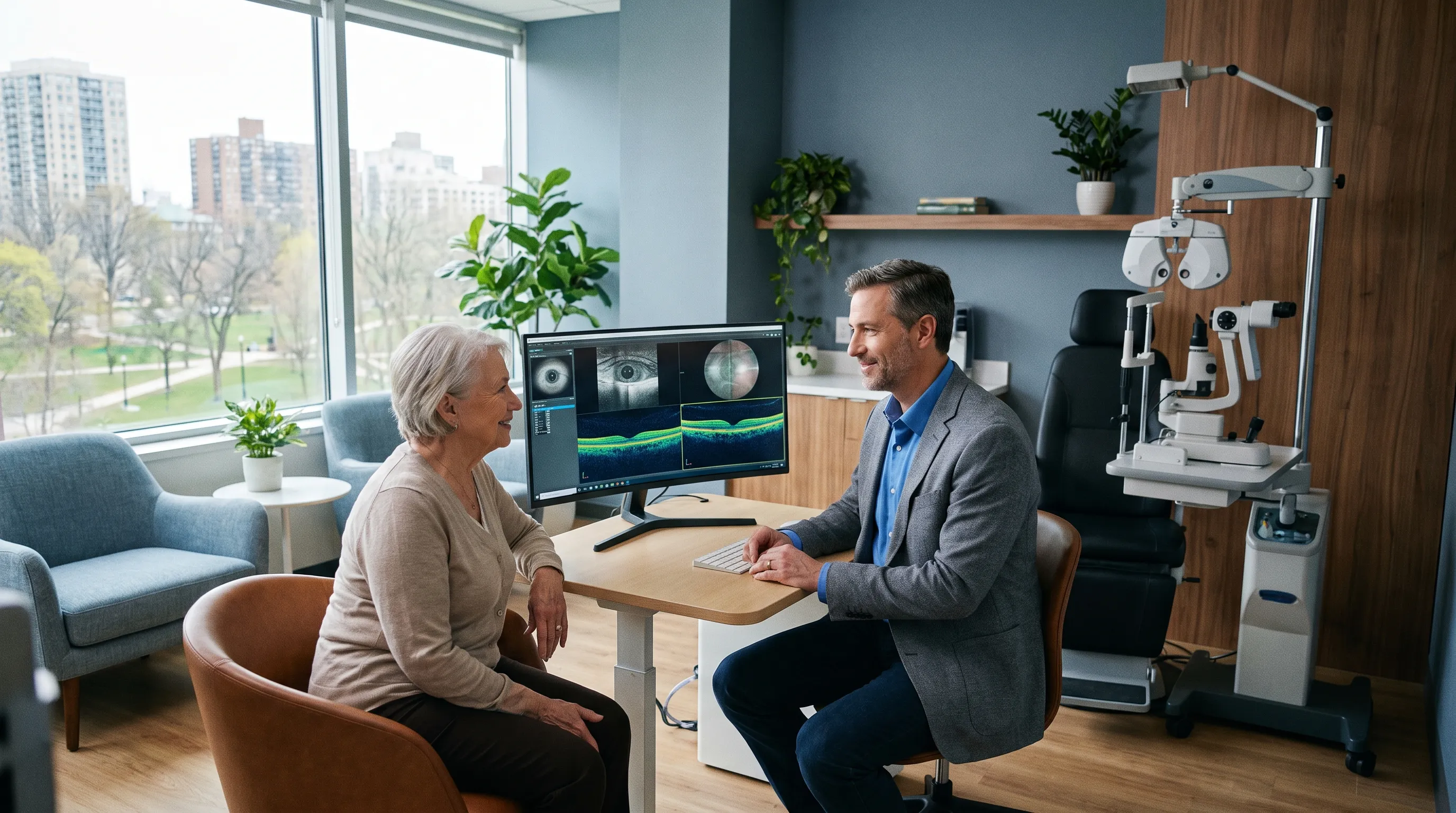
Modern technology has revolutionized how clinicians evaluate anterior segment structures. Anterior segment optical coherence tomography provides high-resolution, cross-sectional images without requiring physical contact. This noninvasive scan measures anterior chamber depth, lens vault thickness, and angle opening distance with remarkable precision. Ultrasound biomicroscopy serves as another advanced modality, particularly useful when corneal opacities prevent optical penetration. These imaging platforms enable specialists to predict the likelihood of progression, calculate surgical success probabilities, and tailor individualized monitoring schedules. Intraocular pressure tonometry, though unable to diagnose angle configuration directly, remains essential for tracking the functional impact of restricted drainage. Goldmann applanation tonometry continues as the reference standard, though newer rebound and dynamic contour devices offer complementary data. When combined with visual field testing and retinal nerve fiber layer analysis, these diagnostic tools create a comprehensive profile of ocular health. Integrating multiple testing modalities ensures that treatment decisions rest on robust, objective data rather than subjective symptom reporting alone.
Medical and Surgical Treatment Options
The most widely utilized preventive intervention for a narrow angle eye is laser peripheral iridotomy, an outpatient procedure that typically takes less than ten minutes. Using a specialized Nd:YAG laser, the surgeon creates one or two microscopic openings in the peripheral iris, establishing an alternative pathway for aqueous humor to flow from the posterior chamber into the anterior chamber. This pressure equalization causes the iris to flatten backward, immediately widening the drainage angle and significantly reducing the risk of acute closure. Most patients tolerate the procedure well, experiencing only mild transient discomfort and temporary light sensitivity. Post-procedure care involves a short course of anti-inflammatory drops and pressure-lowering medications until the eye stabilizes. The Mayo Clinic notes that early LPI intervention prevents permanent structural damage in the vast majority of cases, though lifelong monitoring remains necessary since some individuals may still develop elevated pressure from other mechanisms. Understanding the full spectrum of available therapies empowers patients to collaborate effectively with their healthcare teams.
Laser Peripheral Iridotomy (LPI)
LPI represents a minimally invasive breakthrough that has transformed preventive ophthalmology. The laser energy precisely disrupts peripheral iris tissue without damaging surrounding structures. Post-procedure protocols typically include topical corticosteroid drops administered four times daily for one week to minimize inflammation and prevent transient pressure spikes. Follow-up visits occur within twenty-four to forty-eight hours to verify laser patency and assess angle widening. In approximately ten percent of cases, laser channels may close spontaneously due to iris inflammation or residual pigment migration, necessitating repeat procedures. Patients often report improved peripheral vision and reduced episodic halos within days of successful treatment. Long-term studies demonstrate that timely LPI reduces acute closure incidence by over ninety percent. Despite these impressive outcomes, clinicians emphasize that laser treatment does not reverse existing optic nerve damage. Therefore, patients with advanced pressure-related changes require additional therapeutic interventions alongside iridotomy. Consistent adherence to post-procedure care guidelines maximizes anatomical benefits and minimizes recovery complications.
Medications for Pressure Management
When laser therapy is contraindicated, insufficient, or used as an adjunctive therapy, pharmacological management plays a central role. Miotic agents, particularly pilocarpine, work by contracting the iris sphincter muscle, mechanically pulling the iris away from the trabecular meshwork. While effective for temporary stabilization, these drops require frequent administration and can induce significant accommodative spasm, leading to brow aches and dim-light vision difficulties. Carbonic anhydrase inhibitors, available in topical and systemic formulations, reduce aqueous production by targeting enzymatic pathways in the ciliary body. Beta-blockers and prostaglandin analogs may be prescribed concurrently to maintain optimal pressure levels, especially if chronic glaucomatous damage has already begun. Medication adherence is critical; skipping doses or abruptly stopping treatment can trigger dangerous rebound pressure spikes. Patients should always maintain an updated medication list and discuss potential systemic side effects, particularly if managing cardiovascular or respiratory conditions alongside ocular therapy. Proper drop administration techniques, including punctal occlusion and eyelid closure after instillation, improve drug absorption while minimizing systemic exposure.
Surgical Interventions
In cases where laser procedures fail to adequately widen the drainage space or when lens-induced crowding dominates the pathology, surgical extraction becomes necessary. Phacoemulsification removes the natural lens, eliminating the primary source of anterior displacement and immediately deepening the anterior chamber. Modern micro-incision techniques allow for rapid recovery and excellent refractive outcomes, with many patients experiencing simultaneous correction of presbyopia or astigmatism through premium intraocular lens implantation. For individuals with established synechial closure, glaucoma drainage device implantation or minimally invasive glaucoma surgeries may be required to bypass damaged outflow pathways entirely. Surgical candidacy depends on comprehensive preoperative evaluations, corneal health status, and overall medical fitness. The Glaucoma Research Foundation highlights that early surgical planning prevents irreversible nerve damage and preserves quality of life, particularly for patients who cannot tolerate prolonged medical therapy. Advanced surgical techniques continue to evolve, offering increasingly safer and more effective alternatives for complex anatomical cases.
| Feature | Open-Angle Glaucoma | Narrow Angle Configuration | Acute Angle Closure | Symptoms | Pain Level | Treatment Priority | Progression Rate |
|---|---|---|---|---|---|---|---|
| Anatomical Status | Wide drainage space | Restricted peripheral angle | Complete blockage | Variable | Generally absent | Intermittent discomfort | Severe, sudden |
Lifestyle Adjustments and Preventative Care
Certain everyday conditions can temporarily exacerbate iris crowding in susceptible individuals. Prolonged exposure to dim lighting causes pupil dilation, which thickens the peripheral iris and increases the risk of contact with the cornea. Individuals with a narrow angle eye should maintain well-lit environments while reading, watching screens, or performing detailed hobbies. Additionally, staying in dark movie theaters or participating in stargazing without proper ambient lighting should be approached cautiously. Emotional stress and anxiety also trigger sympathetic nervous system responses that dilate pupils and elevate intraocular pressure. Practicing mindfulness techniques, controlled breathing exercises, and adequate sleep hygiene helps stabilize autonomic responses that influence ocular fluid dynamics. Furthermore, avoiding prone or face-down sleeping positions reduces gravitational pressure on the anterior segment, promoting healthier overnight fluid circulation. Consistent implementation of these environmental modifications significantly lowers the probability of acute pressure spikes.
Environmental Triggers to Avoid
Beyond lighting and posture, several everyday exposures warrant attention. Extreme temperature fluctuations can temporarily alter vascular perfusion and fluid dynamics within the eye. Patients should avoid prolonged exposure to intense heat, such as saunas or hot yoga studios, without adequate hydration and cooling breaks. Certain recreational activities, like heavy weightlifting or intense isometric exercises, cause temporary systemic pressure surges that transmit to the ocular vasculature. Modifying workout routines to incorporate steady-state aerobic activity, such as brisk walking, cycling, or swimming, supports cardiovascular health without triggering dangerous intraocular spikes. Additionally, patients should remain cautious when traveling to high-altitude locations, where reduced atmospheric pressure and hypoxic conditions may influence fluid balance. Pre-travel consultations with an ophthalmologist allow for proactive medication adjustments and emergency preparedness planning. Awareness of these environmental variables transforms passive risk into manageable lifestyle factors.
Routine Eye Care Practices
Consistency in ocular maintenance dramatically improves long-term outcomes. Patients should schedule comprehensive dilated examinations at least annually, ensuring that drainage angles, optic nerve health, and visual fields remain thoroughly documented. Maintaining a detailed personal health journal that tracks pressure readings, medication adherence, and symptom fluctuations provides clinicians with valuable longitudinal data. Proper hydration supports overall systemic circulation, though excessive fluid consumption in short periods can temporarily elevate intraocular pressure. Individuals should aim for steady, moderate water intake distributed evenly throughout the day rather than consuming large volumes rapidly. Protecting the eyes from trauma with appropriate sports eyewear and workplace safety gear prevents secondary inflammatory responses that could further complicate angle anatomy. Finally, avoiding unnecessary over-the-counter medications containing anticholinergic or sympathomimetic properties, such as certain cold remedies and motion sickness treatments, prevents unintended pharmacological dilation. Building these habits into daily routines creates a sustainable foundation for lifelong vision preservation.
Medication and Supplement Considerations
Many commonly prescribed substances influence pupillary size and ciliary body function. Antidepressants, antihistamines, decongestants, and certain asthma inhalers can dilate pupils or alter aqueous humor dynamics. Before starting any new prescription or over-the-counter regimen, individuals with a known narrow angle eye must consult their prescribing physician and ophthalmologist to evaluate ocular compatibility. Omega-3 fatty acids, lutein, and zeaxanthin supplements support retinal health and may provide mild anti-inflammatory benefits, though they do not alter anatomical angle configuration. Patients should avoid unverified herbal remedies claiming to reverse structural narrowing, as these products lack rigorous clinical validation and may interact dangerously with prescribed pressure-lowering drops. Always prioritize evidence-based therapies and maintain transparent communication between all healthcare providers involved in your care. Nutritional support works best as a complementary strategy rather than a standalone intervention, emphasizing whole foods, antioxidant-rich vegetables, and balanced macronutrients that promote systemic vascular health.
Frequently Asked Questions
Can a narrow angle eye be reversed naturally?
No, anatomical narrowing cannot be corrected through diet, exercise, or natural remedies. The structural crowding results from lens positioning and anterior chamber depth, which require medical or laser intervention to modify safely. However, environmental modifications and strict medication adherence can effectively manage symptoms and prevent acute episodes.
How often should I be monitored if I have narrow angles?
Individuals with diagnosed narrow angles typically require comprehensive evaluations every six to twelve months, depending on the degree of restriction and whether preventive laser treatment has been performed. Those with a history of acute closure may need quarterly monitoring initially to ensure pressure stability and detect early glaucomatous changes.
Does wearing glasses cause or worsen a narrow angle?
Prescription eyewear does not alter internal ocular anatomy or influence drainage pathways. Corrective lenses simply adjust how light focuses on the retina. However, certain contact lens types or poorly fitted specialty lenses could theoretically affect tear film dynamics, so always discuss lens wear with your eye care provider.
Is surgery always necessary for treatment?
Surgery is not mandatory for every case. Many patients achieve excellent long-term stability with timely laser peripheral iridotomy combined with routine monitoring. Surgical extraction or drainage procedures are reserved for advanced crowding, established glaucomatous damage, or cases where laser intervention fails to provide adequate anatomical relief.
Can children be born with a narrow angle eye?
While congenital cases are relatively rare, certain developmental conditions and genetic syndromes can produce shallow anterior chambers in pediatric patients. Infantile glaucoma requires specialized evaluation and often involves different surgical approaches compared to adult management. Regular pediatric vision screenings help detect structural anomalies early.
Conclusion
Navigating the complexities of a narrow angle eye requires a proactive approach, combining regular clinical monitoring, timely interventions, and informed lifestyle choices. The anatomical configuration may predispose individuals to elevated pressure, but modern diagnostic tools and targeted therapies have transformed this once-feared condition into a highly manageable health priority. By recognizing early warning signs, adhering to prescribed treatment protocols, and avoiding environmental triggers that exacerbate pupillary dilation, patients can effectively preserve their visual function for decades. Open communication with ophthalmology specialists ensures that care remains personalized, adaptive, and aligned with the latest evidence-based standards. Ultimately, maintaining optimal eye health depends on consistent vigilance and educated decision-making. When you partner with qualified medical professionals and implement the practical strategies outlined throughout this guide, you take definitive control over your long-term ocular wellness. Prioritize comprehensive screenings, respect your body’s warning signals, and commit to sustainable care habits that protect one of your most precious senses.
How we work
Researched and drafted with AI assistance, then reviewed by a human editor against the cited sources.
This article is general health information, not medical advice. Consult a qualified healthcare professional about your own situation.