Blurry Vision After Cataract Surgery: Recovery Timeline, Causes & Expert Solutions

Undergoing cataract surgery is a highly effective and transformative procedure that restores clarity to millions of lives each year. Yet, the journey from the operating room back to crisp, everyday vision is rarely instantaneous. Many patients experience a period of cloudy or unfocused sight during recovery, which can feel alarming after months of anticipating clear eyesight. Understanding why blurry vision after cataract surgery occurs, how long it typically lasts, and when it signals a need for medical intervention is essential for a smooth healing experience. According to the National Institutes of Health (NIH), the human eye is an incredibly complex organ, and replacing its clouded natural lens with a precise artificial implant triggers a cascade of physiological responses. Fluid shifts, cellular migration, neuroadaptation, and tear film alterations all play a role in your postoperative visual quality. By following evidence-based care protocols and recognizing the boundaries between normal healing and potential complications, you can navigate your recovery with confidence. This comprehensive guide explores the science behind post-surgical vision changes, outlines realistic timelines, and provides actionable strategies to protect and optimize your eyes as they heal.
Understanding the Healing Process
The human cornea and intraocular tissues undergo significant structural changes during cataract extraction, making temporary visual disturbances an expected part of recovery. Surgeons make microscopic incisions, emulsify and remove the clouded natural lens, and insert a folded intraocular lens (IOL) into the capsular bag. Even with advanced microsurgical techniques and laser-assisted precision, the eye requires dedicated time to restore its optical integrity. Recognizing the biological mechanisms at work helps patients differentiate between routine healing and issues requiring intervention.
Free calculatorLab Values Reference ChartComplete reference chart for medical lab valuesOpen the calculatorThe Immediate Post-Operative Phase
During the first twenty-four to seventy-two hours following surgery, the cornea experiences transient edema caused by fluid absorption during phacoemulsification. This microscopic swelling alters the cornea's refractive index, temporarily scattering incoming light and producing a soft-focus or hazy visual effect. The eye also produces a higher volume of inflammatory mediators such as prostaglandins and cytokines as part of the natural wound-healing cascade. These substances increase vascular permeability, allowing plasma proteins and immune cells to migrate into the anterior chamber, which can further contribute to temporary blurriness after cataract surgery. Your surgeon will typically prescribe a combination of topical antibiotics and corticosteroids during this window to suppress excessive inflammation and prevent bacterial colonization. Patients who diligently follow the dosing schedule often notice rapid improvement as the corneal endothelium actively pumps fluid back into the anterior stroma and descemet membrane.
Additionally, pupil dilation and residual viscoelastic material used during surgery can cause light sensitivity and mild visual distortion in the initial days. These substances are naturally absorbed or aspirated, but trace amounts may take a few days to clear completely. Resting the eyes, avoiding strenuous activity, and maintaining a strict medication routine are foundational steps to minimizing early visual fluctuations. Most patients report that their vision sharpens progressively as swelling diminishes and the surgical incisions begin sealing at the cellular level.
How the Eye Adapts to Artificial Lenses
Modern cataract surgery replaces the compromised natural crystalline lens with a biocompatible intraocular implant designed to restore refractive focus. However, the optical characteristics of an artificial lens differ significantly from the biological lens you grew up with. The brain's visual cortex must undergo a process called neuroadaptation to reinterpret the new visual data stream. This cognitive adjustment period is particularly noticeable for patients receiving multifocal, extended depth of focus (EDOF), or toric IOLs. These advanced optics provide exceptional range of vision but often introduce temporary visual phenomena such as halos, glare, or mild blur as the brain learns to filter out competing focal points. Clinical data reviewed by the Mayo Clinic demonstrate that neuroadaptation can take anywhere from two weeks to three months, depending on lens design and individual cortical plasticity. Patients who rush the process or expect overnight perfection often misinterpret this neurological learning curve as a surgical complication. In reality, blurry vision after cataract surgery during this phase is frequently a sign of your visual system recalibrating rather than a structural failure. Wearing your prescribed temporary correction, maintaining consistent lighting conditions, and giving yourself grace during the adaptation window significantly improves long-term satisfaction.
Common Causes of Prolonged Blurry Vision
While transient cloudiness is standard, persistent or worsening blur beyond the expected recovery window warrants clinical evaluation. Several well-documented ocular conditions can delay visual stabilization, each requiring distinct management strategies. Identifying the underlying mechanism ensures targeted treatment rather than unnecessary anxiety.
Posterior Capsule Opacification Explained
The capsular bag that secures your new IOL retains microscopic epithelial cells from your original natural lens. Over time, these cells can proliferate, migrate, and undergo fibrotic transformation on the posterior capsule membrane. This process, known as posterior capsule opacification (PCO) or secondary cataract, affects approximately fifteen to twenty percent of patients within five years of surgery, per clinical data from the Cleveland Clinic. PCO gradually scatters light rays before they reach the retina, producing symptoms nearly identical to the original cataract: faded contrast, increased glare, and persistent blurry vision. Fortunately, PCO is not a surgical complication nor a failure of the original procedure; it is a normal biological response. Ophthalmologists resolve it efficiently with a neodymium-doped yttrium aluminum garnet (Nd:YAG) laser capsulotomy. This non-invasive, painless outpatient procedure creates a precise opening in the cloudy membrane, restoring clear optical pathways within minutes. Most patients experience immediate visual improvement, making PCO one of the most successfully treatable causes of delayed postoperative clarity.
Cystoid Macular Edema and Its Impact
Cystoid macular edema (CME) represents another recognized cause of prolonged visual disturbance following lens extraction. The macula, responsible for sharp central vision, relies on a tightly regulated blood-retinal barrier. Surgical inflammation can disrupt this barrier, causing fluid to leak into the extracellular spaces of the fovea and form microscopic cystoid spaces. This swelling distorts photoreceptor alignment, leading to central blurriness, wavy lines, or color desaturation. Irvine-Gass syndrome typically manifests four to six weeks postoperatively, though early onset can occur with preexisting vascular conditions like diabetes or retinal vein occlusion. Diagnosis requires optical coherence tomography (OCT), which provides cross-sectional retinal imaging to quantify fluid accumulation. Treatment protocols generally involve intensified topical NSAID therapy, oral acetazolamide, or periocular corticosteroid injections to reduce vascular leakage. The National Eye Institute (NIH) emphasizes that early detection and prompt anti-inflammatory intervention typically result in complete anatomical and functional recovery within three to six months.
Dry Eye Syndrome Following Surgery
The corneal surface depends on a stable, multi-layered tear film for smooth light refraction. Cataract surgery temporarily disrupts corneal innervation, particularly the superficial trigeminal nerve fibers that regulate basal tear production. Reduced corneal sensitivity decreases reflex tearing, while increased tear osmolarity and evaporation compromise the mucin and lipid layers. The resulting dry eye syndrome directly causes intermittent or constant blur, foreign body sensation, and burning discomfort. Research indicates that over thirty percent of patients experience clinically significant dry eye in the first three months post-surgery. The Mayo Clinic confirms that comprehensive management requires addressing the underlying tear film instability. Prescribed cyclosporine or lifitegrast eye drops reduce ocular surface inflammation, while punctal occlusion, omega-3 supplementation, and environmental humidity control support long-term tear production. Patients who proactively manage ocular dryness frequently notice dramatic improvements in their postoperative visual quality.
Recovery Timeline: What to Expect Week by Week
Understanding the chronological progression of visual restoration helps patients set realistic expectations and adhere to care protocols. While individual healing varies based on age, systemic health, surgical complexity, and IOL type, clinical studies consistently demonstrate predictable recovery milestones.
The First 24 to 72 Hours
Immediately after surgery, your eye will be covered with a protective shield to prevent accidental rubbing or pressure. Vision during this period is often described as foggy, overly bright, or slightly distorted due to dilating drops, corneal swelling, and residual inflammation. Many patients can already perceive shapes and recognize familiar faces, though fine details remain obscured. It is crucial to avoid water exposure, heavy lifting, or bending below the waist during these initial days to prevent intraocular pressure spikes. Sleeping with the protective shield and adhering strictly to the prescribed antibiotic-steroid drop regimen minimizes infection risk and accelerates epithelial healing. Most individuals notice a measurable improvement in clarity by day two or three as the cornea sheds excess fluid.
One to Four Weeks Post-Surgery
By the second week, inflammation typically subsides enough for visual acuity to improve substantially. Patients often report sharper distance vision, reduced light sensitivity, and more accurate color perception. This phase marks the beginning of active neuroadaptation, where the brain reconciles differences in magnification and focus between the surgical eye and any unoperated eye. Fluctuating blur during this period is frequently tied to inconsistent medication compliance, environmental irritants, or unmanaged dry eye. Follow-up appointments during this window allow your surgeon to measure intraocular pressure, assess wound integrity, and adjust drop dosages as needed. Most people resume routine activities like reading, driving, and light exercise within ten to fourteen days, provided their surgeon clears them based on objective healing markers.
Long-Term Visual Stabilization
The final three to six months represent the period of optical maturation. Residual corneal astigmatism settles, the tear film stabilizes, and the visual cortex completes its adaptation to the new intraocular lens. Patients with premium multifocal or toric implants may continue experiencing subtle fluctuations as the brain fine-tunes depth perception and accommodative ranges. Your ophthalmologist will typically schedule a final refraction evaluation around six to eight weeks postoperatively to determine whether corrective lenses are necessary for specific tasks like night driving or computer work. Persistent blurry vision after cataract surgery beyond this timeframe often indicates secondary conditions like PCO, chronic dry eye, or undiagnosed macular changes that require targeted management.
Evidence-Based Strategies for Faster Recovery
Proactive postoperative care significantly influences healing speed and final visual outcomes. Integrating clinical recommendations with daily lifestyle adjustments creates an optimal environment for ocular restoration.
Strict Adherence to Medication Regimens
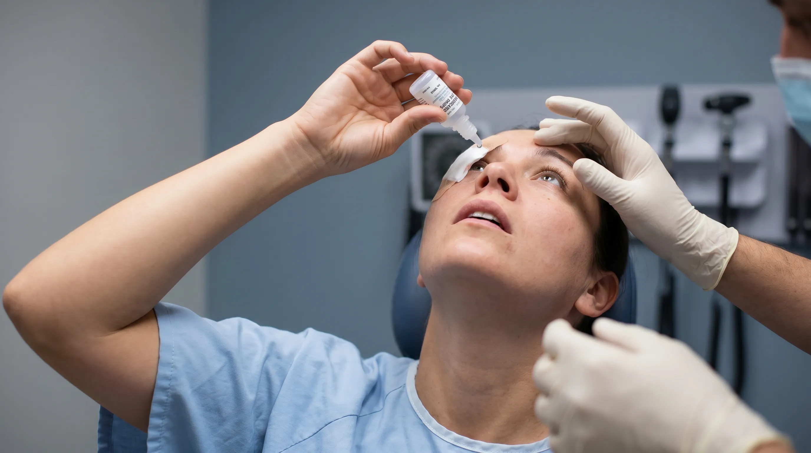
Topical pharmaceutical therapy forms the cornerstone of postoperative recovery. Corticosteroid drops suppress inflammatory cytokines, antibiotic prophylaxis prevents bacterial endophthalmitis, and NSAID drops reduce prostaglandin-mediated vascular leakage and macular swelling. Skipping doses, reducing drop frequency, or stopping medications prematurely disrupts this carefully calibrated biochemical environment. Clinical guidelines emphasize completing the full tapering schedule exactly as prescribed, even if symptoms appear resolved prematurely. Using preservative-free artificial tears at least two hours apart from medicated drops prevents dilution or chemical interaction. Proper instillation technique, including hand hygiene, avoiding bottle tip contact, and gentle nasolacrimal occlusion for thirty seconds, maximizes drug bioavailability while minimizing systemic absorption.
Environmental and Lifestyle Adjustments
Your immediate surroundings directly impact corneal surface integrity and healing efficiency. Dry air, ceiling fans, smoke, and prolonged screen time accelerate tear evaporation and increase ocular strain. Maintaining indoor humidity between forty and sixty percent, using a humidifier during sleep, and practicing the 20-20-20 rule during digital device use preserve tear film stability. Avoiding swimming pools, hot tubs, and dusty environments for at least two weeks prevents microbial contamination and particulate irritation. Wearing wraparound sunglasses outdoors shields healing tissues from ultraviolet radiation, wind, and allergens while reducing squint-related muscular fatigue. Adequate sleep, hydration, and gentle walking improve systemic circulation, delivering oxygen and nutrients essential for cellular repair in the anterior segment.
Nutrition and Systemic Health for Ocular Healing
Systemic wellness profoundly influences localized tissue regeneration. Diets rich in antioxidants, omega-3 fatty acids, vitamins C and E, lutein, and zeaxanthin support retinal health and reduce oxidative stress during the inflammatory phase. Staying well-hydrated maintains aqueous humor production and prevents tear hyperosmolarity. Managing underlying conditions like diabetes, hypertension, or autoimmune disorders is equally critical, as uncontrolled blood glucose or chronic systemic inflammation impairs microvascular perfusion and delays corneal endothelial recovery. Smoking cessation dramatically accelerates healing, as nicotine constricts ocular blood vessels and increases inflammatory mediator production. Integrating these systemic strategies alongside local eye care creates a synergistic environment for optimal visual rehabilitation.
| Recovery Factor | Typical Impact on Vision | Evidence-Based Management | Expected Resolution Window |
|---|---|---|---|
| Corneal Edema | Temporary haze, halos | Topical steroids, elevation, rest | 3-7 days |
| Dry Eye Surface | Fluctuating blur, irritation | Preservative-free tears, humidity, omega-3s | 4-12 weeks |
| Neuroadaptation | Mild distortion, depth perception shifts | Gradual exposure, consistent lighting | 6-12 weeks |
| Posterior Capsule Opacification | Gradual cloudiness, glare | Nd:YAG laser capsulotomy | Immediate post-procedure |
| Macular Edema | Central blurriness, distortion | NSAID drops, OCT monitoring, possible injections | 3-6 months with treatment |
When to Contact Your Surgeon Immediately
While most postoperative visual changes are benign and self-limiting, certain symptoms indicate complications requiring urgent medical evaluation. Prompt intervention prevents irreversible damage and preserves surgical outcomes.
Differentiating Normal Symptoms from Red Flags
Expected postoperative findings include mild scratchiness, tearing, light sensitivity, and gradual blur that steadily improves. Concerning symptoms demand immediate attention: sudden or severe vision loss, intense ocular pain unrelieved by standard analgesics, persistent nausea or vomiting, rapidly increasing floaters or flashing lights, a curtain-like shadow across the visual field, or copious yellow/green discharge accompanied by significant eyelid swelling. These manifestations may indicate retinal detachment, acute infection, severe inflammation, or corneal decompensation. Leading ophthalmic guidelines from the Cleveland Clinic stress that delayed treatment of ocular emergencies drastically reduces the likelihood of full visual recovery.
Emergency Complications to Monitor
Endophthalmitis, though rare (occurring in less than 0.1% of cases), represents a sight-threatening intraocular infection requiring immediate intravitreal antibiotics. Symptoms typically emerge three to five days postoperatively and include profound vision deterioration, severe pain, hypopyon (pus in the anterior chamber), and intense conjunctival injection. Retinal detachment risk increases slightly following cataract extraction, particularly in highly myopic patients or those with intraoperative complications. Macular hole formation, persistent elevated intraocular pressure, and toxic anterior segment syndrome are additional complications that necessitate rapid diagnosis. Maintaining scheduled follow-ups and reporting abnormal changes immediately ensures that specialized interventions like vitrectomy, laser retinopexy, or glaucoma filtration can be deployed before permanent photoreceptor loss occurs.

Vision Correction Options After Surgery
Modern cataract surgery aims for spectacle independence, but anatomical variations and lens selection sometimes require supplemental optical correction. Understanding your available options prevents frustration during the final stabilization phase.
The Role of Prescription Glasses and Contacts
Even with precise biometry and advanced IOL power calculations, mild residual refractive errors frequently remain. Monofocal lenses typically target distance vision, leaving patients dependent on reading glasses for near tasks. Multifocal or EDOF implants reduce dependence but may leave slight astigmatic or myopic residuals that affect night driving contrast. Temporary soft contact lenses can correct irregular astigmatism or anisometropia during the healing period, while permanent glasses are typically prescribed three to six weeks postoperatively when refraction stabilizes. Discussing realistic expectations regarding visual ranges with your surgeon before surgery significantly improves postoperative satisfaction. High-index lenses, anti-reflective coatings, and blue-light filtering options further enhance functional vision quality in varied lighting conditions.
When Additional Procedures Like YAG Laser Capsulotomy Are Needed
As previously discussed, secondary membrane clouding is the most common reason for delayed clarity months or years after initial surgery. The YAG laser capsulotomy is a quick, painless office procedure that restores optical clarity without incisions or hospitalization. After pupil dilation, the ophthalmologist uses focused laser energy to create a central circular opening in the opaque posterior capsule. Visual improvement is often instantaneous or within twenty-four hours. While extremely safe, rare complications include temporary intraocular pressure spikes or retinal traction. Following the procedure, patients continue routine monitoring to ensure stable macular anatomy and optimal retinal health. Understanding that PCO is a normal biological occurrence rather than a surgical failure reduces patient anxiety and promotes timely, effective treatment.
Frequently Asked Questions
How long does blurry vision typically last after cataract surgery?
Most individuals experience significant visual improvement within the first forty-eight hours as corneal swelling resolves and surgical inflammation begins to subside. However, complete optical stabilization typically requires four to eight weeks. During this period, mild fluctuations in clarity, especially in low-light environments or after extended screen use, remain entirely normal. Patients receiving premium multifocal or toric intraocular lenses may experience a slightly longer adaptation period as the brain learns to interpret new focal ranges. Adhering to prescribed drop schedules, protecting the eyes from environmental irritants, and attending scheduled postoperative visits ensure that blurry vision after cataract surgery follows a predictable, healthy resolution curve.
Is it normal to experience blurry vision in both eyes if only one was operated on?
Yes, temporary visual imbalance is common. When one eye delivers crisp, unclouded postoperative input while the fellow eye continues transmitting degraded, cataract-obscured signals, the brain's visual cortex struggles to integrate the mismatched data streams. This binocular rivalry often manifests as overall haziness, depth perception distortion, or mild double vision. The condition typically resolves rapidly once the second eye undergoes successful cataract extraction, usually scheduled several weeks to months after the initial procedure. In the interim, covering the unoperated eye during precision tasks can temporarily reduce visual confusion until surgical rehabilitation of the contralateral eye is complete.
Can dry eye medication resolve blurry vision after cataract surgery?
Frequently, yes. The surgical incisions and phacoemulsification energy temporarily disrupt corneal sensory nerves that regulate basal tear production and reflex blinking. This neurotrophic effect increases tear evaporation and destabilizes the lipid-mucin layers, directly compromising optical smoothness and causing intermittent or persistent blur. Implementing a structured ocular surface management protocol—including frequent preservative-free artificial tears, prescription anti-inflammatory drops like cyclosporine, environmental humidity optimization, and nutritional omega-3 supplementation—often restores tear film integrity. As the ocular surface heals and nerve sensitivity returns over three to six months, the blur associated with dryness typically resolves completely.
What is PCO, and how does it cause blurriness months later?
Posterior capsule opacification occurs when residual lens epithelial cells migrate across the transparent posterior membrane that supports your intraocular implant, proliferate, and undergo fibrotic transformation into a secondary cloudy film. This biological response gradually scatters incoming light before it reaches the retina, recreating symptoms nearly identical to the original cataract: faded contrast, glare, and progressive blurriness. PCO is not a surgical failure but a well-documented, expected tissue response affecting up to twenty percent of patients within several years. It is efficiently treated with an outpatient Nd:YAG laser capsulotomy that painlessly restores optical clarity, typically yielding immediate visual improvement without requiring additional incisions or recovery time.
Should I wear sunglasses while experiencing blurry vision during recovery?
Absolutely. Postoperative eyes remain highly sensitive to ultraviolet radiation, visible blue light, and environmental glare during the initial healing phases. Prolonged exposure increases photophobia, triggers excessive squinting, and accelerates tear film evaporation, all of which exacerbate visual discomfort and blur. Wearing high-quality, wraparound sunglasses with 100% UVA and UVB protection reduces ocular strain, maintains tear film stability, and creates a shield against wind and particulate matter that could compromise corneal healing. Consistent outdoor protection, combined with indoor lighting moderation, significantly supports the comfort and speed of your visual rehabilitation journey.
Conclusion
Navigating the postoperative period requires patience, awareness, and proactive self-care. While experiencing blurry vision after cataract surgery is overwhelmingly a normal and temporary phase of ocular healing, understanding the underlying biological processes empowers you to distinguish routine recovery from conditions requiring clinical attention. By adhering strictly to medication protocols, optimizing your environmental conditions, managing dry eye symptoms, and maintaining open communication with your surgical team, you create the ideal foundation for rapid visual stabilization. Modern lens technology and advanced microsurgical techniques continue to improve outcomes, but individual healing timelines remain highly personal. Trust the evidence-based recovery process, prioritize protective habits, and attend every scheduled follow-up to ensure your journey toward clear, comfortable vision remains safe and successful.
How we work
Researched and drafted with AI assistance, then reviewed by a human editor against the cited sources.
This article is general health information, not medical advice. Consult a qualified healthcare professional about your own situation.