Sperm in Eye: Medical Risks, First Aid, and STI Prevention Guide

Getting semen in your eye is an unexpected situation that can quickly escalate from a moment of confusion to genuine medical concern. While it may sound like a minor annoyance, the ocular surface is one of the most sensitive tissues in the human body, designed specifically to maintain a sterile, balanced environment. When foreign substances breach this delicate barrier, the immune system and physical defense mechanisms spring into action almost instantly. Understanding the physiological reality behind sperm in eye exposure is essential for responding correctly, avoiding unnecessary panic, and protecting your long-term vision. As noted by the Mayo Clinic, proper first aid and awareness of ocular anatomy are crucial for managing exposure to biological fluids. This comprehensive guide will walk you through the anatomy of the eye, the composition of seminal fluid, the genuine risks of infection, and the exact steps you should take if exposure occurs. We will separate evidence-based medical facts from widespread internet myths, ensuring you have the knowledge needed to act swiftly and safely. Whether this happens accidentally during intimacy, through splashing, or as part of an unexpected sexual encounter, knowing how the eye reacts to biological fluids is a crucial component of sexual health and ocular safety. By the end of this article, you will have a clear, science-backed roadmap for managing exposure, recognizing warning signs, and knowing precisely when to consult a healthcare professional.
Understanding Ocular Anatomy and Semen Composition
The Conjunctiva and Ocular Defense Mechanisms
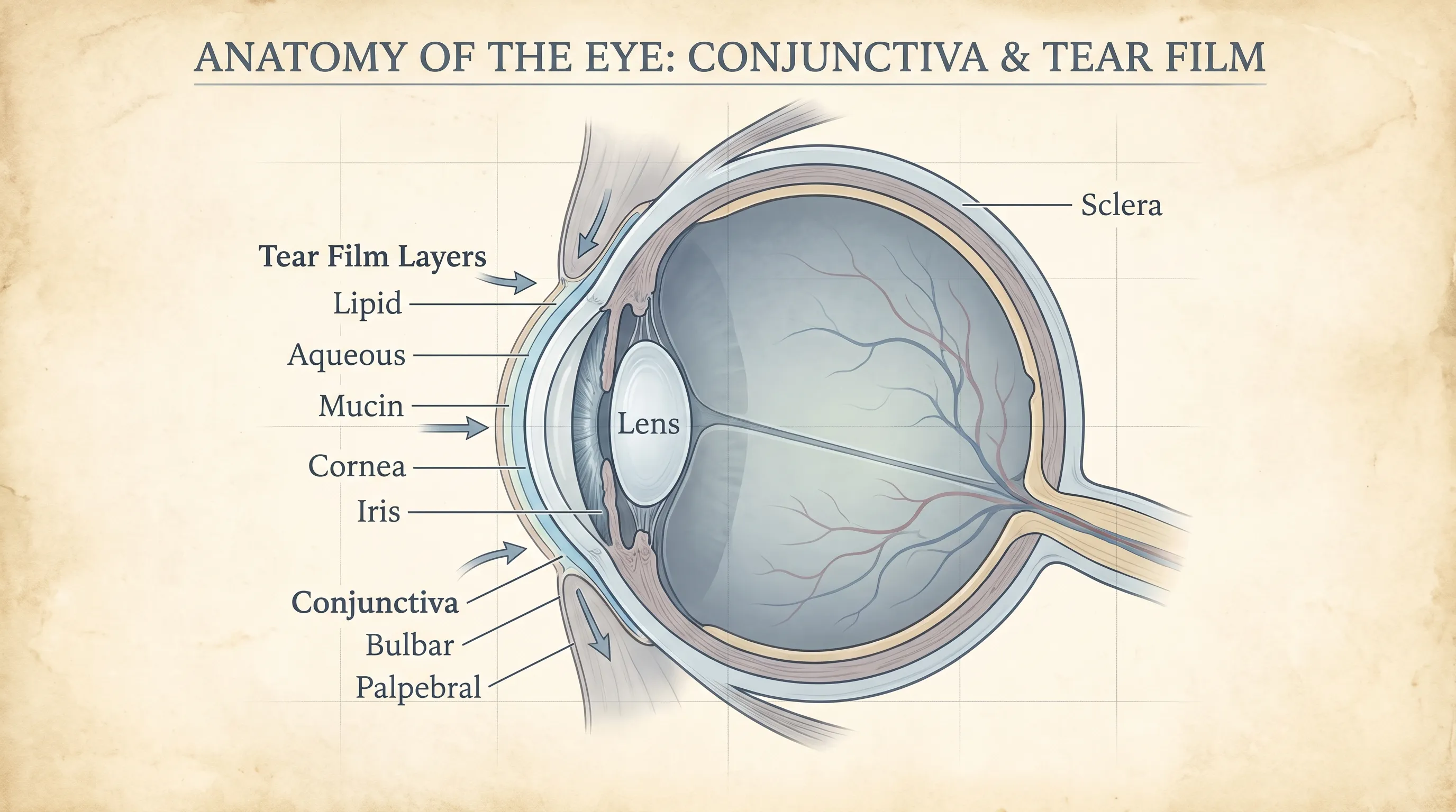
The human eye is protected by several specialized tissue layers, with the conjunctiva serving as the primary defensive interface. This thin, transparent mucous membrane covers the sclera (the white part of the eye) and extends inward to line the palpebral surfaces of the eyelids. Its structural composition includes a stratified columnar epithelium and a vascularized stromal layer rich in goblet cells. These goblet cells continuously secrete mucin, a gel-like glycoprotein that stabilizes the tear film, prevents desiccation, and traps particulate matter before it can scratch the corneal epithelium. Unlike cutaneous tissue, which possesses a thick, keratinized stratum corneum that provides robust chemical and microbial resistance, the conjunctiva lacks this protective barrier. This anatomical distinction makes it exceptionally vulnerable to direct contact with biological fluids. When foreign agents land on the ocular surface, pattern recognition receptors on epithelial cells and resident dendritic cells immediately detect non-self proteins and trigger a localized inflammatory cascade. Pro-inflammatory cytokines such as interleukin-1 (IL-1) and tumor necrosis factor-alpha (TNF-α) are released, recruiting neutrophils and lymphocytes to neutralize potential threats. While highly effective, this immune response inherently disrupts homeostasis, leading to the characteristic symptoms of redness, swelling, and discomfort that accompany ocular irritant exposure. For a detailed breakdown of ocular surface anatomy and defense mechanisms, see resources from the NIH National Eye Institute.
Free calculatorBMI CalculatorCalculate Body Mass IndexOpen the calculatorTear Film Biochemistry and pH Homeostasis
The ocular surface relies on a remarkably precise tear film architecture to maintain optical clarity, corneal hydration, and antimicrobial defense. This three-layered structure consists of an inner mucin layer that adheres tears to the epithelium, a central aqueous layer that supplies nutrients and immune mediators, and an outer lipid layer that prevents rapid evaporation. The aqueous component contains lysozyme, lactoferrin, lipocalin, and secretory IgA, all of which function synergistically to inhibit bacterial and viral colonization. The pH of healthy tears is tightly regulated around 7.4, slightly alkaline but highly stable. Introducing reproductive fluids creates a sudden osmotic and pH imbalance. Semen typically ranges from pH 7.2 to 8.0 due to its fructose, protein, and alkaline buffering systems designed to neutralize vaginal acidity. When this fluid contacts the cornea, it temporarily disrupts the hydrogen ion equilibrium, triggering nociceptors in the superficial nerve plexus. Additionally, the high electrolyte concentration (sodium, potassium, calcium, magnesium) and viscous protein matrix interfere with tear film stability, causing transient refractive changes and blurred vision until natural drainage and reflex tearing restore equilibrium. Clinical perspectives on tear film composition and pH regulation are extensively covered by the Cleveland Clinic.
What Is in Semen That Triggers a Reaction?
Seminal plasma is a complex biochemical mixture synthesized primarily in the seminal vesicles, prostate gland, and bulbourethral glands. It contains spermatozoa suspended in a nutrient-rich medium comprising fructose, citric acid, prostaglandins (particularly PGE1 and PGE2), proteolytic enzymes like prostate-specific antigen (PSA) and acid phosphatase, polyamines, zinc, and various immunomodulatory cytokines. Each component plays a specific reproductive role, but none are formulated for ocular compatibility. PSA, for example, cleaves semenogelin proteins to liquefy coagulated semen post-ejaculation, but when applied to mucous membranes, it acts as a foreign enzymatic irritant. Prostaglandins are potent vasodilators that increase vascular permeability, directly contributing to hyperemia (redness) and edema. Fructose and other carbohydrates provide metabolic substrates that can theoretically support microbial proliferation if pathogenic organisms are present. The cumulative effect of these compounds on the delicate corneal epithelium initiates a rapid neurogenic and inflammatory response, manifesting as burning, stinging, photophobia, and reflex lacrimation. Recognizing that these physiological reactions are normal defense mechanisms rather than indicators of permanent damage helps reduce anxiety and encourages appropriate management.
Immediate Effects and Physiological Responses
Chemical and Mechanical Irritation Explained
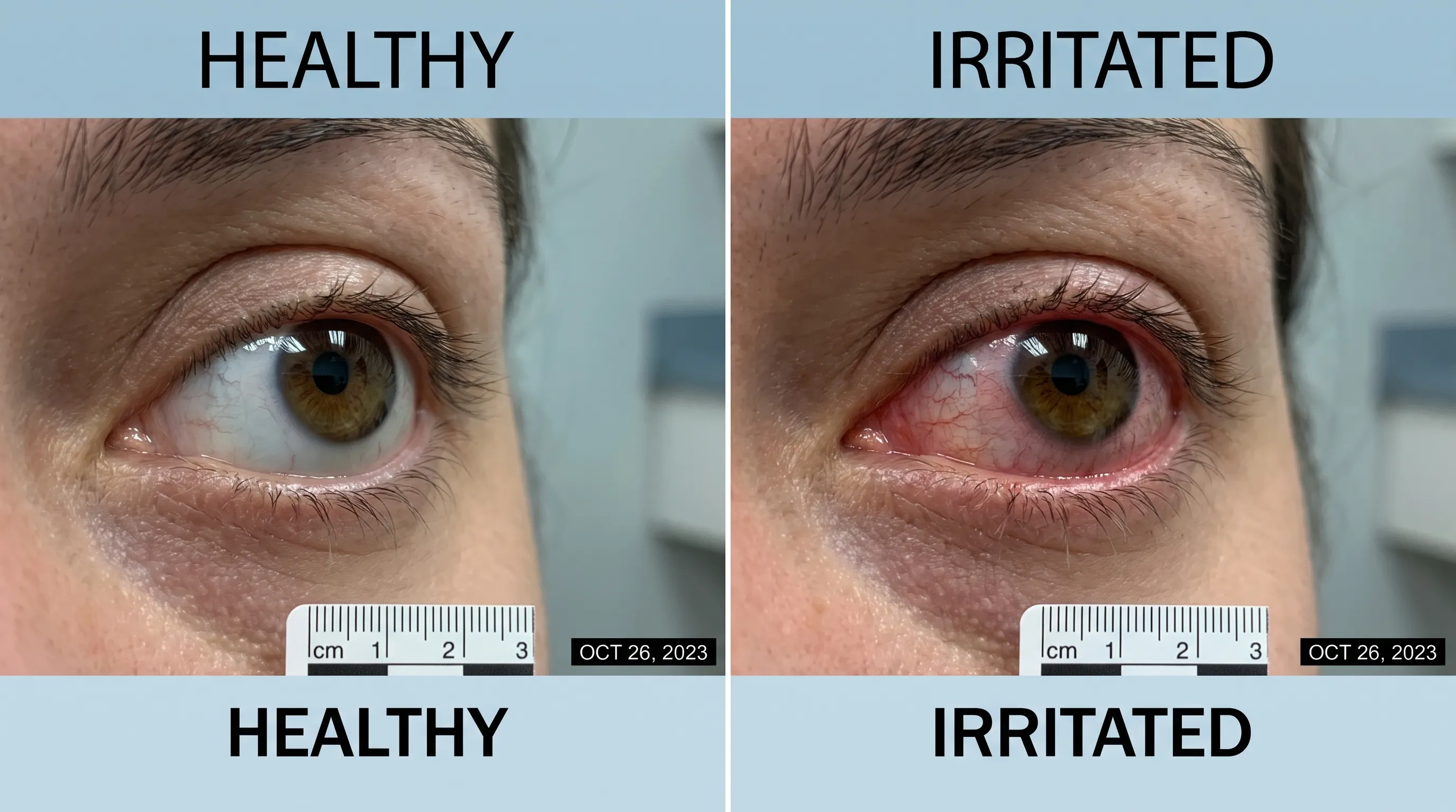
Upon contact, the ocular surface registers both chemical and mechanical stimuli simultaneously. The chemical irritation stems from the abrupt pH shift and the denaturation of epithelial surface glycoproteins by foreign proteolytic enzymes. Nerve endings in the corneal stroma, which are approximately 500 times more sensitive than cutaneous receptors, transmit sharp pain and foreign-body sensations via the nasociliary branch of the trigeminal nerve. Mechanically, the higher viscosity of seminal fluid compared to natural tears creates a temporary film that disrupts normal blinking dynamics. This altered friction coefficient causes micro-trauma to the superficial epithelial cells, particularly during rapid blinking attempts. The combination of enzymatic activity, pH disruption, and altered surface tension results in immediate discomfort that typically peaks within the first two to five minutes. While distressing, this initial phase is largely self-limiting if the substance is promptly removed. Failure to irrigate adequately prolongs contact time, allowing deeper epithelial penetration and increasing the likelihood of secondary inflammation or microbial adhesion.
The Role of Tears and Reflex Blinking
Reflex tearing is the eye’s fastest physiological countermeasure against ocular surface threats. Triggered by afferent trigeminal nerve signaling, the lacrimal gland and accessory lacrimal glands (glands of Krause and Wolfring) rapidly secrete aqueous tears enriched with antimicrobial proteins and electrolytes. This surge in tear volume increases intraocular surface pressure, physically displacing the irritant toward the puncta and nasolacrimal drainage system. Concurrently, blink rate increases from the baseline of 12–15 per minute to 25–30 per minute, enhancing mechanical clearance. However, reflex tearing is quantitatively insufficient for thick or protein-dense fluids. The nasolacrimal duct has a finite flow rate, and excessive tearing can cause overflow onto the face rather than efficient drainage. Furthermore, reflex tears contain a higher proportion of water and a lower concentration of protective mucins and lipids, temporarily compromising tear film stability. This paradoxical effect underscores why relying solely on natural flushing is inadequate and why structured, prolonged irrigation remains the clinical standard for decontamination.
Differentiating Irritation from True Allergic Response
It is crucial to distinguish between direct chemical irritation and an allergic conjunctival reaction. True ocular allergy involves type I hypersensitivity, mediated by mast cell degranulation and histamine release, typically occurring upon re-exposure to an allergen after prior sensitization. Initial exposure to sperm in eye scenarios usually triggers non-specific chemical inflammation, characterized by immediate burning, clear tearing, and transient redness that resolves within hours after flushing. Allergic responses, by contrast, feature intense pruritus (itching), stringy mucus, and bilateral involvement that may persist or recur with subsequent exposures. In rare cases, individuals with known atopic conditions may develop localized IgE-mediated conjunctival hyperreactivity upon contact with seminal proteins. Management strategies differ significantly: chemical irritation responds well to irrigation and supportive lubrication, whereas allergic inflammation often requires topical antihistamines or mast cell stabilizers under medical supervision. Proper differentiation prevents unnecessary medication use and ensures appropriate follow-up.
The Real Risk: Sexually Transmitted Infections
Chlamydia and Gonorrhea: Bacterial Threats
While acute chemical irritation dominates the initial presentation, the most clinically consequential risk of semen in eye exposure is the transmission of bacterial STIs. The conjunctiva’s mucosal architecture provides an ideal environment for obligate intracellular and extracellular pathogens to attach, invade, and replicate. Chlamydia trachomatis, an obligate intracellular bacterium, binds to conjunctival epithelial cells via specific adhesion proteins, initiating a slow but progressive infection known as adult inclusion conjunctivitis. Clinical features typically emerge three to ten days post-exposure and include conjunctival follicles, mucopurulent discharge, eyelid swelling, and preauricular lymphadenopathy. Without appropriate antibiotic therapy, chronic chlamydial infection can induce conjunctival scarring, symblepharon formation, and corneal pannus, permanently compromising ocular surface health. Neisseria gonorrhoeae presents a far more urgent clinical scenario. Gonococcal conjunctivitis is classified as hyperacute due to its aggressive epithelial invasion and potent protease production. Symptoms appear within 12 to 48 hours and are marked by copious, thick purulent discharge that can reaccumulate within minutes of wiping, severe chemosis, and intense pain. The bacterium rapidly penetrates the intact corneal epithelium, leading to ulceration, stromal infiltrates, and potential perforation within 72 hours. Immediate ophthalmic consultation and systemic therapy are mandatory to preserve vision. For comprehensive clinical data on these pathogens, consult the CDC Sexually Transmitted Infections guidelines.
Viral Risks: Herpes and Other Pathogens
Viral transmission to the conjunctiva represents another significant threat. Herpes simplex virus (HSV) type 1 or 2 can be inoculated into the ocular surface during direct fluid contact. HSV preferentially infects epithelial keratinocytes, utilizing neuronal retrograde transport to establish latency in the trigeminal ganglion. Upon reactivation or primary infection, it manifests as herpetic keratoconjunctivitis, often presenting with pathognomonic dendritic corneal ulcers visible under fluorescein staining. Patients report severe photophobia, tearing, and foreign-body sensation. Unlike bacterial infections, HSV requires antiviral nucleoside analogs (acyclovir, valacyclovir) to halt viral replication; topical corticosteroids are strictly contraindicated during active epithelial disease as they accelerate viral proliferation and corneal melting. Syphilis, caused by Treponema pallidum, rarely transmits via direct conjunctival contact but can manifest as ocular syphilis in secondary or tertiary stages, causing interstitial keratitis, posterior uveitis, or optic neuritis. Human immunodeficiency virus (HIV) poses a theoretical mucosal transmission risk, but epidemiological studies consistently demonstrate exceedingly low transmission rates via ocular exposure. The CDC classifies ocular mucous membrane exposure to HIV-positive fluids as low risk, though post-exposure prophylaxis (PEP) evaluation remains standard when partner status is confirmed positive.
Epidemiological Context of Ocular STI Transmission
Sexually transmitted ocular infections account for a notable percentage of adult conjunctivitis cases in sexually active populations. In the United States, bacterial conjunctivitis generates approximately one million primary care visits annually, with a subset directly attributable to sexual exposure or autoinoculation from genital infections. The World Health Organization (WHO)) reports that chlamydia and gonorrhea remain among the most prevalent bacterial STIs globally, with asymptomatic carriers facilitating silent transmission. Ocular manifestations are more frequently diagnosed in young adults, men who have sex with men, and individuals with inconsistent barrier protection or multiple partners. Neonatal conjunctivitis from maternal STI transmission is extensively documented and routinely prevented with prophylactic ocular ointment at birth, highlighting the established pathogenic pathway from genital secretions to the conjunctiva. In adults, however, awareness is often lacking, leading to delayed diagnosis. Public health initiatives emphasize integrating ocular symptom screening into standard sexual health assessments, particularly when patients present with unexplained conjunctivitis that fails to respond to conventional empirical therapy.
Debunking Popular Myths and Misconceptions
The "Natural Remedy" Fallacy
Over the past decade, various unregulated wellness communities and social media platforms have propagated pseudoscientific claims that seminal fluid applied to the eyes offers anti-aging, vision-enhancing, or immune-boosting properties. These assertions consistently lack empirical validation, randomized controlled trials, or biochemical plausibility. The ocular surface does not absorb systemic nutrients through topical application; instead, the cornea relies on diffusion from the aqueous humor and limbal vasculature for metabolic support. Introducing complex reproductive proteins disrupts the tear film's lipid-aqueous-mucin equilibrium, compromising epithelial integrity rather than enhancing it. No peer-reviewed dermatological, ophthalmological, or immunological journal has published data supporting ocular rejuvenation or visual acuity improvement from seminal exposure. The propagation of such myths often stems from misinterpretation of seminal composition studies or anecdotal reporting, neither of which meets scientific standards for clinical recommendation.
Scientific Consensus on Ocular Health
Authoritative medical organizations uniformly reject claims of ocular benefits from reproductive fluid exposure. The American Academy of Ophthalmology (AAO) and Mayo Clinic explicitly state that the tear film requires precise sterile conditions and homeostatic regulation to prevent desiccation, infection, and refractive error. Seminal plasma contains no growth factors, antioxidants, or anti-inflammatory compounds that interact beneficially with corneal or conjunctival tissue. In fact, prolonged exposure increases the risk of toxic conjunctivitis, epithelial micro-abrasions, and secondary bacterial overgrowth due to disrupted tear film dynamics. Evidence-based ocular health prioritizes hygiene, protective eyewear, nutritional adequacy (vitamins A, C, E, zinc, and omega-3 fatty acids), and routine clinical monitoring. Substituting proven preventive measures with unverified biological applications not only fails to deliver promised outcomes but actively introduces preventable risks to a highly specialized sensory organ.
Why Pseudoscientific Claims Persist
The endurance of ocular semen myths can be traced to several sociocultural and cognitive factors. First, the human brain exhibits confirmation bias and anecdotal fallacy, where isolated subjective reports of temporary symptom relief are misinterpreted as therapeutic effects. Second, the commercial wellness industry frequently capitalizes on unregulated alternative therapies, marketing complex biological mixtures as "natural elixirs" without rigorous clinical oversight. Third, limited public education regarding ocular anatomy and tear film physiology creates information vacuums that pseudoscience readily fills. Medical professionals counter this by promoting transparent, evidence-based patient education, emphasizing that vision preservation relies on sterile practices, prompt infection management, and adherence to established ophthalmic guidelines rather than experimental topical applications.
First Aid and Immediate Action Steps
Step-by-Step Eye Flushing Protocol
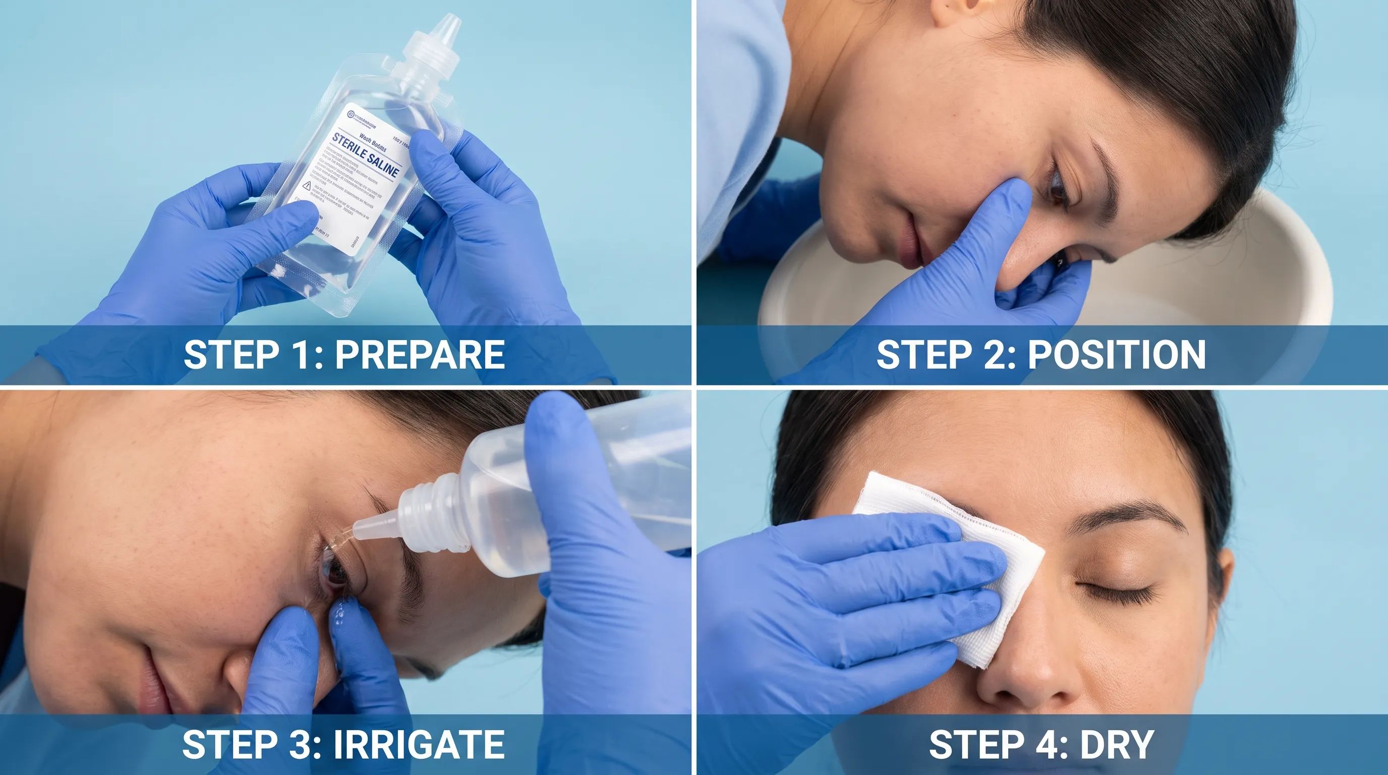
When semen contacts the eye, immediate decontamination is the single most effective intervention. Medical guidelines, including those from the Mayo Clinic, recommend a standardized irrigation procedure to minimize tissue damage and pathogen retention. Locate a source of clean, lukewarm water, sterile saline, or commercially formulated ocular wash. Avoid ice-cold water, which induces vasospasm and increases discomfort. Position your head over a sink or shower, tilt it slightly to the affected side, and use your index finger and thumb to gently hold the upper and lower eyelids open. Begin irrigation at the medial canthus (inner corner), allowing the fluid stream to flow laterally across the globe to prevent cross-contamination. Maintain continuous flow for 10 to 15 minutes, ensuring complete coverage of the corneal and conjunctival surfaces. Do not increase water pressure beyond a gentle stream, as high-velocity jets can cause epithelial stripping. After irrigation, blink normally to redistribute the tear film, and gently blot the periocular skin with a sterile gauze or clean tissue. Do not reuse towels or apply compresses until a healthcare provider confirms the absence of infectious pathology.
What to Avoid (Rubbing, Contact Lenses, OTC Drops)
Several common post-exposure behaviors significantly worsen outcomes. Rubbing the eye is strongly contraindicated; mechanical friction drives residual proteins and potential pathogens deeper into the fornices, increases epithelial shear stress, and introduces cutaneous flora to the ocular surface. If contact lenses were in place at the time of exposure, remove them immediately prior to irrigation. Lenses trap irritants against the cornea, creating a high-concentration microenvironment that exacerbates toxicity and infection risk. Discard exposed lenses and never attempt to disinfect and reuse them. Additionally, avoid over-the-counter vasoconstricting drops (e.g., tetrahydrozoline, naphazoline) immediately after exposure. These agents temporarily reduce redness by constricting superficial vessels but mask clinical signs, delay healing, and can cause rebound hyperemia. If lubrication is necessary post-flushing, use only preservative-free artificial tears until a physician evaluates the eye. Avoid applying home remedies, herbal solutions, or unpasteurized products, as they introduce unpredictable chemical loads and microbial contaminants.
Post-Exposure Monitoring Timeline
After proper irrigation, the acute irritation phase typically subsides within 2 to 4 hours. Monitor the eye closely during the first 24 hours for symptom resolution. Mild tearing and slight pinkness may persist but should steadily improve. Document any changes in visual acuity, pain intensity, or discharge characteristics. If symptoms plateau or worsen after 12 hours, initiate medical consultation. Keep a log of onset time, exposure circumstances, partner STI status (if known), and flushing duration. This information accelerates clinical triage and informs diagnostic testing. Do not cover the eye with an occlusive patch, as trapped heat and moisture create an optimal environment for bacterial proliferation. Maintain strict hand hygiene and avoid sharing bedding, towels, or eye makeup until cleared by a professional.
When to Seek Professional Medical Care
Red Flags That Require an Ophthalmologist
Certain clinical presentations mandate immediate ophthalmic evaluation regardless of perceived exposure severity. Seek urgent care if you experience: purulent, yellow-green, or copious discharge; severe photophobia or inability to keep the eye open; rapid vision blurring or halos around lights; intense, unrelenting pain that does not respond to flushing; marked eyelid swelling or periorbital cellulitis signs; preauricular lymph node enlargement; or a history of known STI-positive partner exposure. These indicators suggest bacterial invasion, viral keratitis, or significant epithelial compromise that exceeds home management capabilities. Delaying professional intervention increases the risk of corneal ulceration, stromal necrosis, and permanent refractive changes. Ophthalmologists utilize slit-lamp biomicroscopy to assess epithelial defects, stromal integrity, and anterior chamber inflammation, enabling precise diagnosis and targeted therapy.
Diagnostic Tests and Clinical Treatments
Clinical evaluation begins with a comprehensive ocular surface examination, often supplemented by fluorescein staining to identify corneal abrasions or dendritic ulcers. If an infectious etiology is suspected, the provider will perform conjunctival swabs for Gram stain, bacterial culture, and nucleic acid amplification testing (NAAT) targeting C. trachomatis and N. gonorrhoeae. Viral PCR assays may be ordered for HSV or adenovirus screening. The CDC Sexually Transmitted Infections Treatment Guidelines recommend empiric dual therapy for suspected gonococcal and chlamydial ocular exposure while awaiting results. This typically involves intramuscular ceftriaxone 500mg (1g if >150kg) plus oral doxycycline 100mg twice daily for 7 days, or azithromycin 1g single dose. Topical fluoroquinolone drops may be prescribed as adjunctive therapy for severe bacterial load. HSV infections require systemic antivirals such as acyclovir 400mg five times daily for 10–14 days. Corticosteroids are avoided in active epithelial HSV but may be introduced cautiously later to manage immune-mediated stromal inflammation. Follow-up visits at 48 hours and one week ensure therapeutic response, monitor corneal healing, and adjust regimens based on culture sensitivities.
Navigating Healthcare Privacy and STI Disclosure
Patients often express concern about privacy and stigma when seeking care for ocular STI exposure. Healthcare providers are legally bound by HIPAA and professional ethics to maintain strict confidentiality. Disclosing exposure circumstances, including sexual contact and partner history, is medically essential for accurate diagnosis and appropriate prescribing but remains protected health information. Many clinics offer confidential STI screening, anonymous testing codes, and integrated sexual health counseling. Patients should feel empowered to communicate openly about exposure without fear of judgment, as transparent reporting directly impacts treatment efficacy and public health tracking. Telemedicine platforms and community health centers also provide accessible, discreet consultation options for those uncomfortable with in-person visits.
Prevention and Safe Practices
Barrier Protection and Partner Communication
The most reliable strategy to prevent sperm in eye exposure involves proactive communication and physical precautions. Discuss sexual boundaries, fluid contact preferences, and STI testing status with partners before intimacy. If ocular exposure is a concern, consider positioning adjustments, using towels for incidental splash, or wearing well-fitted protective eyewear during activities with higher fluid projection risk. Consistent condom use reduces genital pathogen load and minimizes fluid exchange. Establish mutual agreements regarding consent, testing frequency, and safe practices to create an environment where both parties prioritize health and comfort. Remember that prevention is always safer, more effective, and less anxiety-inducing than post-exposure management.
Routine STI Screening and Sexual Health
Regular sexual health screenings form the foundation of STI prevention. Sexually active individuals should undergo annual testing for chlamydia, gonorrhea, syphilis, and HIV, with increased frequency for those with multiple partners, inconsistent barrier use, or high-prevalence community exposure. The CDC and WHO emphasize that asymptomatic carriers frequently transmit pathogens unknowingly, making routine NAAT testing essential. Vaccination against HPV and Hepatitis B further reduces viral transmission risks. Integrating ocular symptom awareness into sexual health education ensures individuals recognize early warning signs and seek prompt care. Maintaining a relationship with a primary care provider or sexual health clinic streamlines testing, treatment, and preventive counseling.
Environmental and Occupational Safety Considerations
While most semen in eye incidents occur in private settings, healthcare workers, laboratory personnel, and first responders may encounter biological fluid splashes professionally. OSHA guidelines mandate use of ANSI-approved splash goggles, face shields, and engineering controls to prevent ocular exposure during procedures involving bodily fluids. Strict adherence to standard precautions, immediate eyewash station availability, and post-exposure reporting protocols minimize occupational transmission risks. These institutional safety frameworks mirror personal hygiene principles: immediate decontamination, barrier protection, and prompt medical follow-up are universally applicable across clinical, domestic, and professional environments.
Frequently Asked Questions
What should I do immediately if I get sperm in eye?
The most critical step is initiating thorough irrigation within seconds of exposure. Flush the affected eye with clean, lukewarm water or sterile saline for a full 10 to 15 minutes. Keep the eyelid open, tilt your head to allow drainage away from the unaffected eye, and avoid rubbing. Remove contact lenses immediately, as they trap irritants against the cornea. After flushing, monitor symptom progression and seek medical evaluation if redness, pain, or discharge persists beyond 24 hours.
Can semen in the eye cause a sexually transmitted infection?
Yes, transmission is possible. The conjunctiva functions as a mucous membrane capable of absorbing pathogens. If the source partner carries Chlamydia trachomatis, Neisseria gonorrhoeae, herpes simplex virus, or syphilis, direct ocular exposure can inoculate these organisms into the eye. This may result in bacterial conjunctivitis, viral keratitis, or more severe complications if left untreated. Prompt clinical assessment is strongly recommended when STI status is unknown or confirmed positive.
Is there any medical benefit to putting semen in the eyes?
No. Comprehensive clinical research and peer-reviewed literature confirm zero health benefits from ocular semen exposure. Claims suggesting anti-aging, vision enhancement, or immune support are scientifically unfounded and contradict established ophthalmological principles. The eye's tear film requires precise sterility and biochemical balance; introducing foreign proteins disrupts this environment, increasing the likelihood of chemical irritation, allergic reaction, and microbial infection.
How long does it take for symptoms to appear if an infection develops?
Timeline depends on the specific pathogen involved. Chemical irritation begins immediately. Gonococcal conjunctivitis typically manifests within 12 to 48 hours with severe purulent discharge and pain. Chlamydial infection usually presents within 3 to 10 days with follicular conjunctivitis and mucopurulent discharge. HSV keratitis may emerge within 2 to 12 days, often accompanied by dendritic ulcers and photophobia. Any symptom progression beyond 24 hours warrants professional evaluation to rule out infectious etiology.
Should I take antibiotics if my partner's STI status is unknown?
Only a licensed healthcare professional can determine appropriate pharmacological intervention. If exposure to an infected partner is suspected or confirmed, clinicians frequently initiate empiric antibiotic regimens for chlamydia and gonorrhea while awaiting NAAT results. Self-prescribing or delaying care increases the risk of corneal scarring, stromal involvement, and systemic dissemination. Always complete prescribed courses and attend follow-up appointments to verify pathogen clearance and ocular surface recovery.
Conclusion
Encountering sperm in eye exposure can be unsettling, but a clear understanding of ocular physiology, evidence-based first aid, and infection risks empowers you to respond effectively. The conjunctiva is highly sensitive to chemical shifts and foreign proteins, making immediate, prolonged irrigation the most critical intervention to prevent epithelial damage and pathogen retention. While transient irritation is common and typically resolves quickly, the potential for sexually transmitted ocular infections demands vigilance and prompt medical consultation when red flags appear. By dispelling pseudoscientific myths, adhering to clinical guidelines, and prioritizing open communication and routine STI screening, individuals can significantly reduce exposure risks and safeguard long-term visual health. Always consult a qualified ophthalmologist or healthcare provider for personalized evaluation, particularly when symptoms persist, partner STI status is uncertain, or vision changes occur. Protecting your eyes through informed, science-backed practices remains the most reliable pathway to maintaining optimal ocular wellness and overall sexual health.
How we work
Researched and drafted with AI assistance, then reviewed by a human editor against the cited sources.
This article is general health information, not medical advice. Consult a qualified healthcare professional about your own situation.