Can Dry Eyes Cause Blindness? Understanding the Risks, Treatments, and Vision Protection

The question of whether chronic ocular surface discomfort can progress to permanent vision loss is one of the most pressing concerns for millions of individuals experiencing persistent irritation. Millions of people worldwide manage dry eye syndrome, a condition characterized by an unstable tear film that fails to adequately lubricate and protect the front surface of the eye. While occasional dryness is a common, manageable nuisance, many individuals rightly wonder: can dry eyes cause blindness? The short answer is that while complete blindness is exceptionally rare, severe and untreated ocular surface disease can absolutely lead to irreversible corneal damage, significant visual impairment, and in extreme cases, functional vision loss. Understanding the biological mechanisms behind tear film dysfunction, recognizing the early warning signs of corneal compromise, and implementing evidence-based management strategies are essential steps in protecting your long-term ocular health.
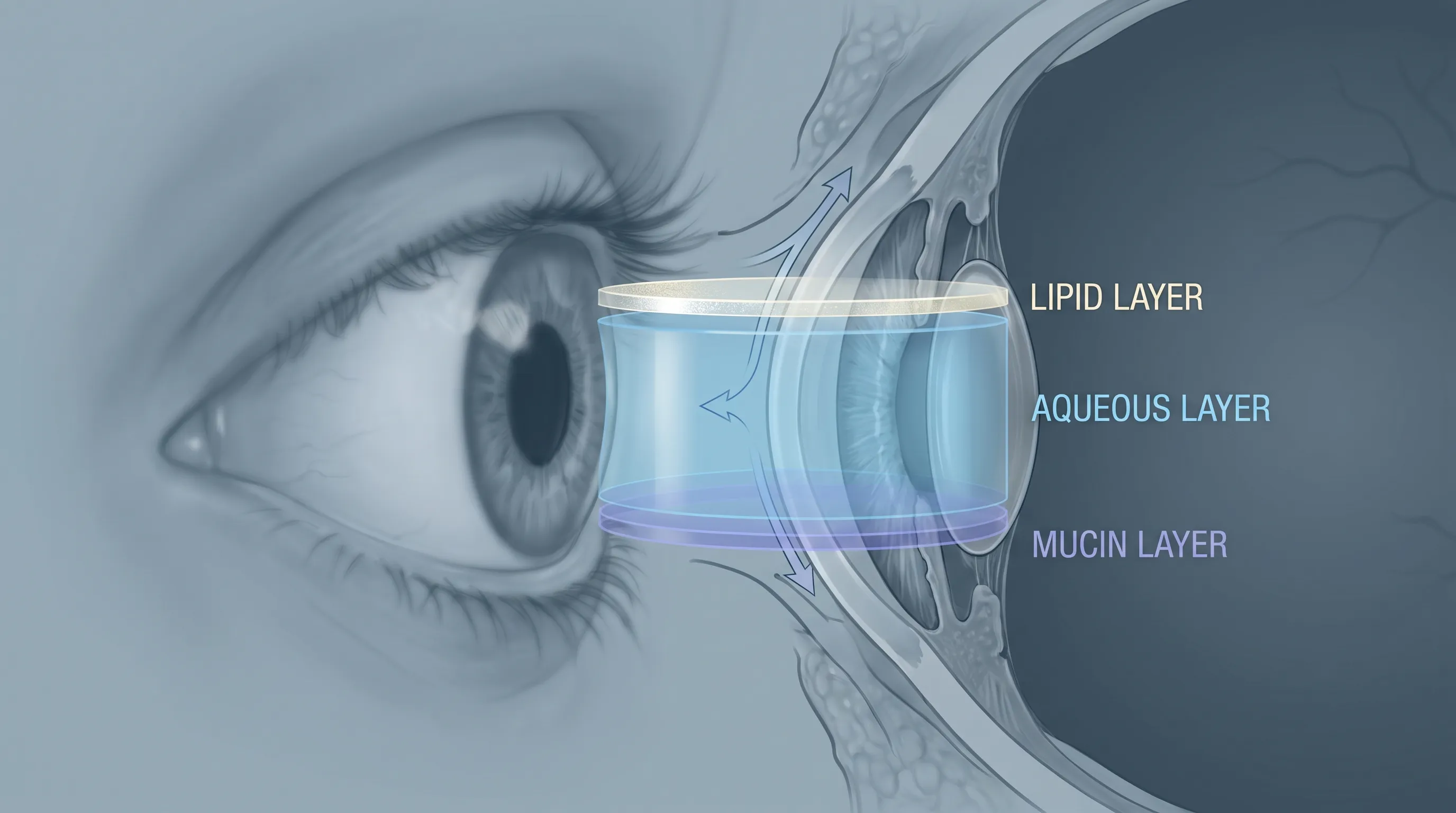
The tear film is far more than a simple layer of moisture. It is a sophisticated, three-layered biological barrier composed of lipids, aqueous fluid, and mucins, working in precise harmony to nourish the cornea, flush away debris, neutralize pathogens, and provide a perfectly smooth optical surface for light refraction. When any component of this delicate system fails, the consequences extend far beyond temporary discomfort. Chronic dryness initiates a cascade of inflammatory processes that degrade the corneal epithelium, disrupt nerve signaling, and compromise the eye’s natural defense mechanisms. This article provides a comprehensive, medically grounded examination of how dry eye disease progresses, the actual risks it poses to permanent vision, and the clinically proven interventions that can halt or reverse damage before it reaches a critical threshold. By understanding the pathophysiology and adopting proactive lifestyle and therapeutic measures, you can maintain clear, comfortable vision for decades to come.
Understanding Dry Eye Disease and Its Mechanisms
What Is Dry Eye Syndrome?
Dry eye syndrome, clinically referred to as keratoconjunctivitis sicca, is a multifactorial disease of the tears and ocular surface that results in symptoms of discomfort, visual disturbance, and tear film instability with potential damage to the ocular surface. It is frequently accompanied by increased osmolarity of the tear film and inflammation of the ocular surface. The Tear Film & Ocular Surface Society (TFOS) Dry Eye Workshop II (DEWS II) defines it as a condition where the eye either does not produce enough tears or produces poor-quality tears that evaporate too quickly. This distinction is crucial because treatment strategies differ fundamentally depending on the underlying mechanism. Patients experiencing frequent foreign body sensations, burning, fluctuating vision, or paradoxical watery eyes are often experiencing the complex pathophysiology of ocular surface disease rather than simple dehydration.
Free calculatorBMI CalculatorCalculate Body Mass IndexOpen the calculatorThe condition affects approximately 15 to 30 percent of the global adult population, with prevalence increasing significantly after the age of 50 and among women due to hormonal fluctuations. Modern environmental stressors, including prolonged digital screen use, air-conditioned indoor environments, contact lens wear, and certain systemic medications, have accelerated the incidence rates worldwide. Recognizing dry eye as a chronic medical condition rather than a temporary inconvenience is the first step toward effective long-term management.
The Tear Film: A Delicate Balance
The tear film is structured into three primary layers that work synergistically. The outermost lipid layer is secreted by the meibomian glands along the eyelid margins and prevents rapid evaporation of the underlying aqueous layer. The middle aqueous layer, produced by the lacrimal and accessory glands, provides hydration, oxygen, and antimicrobial proteins like lysozyme and lactoferrin. The innermost mucin layer, secreted by goblet cells in the conjunctiva, transforms the hydrophobic corneal surface into a hydrophilic environment, allowing tears to spread evenly across the eye.
When any of these layers are compromised, tear breakup time decreases dramatically. A healthy tear film remains stable for 10 seconds or more between blinks. In dry eye disease, this stability often drops below 5 seconds, leading to rapid desiccation of the corneal epithelium. This exposes nerve endings, triggers inflammatory cytokine release, and creates a vicious cycle of surface damage and reflex tearing that fails to properly rehydrate the tissue. Understanding this delicate balance is critical when evaluating whether can dry eyes cause blindness, as the breakdown of these protective layers directly correlates with the risk of structural ocular damage.
Evaporative vs. Aqueous-Deficient Dry Eye
Clinical classification of dry eye disease hinges on identifying whether the primary issue is insufficient tear production or excessive tear evaporation. Aqueous-deficient dry eye occurs when the lacrimal glands fail to produce adequate fluid volume. This is commonly associated with aging, hormonal changes, and autoimmune conditions such as Sjögren’s syndrome, rheumatoid arthritis, or lupus. Patients with this subtype typically experience profound dryness, stringy mucus discharge, and an inability to produce reflex tears even in windy or smoky environments.
Evaporative dry eye, accounting for roughly 80 percent of clinical cases, stems primarily from Meibomian Gland Dysfunction (MGD). When these tiny eyelid glands become obstructed by thickened, waxy secretions, the lipid layer thins, causing rapid tear evaporation. Even patients with normal aqueous tear production will experience severe dryness if the lipid barrier is compromised. Blepharitis, chronic eyelid inflammation, and bacterial overgrowth further exacerbate gland dysfunction. Distinguishing between these subtypes requires specialized diagnostic testing, including tear osmolarity measurement, meibography, and tear breakup time evaluation. Proper classification ensures targeted treatment, which is essential for preventing progressive corneal compromise.
The Direct Link: Can Dry Eyes Cause Blindness?
Corneal Complications from Chronic Dryness
To accurately answer the question of whether can dry eyes cause blindness, it is essential to examine the structural impact of prolonged ocular surface dehydration. The cornea relies entirely on the tear film for oxygen, nutrients, and epithelial cell regeneration. Chronic dryness initiates micro-trauma to the corneal epithelium, the transparent outermost layer of the cornea. Over time, repeated abrasion, delayed wound healing, and sustained inflammatory mediator exposure lead to epithelial breakdown.
When the epithelium becomes compromised, the underlying stromal layer becomes vulnerable to infection and mechanical irritation. Persistent dry eye can lead to superficial punctate keratitis, characterized by tiny spots of epithelial loss that stain positively with fluorescein dye. While these early lesions are typically reversible with proper lubrication and anti-inflammatory therapy, ignoring them allows the condition to progress to more severe structural complications. The inflammatory cascade releases matrix metalloproteinases that degrade corneal extracellular matrix components, weakening the tissue integrity and increasing susceptibility to ulceration.
Ulceration, Scarring, and Vision Loss
Corneal ulceration represents a critical escalation in dry eye disease progression. When the epithelial barrier is severely compromised, bacteria, fungi, or environmental pathogens can penetrate the stromal layer, initiating an infectious or sterile ulcer. These lesions appear as opaque, white, or yellowish spots on the cornea and are frequently accompanied by intense pain, photophobia, purulent discharge, and significant vision blurring. Sterile corneal ulcers, driven by severe dryness and inflammation rather than active infection, can still cause substantial stromal melting and thinning if left untreated.
Healing of corneal ulcers typically results in fibrotic scarring. Because the cornea must remain perfectly transparent to refract light accurately onto the retina, any scar tissue disrupts the optical pathway and causes permanent vision impairment. Depending on the location and density of the scar, patients may experience glare, halos, reduced visual acuity, or distorted vision. In extreme, neglected cases involving autoimmune conditions or severe neurotrophic keratopathy, the cornea can perforate, leading to catastrophic vision loss. This severe endpoint is why medical professionals emphasize that while complete blindness from routine dry eye is rare, the progression can absolutely cause functionally significant, irreversible damage.
When Dry Eye Becomes an Ocular Emergency
Certain clinical presentations require immediate ophthalmologic intervention to prevent permanent structural compromise. Patients experiencing sudden, severe eye pain that does not improve with artificial tears, marked light sensitivity that forces eye closure, sudden onset of significant vision blur, or the appearance of a visible white spot on the cornea should seek emergency care. These symptoms indicate advanced epithelial breakdown, possible ulceration, or secondary infection.
Neurotrophic keratopathy represents another emergency scenario where corneal nerve damage (often from diabetes, herpes simplex zoster, or prior surgery) eliminates protective blink reflexes and pain signaling. The eye dries out silently, and patients may not realize the severity of the damage until vision deteriorates significantly. In these cases, aggressive lubrication, autologous serum tears, amniotic membrane transplantation, or temporary tarsorrhaphy may be required to salvage the cornea. Recognizing these red flags is vital when considering the broader implications of can dry eyes cause blindness, as timely intervention almost always halts disease progression before irreversible scarring occurs.
Risk Factors and Underlying Conditions
Environmental and Lifestyle Triggers
Modern lifestyle and environmental factors significantly accelerate dry eye progression and increase the likelihood of corneal complications. Prolonged digital screen use reduces blink rate by up to 60 percent, allowing tear film evaporation to outpace replenishment. Air-conditioned and heated indoor environments drastically reduce ambient humidity, while direct exposure to ceiling fans, car ventilation, or industrial drafts increases tear film turnover rates beyond physiological limits.
Outdoor pollutants, including particulate matter, ozone, and industrial smoke, trigger ocular surface inflammation and disrupt meibomian gland function. Contact lens wear compounds these issues by altering tear film dynamics, increasing oxygen demand, and serving as a surface for protein and lipid deposition. Smoking tobacco exposes the ocular surface to thousands of toxic compounds that impair mucin production, reduce tear stability, and accelerate meibomian gland atrophy. Individuals working in aviation, manufacturing, healthcare, or information technology face disproportionately higher risks of chronic ocular surface disease.
Mitigating these triggers requires intentional environmental modifications. Installing desktop humidifiers, utilizing anti-glare screens, positioning computer monitors slightly below eye level to encourage partial eyelid closure, and taking structured visual breaks are evidence-based strategies that reduce evaporative stress on the tear film. Wearing moisture chamber glasses outdoors and in windy conditions creates a localized microenvironment that slows tear evaporation and protects the ocular surface from airborne irritants.
Autoimmune Disorders and Systemic Diseases
Systemic inflammatory conditions represent a major driver of severe, treatment-resistant dry eye disease. Sjögren’s syndrome, an autoimmune disorder targeting moisture-producing glands, profoundly suppresses lacrimal and salivary secretion. Patients often experience profound oral and ocular dryness, requiring coordinated management between rheumatologists and ophthalmologists. Rheumatoid arthritis, systemic lupus erythematosus, scleroderma, and Graves’ disease similarly trigger immune-mediated attack on the ocular surface, leading to accelerated goblet cell loss and meibomian gland destruction.
Diabetes mellitus causes both lacrimal gland neuropathy and reduced corneal sensitivity, impairing the natural feedback loops that stimulate tear production and blinking. Thyroid eye disease alters eyelid positioning and increases corneal exposure, while Parkinson’s disease and facial nerve palsies reduce blink frequency and completeness. Patients with these systemic conditions require proactive, aggressive ocular surface management, as their baseline inflammation and reduced healing capacity significantly increase the risk of rapid corneal deterioration. Addressing the underlying autoimmune activity through targeted immunosuppression or biologic therapy often yields substantial improvements in tear stability and surface integrity.
Age, Hormones, and Medication Side Effects
Physiological aging naturally reduces lacrimal gland function and alters meibomian gland lipid composition. Women are significantly more susceptible to dry eye due to the profound impact of androgens and estrogens on meibomian gland activity and tear film homeostasis. Pregnancy, menopause, and oral contraceptive use can trigger sudden onset or exacerbation of symptoms due to rapid hormonal shifts that destabilize the lipid layer.
Numerous medications directly impair tear production or alter tear composition. Antihistamines, decongestants, antidepressants (particularly SSRIs and SNRIs), antihypertensives, acne medications like isotretinoin, anticholinergics, and diuretics all carry documented risks of ocular desiccation. Long-term use of these medications requires regular ocular surface monitoring. Patients managing chronic conditions should never discontinue prescribed medications without consulting their physician, but they should proactively discuss dry eye mitigation strategies, including scheduled preservative-free lubrication and regular ophthalmic evaluation, to prevent cumulative surface damage.
Signs That Dry Eyes Are Progressing to Serious Vision Problems
Early Warning Signals You Shouldn’t Ignore
Recognizing the transition from manageable dryness to sight-threatening complications requires vigilance for specific clinical markers. Early warning signs include persistent morning dryness that does not resolve with overnight rest, fluctuating visual clarity that temporarily improves after blinking, and a gritty, sandpaper sensation that intensifies throughout the day. Patients may notice increased mucus accumulation, redness localized to the lower conjunctiva, or difficulty wearing contact lenses due to rapid drying and adhesion to the ocular surface.
As surface compromise advances, symptoms escalate to burning that disrupts reading or screen work, sensitivity to bright sunlight or fluorescent lighting, and episodes of blurred vision that persist despite artificial tear use. Paradoxical watery eyes often indicate severe reflex tearing attempting to compensate for underlying instability, but this watery fluid lacks the necessary mucin and lipid components to stabilize the tear film. Tracking these symptoms in a daily journal helps clinicians differentiate between transient environmental irritation and progressive ocular surface disease, ensuring timely escalation of care before structural damage occurs.
Differentiating Between Irritation and Structural Damage
Understanding the distinction between symptomatic irritation and actual anatomical damage is crucial for appropriate triage and management. Mild irritation typically presents as intermittent burning, mild redness, and foreign body sensation that responds predictably to over-the-counter lubricants and environmental adjustments. The cornea remains transparent, visual acuity stays stable, and symptoms fluctuate with environmental triggers.
Structural damage manifests as consistent, localized pain that resists topical lubrication, visible white or gray opacities on the corneal surface, significant light sensitivity that forces protective squinting, and persistent visual distortion that does not clear with blinking. Clinically, this corresponds to fluorescein staining patterns revealing epithelial defects, filamentary keratitis where strands of degenerated epithelial cells adhere to the cornea, or early stromal thinning. Patients experiencing these progressive symptoms require comprehensive slit-lamp examination, tear osmolarity testing, and potentially corneal topography or confocal microscopy to assess nerve density and tissue integrity. Differentiating these stages accurately prevents delayed treatment of sight-threatening conditions.
When to Seek Immediate Ophthalmologic Care
Certain clinical presentations demand urgent professional evaluation to prevent irreversible vision loss. Patients should schedule an immediate appointment or visit urgent eye care if they experience sudden, severe eye pain unresponsive to artificial tears, noticeable decrease in visual acuity that lasts beyond a few minutes, visible white spots or lesions on the colored portion of the eye, significant eye discharge that is yellow or green in color, or difficulty keeping the eye open due to extreme light sensitivity.
These symptoms suggest corneal ulceration, secondary bacterial or fungal infection, or severe neurotrophic compromise. Delaying treatment beyond 24 to 48 hours can allow infections to penetrate deeper into the stroma, increasing the risk of scarring and permanent refractive errors. Additionally, patients with a history of corneal surgery, chemical exposure, or severe autoimmune disease should maintain a low threshold for urgent care, as their baseline healing capacity is reduced. Prompt initiation of targeted antibiotic, anti-inflammatory, or regenerative therapies significantly improves prognosis and prevents long-term visual impairment.
Evidence-Based Treatments and Preventive Strategies
Medical Interventions and Prescription Therapies
Modern ophthalmology offers a robust arsenal of prescription treatments targeting the inflammatory pathways that drive chronic dry eye and corneal damage. Immunomodulatory eye drops, such as cyclosporine (Restasis, Cequa) and lifitegrast (Xiidra), work by suppressing T-cell activation and reducing pro-inflammatory cytokine production on the ocular surface. These medications typically require 3 to 6 months of consistent use to achieve full therapeutic effect, but they significantly improve goblet cell density, tear film stability, and subjective comfort.
Short-term topical corticosteroids (loteprednol, fluorometholone) provide rapid inflammation control during acute exacerbations, bridging patients to long-term immunomodulation without the steroid-induced side effects of prolonged use. Secretagogues like pilocarpine or cevimeline stimulate tear production through parasympathetic pathways, particularly beneficial for autoimmune-related aqueous deficiency. Varenicline nasal spray (Tyrvaya) activates the trigeminal nerve to enhance natural tear production systemically. These targeted pharmacological approaches address the root pathophysiology rather than merely masking symptoms, fundamentally altering disease progression and protecting the cornea from inflammatory degradation.
Over-the-Counter Drops and Advanced Lubrication
Artificial tears remain the cornerstone of daily dry eye management, but not all formulations are equally effective. Preservative-free single-use vials are essential for patients applying drops more than four times daily, as benzalkonium chloride (BAK) and other preservatives disrupt the lipid layer, induce corneal epithelial toxicity, and worsen long-term surface health. Lipid-enhanced drops containing mineral oil, phospholipids, or omega fatty acids specifically target evaporative dry eye by reinforcing the meibomian-derived barrier.
Gel and ointment formulations provide extended overnight protection, particularly useful for patients with nocturnal exposure keratopathy or incomplete eyelid closure. Hyaluronic acid-based drops offer superior moisture retention and promote corneal wound healing through viscoelastic properties that maintain hydration between applications. When evaluating over-the-counter options, patients should prioritize preservative-free formulations with pH-balanced, isotonic compositions that closely mimic natural tear osmolarity. Consistent, scheduled application—rather than waiting for symptoms to appear—maintains a stable tear film and prevents the micro-trauma that accumulates toward significant corneal damage.
In-Office Procedures and Long-Term Management
When conservative therapies fail to restore adequate ocular surface hydration, minimally invasive procedures offer targeted, long-lasting solutions. Punctal plugs, tiny silicone or collagen devices inserted into the tear drainage canals, reduce tear clearance and extend tear film residence time on the ocular surface. Thermal pulsation systems like LipiFlow, TearCare, or iLux apply controlled heat and gentle mechanical expression to the eyelids, liquefying obstructed meibomian secretions and restoring healthy lipid flow. These treatments typically require maintenance sessions every 6 to 12 months.
Intense pulsed light (IPL) therapy reduces periocular inflammation, targets abnormal telangiectatic blood vessels around the meibomian glands, and improves gland function through photothermal mechanisms. Amniotic membrane transplantation, applied in-office for severe epithelial defects, delivers anti-inflammatory cytokines, growth factors, and structural scaffolding that accelerates corneal healing and prevents scarring. For patients with complete lid closure failure or severe exposure, temporary or permanent tarsorrhaphy partially sutures the eyelids together to protect the cornea. Combining these procedural interventions with consistent home care creates a comprehensive management strategy that effectively halts disease progression and preserves visual function.

Daily Habits to Protect Your Vision
Environmental Modifications and Workspace Setup
Optimizing your immediate environment significantly reduces evaporative stress and supports long-term tear film stability. Positioning your workstation away from direct HVAC vents, ceiling fans, or open windows prevents rapid airflow across the ocular surface. Desktop or personal humidifiers maintaining ambient humidity between 40 and 60 percent dramatically slow tear evaporation, particularly during winter months or in arid climates. Using high-quality air purifiers reduces airborne particulate matter that triggers inflammatory responses on the ocular surface.
Ergonomic adjustments play a surprising role in dry eye prevention. Setting computer monitors 20 to 28 inches from your face and slightly below eye level encourages a naturally lower eyelid position, reducing exposed surface area and tear evaporation by up to 40 percent. Anti-reflective coatings on eyeglasses and screens minimize glare-induced squinting, which disrupts natural blink mechanics. Implementing blue light filtering or amber-tinted lenses may reduce photostress in highly sensitive individuals, though clinical evidence remains mixed. Creating a controlled microenvironment around your eyes is one of the most effective, cost-free interventions for preventing chronic desiccation and subsequent corneal compromise.
Nutrition and Supplements for Ocular Health
Systemic nutrition profoundly influences tear production, meibomian gland function, and ocular surface inflammation. Omega-3 fatty acids, particularly eicosapentaenoic acid (EPA) and docosahexaenoic acid (DHA), are essential components of meibum lipids and possess potent anti-inflammatory properties that reduce matrix metalloproteinase activity on the corneal surface. Clinical trials demonstrate that daily supplementation with 1,500 to 2,000 mg of high-quality, triglyceride-form omega-3s significantly improves tear breakup time, reduces OSDI symptom scores, and stabilizes meibomian gland secretion.
Hydration status directly correlates with lacrimal gland output. Consuming 2 to 3 liters of water daily ensures adequate systemic hydration for tear production. Antioxidant-rich foods containing vitamins A, C, and E protect ocular surface cells from oxidative stress. Lutein, zeaxanthin, and astaxanthin accumulate in ocular tissues and reduce inflammatory cytokine expression. Probiotic and fermented foods support gut microbiome balance, which emerging research links to reduced autoimmune-driven ocular surface inflammation. Prioritizing a whole-food diet rich in leafy greens, fatty fish, nuts, and seeds while minimizing processed carbohydrates and trans fats creates a systemic environment that naturally supports tear film integrity and accelerates epithelial healing.
Screen Time Management and the 20-20-20 Rule
Digital eye strain exacerbates dry eye pathology primarily through reduced blink frequency and incomplete eyelid closure during prolonged visual concentration. Studies show that individuals focusing on screens blink approximately 12 to 15 times per minute, compared to 22 to 25 times during normal conversation. Many of these digital blinks are incomplete, failing to spread the full lipid layer across the corneal surface. This mechanical deficiency allows rapid tear film breakup and subsequent epithelial exposure.
The 20-20-20 rule—looking at an object 20 feet away for 20 seconds every 20 minutes—provides critical physiological reset periods. During these breaks, consciously performing 5 to 10 complete, deliberate blinks fully engages the orbicularis oculi muscle, redistributes the lipid layer, and stimulates lacrimal reflex secretion. Incorporating scheduled screen breaks reduces cumulative evaporative stress and prevents the progressive epithelial micro-trauma that leads to significant ocular surface disease. Using screen reminders, apps, or smartwatch alerts ensures compliance, particularly for professionals working remotely or in high-intensity digital environments. Combined with proper ergonomics and environmental humidity, these behavioral modifications form a powerful, non-pharmacological defense against vision-threatening complications.
Frequently Asked Questions
Can dry eyes cause blindness if left completely untreated?
While typical dry eye syndrome rarely progresses to total blindness, severe and completely untreated cases can absolutely cause corneal ulceration, stromal melting, scarring, and permanent visual impairment. The inflammatory cascade initiated by chronic tear film instability degrades the corneal epithelium and compromises structural integrity. Prompt medical evaluation and consistent adherence to prescribed treatments almost always prevent irreversible damage, making early intervention critical for long-term vision preservation.
How long does it take for dry eyes to damage the cornea?
The timeline for corneal damage varies dramatically based on disease severity, underlying etiology, and environmental exposure. Mild evaporative dry eye may cause only superficial, reversible epithelial changes over months or years. In contrast, severe autoimmune-related aqueous deficiency, neurotrophic keratopathy, or untreated meibomian gland dysfunction can precipitate epithelial breakdown and ulceration within weeks. Consistent lubrication, anti-inflammatory therapy, and environmental control significantly extend the window before structural damage occurs.
Are over-the-counter eye drops enough to prevent serious complications?
Preservative-free artificial tears effectively manage mild to moderate symptoms and provide temporary surface protection. However, they do not address the underlying inflammatory pathways, meibomian gland obstruction, or lacrimal dysfunction driving progressive disease. Chronic or worsening dry eye typically requires prescription immunomodulators, punctal occlusion, thermal pulsation therapy, or targeted nutritional intervention to halt surface degradation. Relying solely on temporary lubricants without addressing root causes may delay necessary treatment and allow subclinical damage to accumulate.
Can dry eyes worsen after LASIK or refractive surgery?
Temporary post-LASIK dry eye is extremely common due to transient disruption of corneal sensory nerves that regulate tear production and blinking. In most patients, nerve regeneration restores normal function within 6 to 12 months, and symptoms gradually resolve. However, individuals with pre-existing dry eye disease or significant meibomian gland dysfunction face prolonged recovery and increased risk of surface compromise. Comprehensive pre-operative screening, prophylactic anti-inflammatory drops, and aggressive post-operative lubrication protocols minimize risks and protect visual outcomes.
What specific symptoms indicate that dry eye is progressing to corneal damage?
Warning signs requiring immediate professional evaluation include persistent severe pain unresponsive to artificial tears, significant light sensitivity that forces protective squinting, sudden or lasting blurred vision that does not clear with blinking, visible white or gray opacities on the corneal surface, thick yellow or green discharge, and a sensation of glass or sharp debris in the eye. These indicators suggest epithelial breakdown, sterile ulceration, or secondary infection. Delaying treatment beyond 48 hours increases the risk of stromal scarring and permanent refractive errors, making urgent ophthalmologic assessment essential.
Conclusion
The progression from chronic ocular surface dryness to permanent vision impairment is a medically documented reality, but it is largely preventable with timely intervention and consistent management. Understanding the biological mechanisms of tear film instability, recognizing the inflammatory pathways that degrade the corneal epithelium, and implementing targeted treatments transforms dry eye from a frustrating daily nuisance into a highly manageable condition. While the question of can dry eyes cause blindness highlights legitimate concerns, the clinical evidence overwhelmingly demonstrates that complete vision loss is rare when patients adhere to evidence-based care protocols, maintain regular ophthalmologic monitoring, and address underlying systemic or environmental triggers.
Protecting your ocular health requires a proactive, multifaceted approach combining prescription therapies, in-office procedures, nutritional optimization, environmental modifications, and disciplined digital hygiene. By prioritizing tear film stability, controlling inflammation, and seeking professional evaluation at the first signs of corneal compromise, you can preserve clear, comfortable vision for the long term. Schedule a comprehensive dry eye evaluation with a qualified optometrist or ophthalmologist, discuss your specific symptoms and risk factors, and commit to a personalized management plan that safeguards your sight and enhances your daily quality of life. For additional clinical guidance and evidence-based resources, consult the American Academy of Ophthalmology and the National Eye Institute’s dry eye research portal.
How we work
Researched and drafted with AI assistance, then reviewed by a human editor against the cited sources.
This article is general health information, not medical advice. Consult a qualified healthcare professional about your own situation.