Understanding the Eye Condition GPC: Symptoms, Diagnosis, and Evidence-Based Treatment
Understanding and Managing Giant Papillary Conjunctivitis (GPC)
If you wear contact lenses and have recently experienced persistent itching, a gritty foreign-body sensation, or an unusual buildup of thick, ropy mucus, you may be encountering one of the most common complications in modern contact lens practice and ocular surface disease. The eye condition GPC, clinically known as Giant Papillary Conjunctivitis, is a bilateral inflammatory reaction that primarily affects the inner surface of the upper eyelid. While the terminology sounds alarming, this condition is highly manageable when identified early and treated according to established clinical guidelines. Understanding the precise triggers, recognizing the subtle progression of symptoms, and adopting rigorous lens hygiene practices are the cornerstones of restoring ocular comfort and preserving long-term contact lens tolerance.
Free calculatorLab Values Reference ChartComplete reference chart for medical lab valuesOpen the calculatorMillions of individuals worldwide rely on vision correction, yet many remain unaware that prolonged lens wear without proper maintenance can trigger a cascade of immune responses that fundamentally alter the ocular surface. Studies suggest that GPC affects approximately 1% to 5% of rigid gas permeable (RGP) wearers and up to 10% to 15% of soft contact lens users, depending on wearing schedule, lens material, and replacement modality. This comprehensive guide explores the anatomical foundations, immunological pathways, diagnostic criteria, and evidence-based treatment protocols necessary to navigate this condition safely and effectively.
Understanding the Eye Condition GPC
Giant Papillary Conjunctivitis represents a distinct clinical entity that bridges mechanical irritation and localized allergic hypersensitivity. To manage it effectively, patients and practitioners must first understand the precise anatomical and immunological mechanisms at play. The conjunctiva is a thin, transparent, and highly vascularized mucous membrane that lines the inner eyelids (palpebral conjunctiva) and covers the anterior sclera (bulbar conjunctiva). The superior tarsal conjunctiva, which lies against the upper eyelid, is particularly vulnerable due to its direct contact with the posterior surface of a contact lens during every blink cycle. When this membrane is subjected to chronic mechanical friction or exposed to accumulated allergens, the tissue undergoes structural and cellular changes that define this condition.
What Exactly Is Giant Papillary Conjunctivitis?
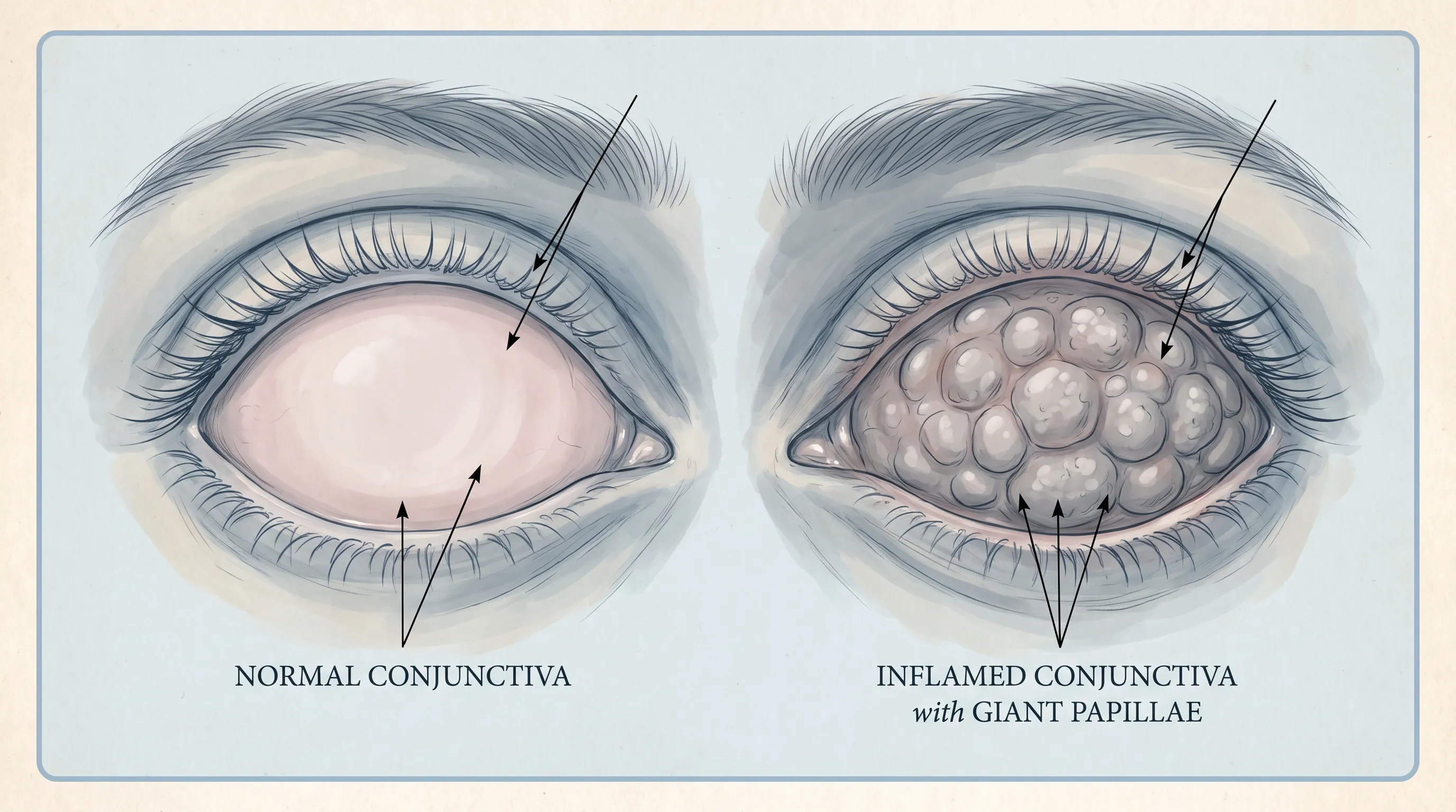
GPC is characterized by the development of elevated, flattened conjunctival papillae on the superior tarsal conjunctiva. Unlike typical allergic conjunctivitis, which primarily involves diffuse redness, chemosis (swelling), and watery discharge, GPC presents with distinct structural remodeling. The hallmark diagnostic criterion is the presence of papillae measuring greater than 1.0 millimeters in diameter. These raised formations are not blisters, cysts, or active infections; rather, they are localized areas of conjunctival epithelial hyperplasia, goblet cell proliferation, and dense inflammatory cell infiltration. The underlying stroma becomes edematous and fibrotic, creating a cobblestone-like appearance that disrupts the normal ocular surface dynamics.
The condition is almost always bilateral, though it may present asymmetrically depending on lens fit, individual eyelid anatomy, and dominance of lens-related factors on one eye. Historically referred to as Contact Lens-Induced Papillary Conjunctivitis (CLPC), the terminology evolved to reflect the fact that similar inflammatory responses can be triggered by non-lens factors. These include ocular prostheses (artificial eyes), exposed surgical sutures following cataract or glaucoma procedures, scleral buckles, trichiasis (misdirected eyelashes), and retained foreign bodies. Regardless of the inciting trigger, the underlying tissue reaction follows a remarkably consistent clinical pathway that requires targeted, multi-faceted intervention.
The Pathophysiology and Immune Response
The development of this eye condition GPC is driven by a complex interplay between mechanical stress, tear film biochemistry, and both Type I and Type IV hypersensitivity reactions. When a contact lens rests against the ocular surface, it moves approximately 10 to 15 millimeters with each blink. Over time, microscopic deposits of tear proteins (such as lysozyme, lactoferrin, and lipocalin), lipids, calcium, mucin, and environmental debris accumulate on the lens matrix. If the lens is not properly cleaned, replaced on schedule, or if the wearer uses solutions containing certain preservatives, these deposits denature and act as persistent, foreign antigens. Simultaneously, the physical friction of the roughened or aged lens surface against the delicate tarsal conjunctiva causes microtrauma to the epithelial layer, compromising the protective barrier.
This dual assault triggers a robust, self-perpetuating immune cascade. Mast cells embedded within the conjunctival stroma recognize the accumulated denatured proteins and mechanical stress, leading to immediate degranulation and the release of preformed and newly synthesized mediators, including histamine, tryptase, leukotrienes (LTB4, LTC4), and prostaglandins. These inflammatory mediators cause localized vasodilation, increased vascular permeability, nerve sensitization, and intense pruritus (itching). As the inflammatory process chronicizes, chemokines recruit secondary inflammatory cells. Eosinophils release major basic protein and eosinophil cationic protein, which are directly cytotoxic to epithelial cells. Lymphocytes, particularly Th2 cells, release interleukins (IL-4, IL-5, IL-9, IL-13) that promote IgE production, eosinophil survival, and goblet cell hyperplasia.
Fibroblasts within the substantia propria are activated by cytokines and growth factors (such as TGF-β and PDGF), leading to excessive collagen synthesis and extracellular matrix deposition. Epithelial cells proliferate outward to form the characteristic giant papillae. This structural remodeling creates a vicious pathological cycle: the enlarged, irregular papillae increase surface friction against the contact lens and cornea, which amplifies mechanical trauma during blinking, further stimulating mast cell activation and cytokine release. Additionally, the altered conjunctival topography disrupts normal tear film distribution and mucin spreading, leading to focal dryness and further epithelial stress. Understanding this pathophysiological loop is essential for appreciating why simply removing the lens temporarily is rarely sufficient without comprehensive anti-inflammatory and barrier-restoring therapy.
Recognizing the Clinical Stages
Giant Papillary Conjunctivitis rarely appears overnight. Instead, it progresses through three well-documented clinical stages, each with distinct symptomatology, histological changes, and therapeutic implications. Recognizing these stages early is the most effective strategy for preventing long-term contact lens intolerance, corneal complications, and chronic ocular surface disease. Clinicians often utilize standardized grading systems, such as the Efron Grading Scale, which categorizes papillary severity from Grade 1 (minimal, barely visible changes) to Grade 4 (severe, cobblestone appearance with papillae >1.0 mm and significant hyperemia).
Early Stage (Grade 1-2): Initial Irritation
In the early phase, the eye condition GPC manifests as mild, intermittent symptoms that patients frequently mistake for seasonal allergies, mild dry eye, or normal lens fatigue. Individuals typically report occasional itching, a slight foreign-body sensation upon blinking, and minor lens awareness that fluctuates throughout the day, often worsening by late afternoon. Upon careful clinical examination, the superior tarsal conjunctiva may show minimal hyperemia (redness) and the initial formation of micro-papillae measuring 0.3 to 0.5 millimeters. Mucus production remains minimal or absent, and visual acuity is generally unaffected. Because these symptoms are subtle and easily masked by over-the-counter artificial tears or seasonal antihistamine drops, many patients continue wearing their lenses as usual. This continuation inadvertently accelerates antigen exposure and mechanical trauma, pushing the tissue into the next inflammatory phase. Early intervention during this window yields the fastest recovery and prevents permanent structural conjunctival changes.
Moderate Stage (Grade 2-3): Progressive Inflammation
As the immune response intensifies and chronic inflammatory mediators accumulate, the condition advances to the moderate stage. The conjunctival papillae enlarge, typically reaching 0.5 to 1.0 millimeters in diameter, and multiply across the tarsal plate. Patients experience noticeably increased lens movement during blinking because the enlarged papillae act as a rough surface, disrupting the smooth lens-to-cornea interface and compromising the tear lens. Itching becomes more persistent and pronounced, particularly immediately following lens removal when histamine receptors are suddenly exposed to airflow and tear film changes.
The most distinguishing feature of this stage is the development of a moderate, stringy mucous film that coats the anterior eye, leading to intermittent blurred vision that temporarily clears with blinking. Early signs of contact lens intolerance emerge, as patients feel compelled to remove lenses earlier than their prescribed wearing schedule, often experiencing rebound itching and redness after several hours out of lenses. Clinical evaluation reveals more pronounced hyperemia, dilated conjunctival vessels, and visible vascular loops extending into the papillary bases. Daily activities, including prolonged screen use, reading, and driving, may become uncomfortable due to fluctuating vision and persistent irritation.
Advanced Stage (Grade 4): Severe Intolerance and Tissue Remodeling
If left unaddressed, GPC reaches an advanced state characterized by profound tissue remodeling, fibroblast hyperactivity, and severe functional impairment. Papillae consistently exceed 1.0 millimeter, creating a highly irregular, cobblestone-like ocular surface. Patients report intolerable foreign-body sensation, stinging pain upon lens insertion or blinking, significant photophobia, and excessive tearing as a reflexive response to surface disruption. Mucus production becomes thick, tenacious, and ropy, frequently causing the eyelids to adhere firmly upon waking.
Contact lens deposits accelerate dramatically due to the compromised tear film and increased mucin interaction with the lens material, leading to rapid clouding and severely compromised optical performance. Lens mobility becomes excessive and erratic, with noticeable rotation (in toric lenses) and decentration, severely disrupting vision quality and astigmatism correction. At this stage, continued lens wear is medically contraindicated. The conjunctival stroma has undergone significant fibrotic changes, making the tissue less responsive to standard anti-inflammatory therapies. Patients often require extended pharmacological management, including tapering topical corticosteroids or long-term immunomodulators, to reverse fibroblast proliferation and restore conjunctival homeostasis. Delayed treatment at this stage increases the risk of secondary corneal complications, including epithelial defects, scarring, and permanent contact lens intolerance.
Diagnosis and Clinical Evaluation
Accurate diagnosis requires a systematic, evidence-based clinical approach to distinguish GPC from other inflammatory, infectious, and toxic ocular conditions. Ophthalmologists and optometrists rely on specialized diagnostic equipment, standardized grading scales, and comprehensive patient histories to confirm the presence, etiology, and severity of the condition.
The Slit-Lamp Examination Process
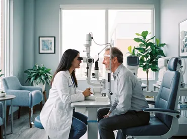
The definitive diagnostic tool for GPC is the slit-lamp biomicroscope combined with proper upper eyelid eversion. During the examination, the clinician uses a cotton-tipped applicator or the edge of a sterile instrument to gently retract the upper eyelid, exposing the full extent of the tarsal conjunctiva. Using high-magnification (16x to 40x) and a focused, narrow light beam, the practitioner evaluates the conjunctival surface for the presence, size, density, and distribution of papillae. The use of vital dyes, such as sodium fluorescein or lissamine green, helps highlight epithelial microtrauma, pinpoint areas of mucous adhesion, and assess tear film stability.
Clinicians assess vascularization patterns, quantifying whether vessels merely skirt the base of papillae or actively penetrate into the tips, which indicates more advanced inflammation. Mucous discharge quantity and viscosity are noted, and the cornea is carefully examined for associated changes such as punctate epithelial erosions (PEE), superior epithelial arcuate lesions (SEALs), or contact lens-induced peripheral ulceration (CLPU). Grading scales typically classify severity objectively, guiding subsequent treatment decisions and establishing a reproducible baseline for monitoring therapeutic progress across multiple visits.
Differential Diagnosis and Ruling Out Mimics
Several ocular conditions share overlapping symptoms with GPC, making differential diagnosis essential to avoid mismanagement. Allergic conjunctivitis typically presents with diffuse conjunctival injection, significant chemosis, and watery discharge but lacks the discrete, large papillae confined to the superior tarsal plate. Vernal keratoconjunctivitis (VKC) and atopic keratoconjunctivitis (AKC) can produce giant papillae but are typically associated with severe systemic atopy, occur in younger patients (VKC), and often involve the cornea with shield ulcers or Horner-Trantas dots.
Superior Limbic Keratoconjunctivitis (SLK) involves inflammation and redundancy of the superior bulbar conjunctiva near the limbus, often associated with thyroid dysfunction or mechanical friction, and requires distinct therapeutic approaches. Dry Eye Disease (DED) can cause significant foreign-body sensation, reflex tearing, and fluctuating vision but does not produce the characteristic conjunctival hyperplasia seen in this eye condition GPC, though the two conditions frequently coexist and exacerbate one another. Toxic conjunctivitis from chronic use of preserved eye drops can mimic GPC but typically shows diffuse conjunctival toxicity rather than discrete tarsal papillae.
Additionally, microbial keratitis, viral conjunctivitis (adenoviral or herpes), and bacterial blepharitis must be ruled out if purulent discharge, corneal infiltrates, or follicular reactions are present, as these require urgent antimicrobial or antiviral therapy rather than immunomodulatory protocols. Comprehensive patient history regarding lens wear schedule, solution brand, systemic allergies, medication history, occupational exposures, and prior ocular surgeries further refines diagnostic accuracy.
| Condition | Primary Location | Key Clinical Feature | Primary Trigger |
|---|---|---|---|
| Giant Papillary Conjunctivitis | Superior tarsal conjunctiva | Papillae >1.0 mm, thick stringy mucus, lens intolerance | Contact lenses, mechanical friction, denatured proteins |
| Allergic Conjunctivitis | Bulbar & palpebral conjunctiva | Diffuse redness, chemosis, watery discharge | Pollen, dust, pet dander, molds |
| Vernal Keratoconjunctivitis | Superior tarsal conjunctiva | Cobblestone papillae, corneal shield ulcers, severe itch | Severe atopy, Th2 hypersensitivity |
| Dry Eye Disease | Corneal surface & tear film | Break-up time <5 sec, punctate staining, burning | Meibomian gland dysfunction, autoimmune, screen time |
| Toxic Conjunctivitis | Diffuse conjunctiva | Follicular reaction, diffuse toxicity, chronic redness | Preservatives (BAK), medication overuse |
| Superior Limbic Keratoconjunctivitis | Superior limbus & bulbar conjunctiva | Thickened redundant superior bulbar conjunctiva | Mechanical friction, thyroid eye disease |
| Viral Conjunctivitis | Diffuse conjunctiva | Follicles, preauricular lymphadenopathy, watery discharge | Adenovirus, enterovirus, HSV |
Evidence-Based Treatment Protocols
Management of GPC follows a structured, phased approach endorsed by major ophthalmological and optometric bodies, including the American Academy of Ophthalmology and the Tear Film & Ocular Surface Society (TFOS). The primary goals are to halt the inflammatory cascade, promote epithelial and stromal healing, and establish a sustainable lens-wearing regimen that prevents recurrence while maintaining visual function.
Phase One: The Essential Contact Lens Holiday
The absolute first step in treating GPC is immediate and complete cessation of contact lens wear. Continuing to wear lenses over inflamed, irregular tissue perpetuates mechanical friction, maintains antigen exposure, and renders pharmacological treatments significantly less effective. Clinical guidelines typically recommend a strict lens-free period of two to four weeks for mild to moderate cases, and up to six to eight weeks for severe or refractory presentations. This holiday allows the conjunctival epithelium to regenerate, mast cell granules to replenish, and fibroblast proliferation to subside.
During this period, patients should switch exclusively to prescription eyeglasses. It is crucial to dispose of the triggering contact lenses and replace the lens case to eliminate residual biofilm. Patients often experience significant discomfort during the first few days of cessation due to sudden exposure of hypersensitive nerve endings. Cold compresses applied for 10 minutes, 3-4 times daily, can provide substantial symptomatic relief by inducing localized vasoconstriction and reducing nerve conduction velocity. Preservative-free artificial tears with hyaluronic acid or carboxymethylcellulose help restore the tear film, flush inflammatory mediators, and improve epithelial hydration. Strict compliance with the lens holiday is the single strongest predictor of rapid symptom resolution. Patients who attempt to "push through" discomfort or wear lenses intermittently during this phase frequently experience prolonged recovery, rebound inflammation, and increased risk of permanent conjunctival fibrosis.
Phase Two: Targeted Pharmacological Management
While the conjunctiva heals, targeted topical therapies are prescribed to control inflammation, modulate immune response, and alleviate symptoms. First-line therapy typically involves dual-action mast cell stabilizer/antihistamine combinations such as olopatadine HCl 0.1%-0.2%, ketotifen fumarate 0.025%, or azelastine HCl 0.05%. These medications simultaneously block histamine H1 receptors (providing rapid itch relief) and inhibit mast cell degranulation (preventing further release of inflammatory mediators), offering both immediate and long-term symptom control. For patients experiencing significant ocular surface discomfort or pain, nonsteroidal anti-inflammatory drug (NSAID) drops like ketorolac tromethamine 0.5% or diclofenac sodium 0.1% may be added for 1-2 weeks to reduce prostaglandin-mediated inflammation, though they must be used cautiously due to potential corneal epithelial toxicity with prolonged use.
In moderate to severe cases where papillae exceed 0.5-1.0 mm and symptoms significantly impair daily function, short-course topical corticosteroids may be prescribed. Loteprednol etabonate 0.5% or fluorometholone 0.1% are frequently favored for their ester-based structures, which offer potent anti-inflammatory effects with significantly reduced risks of intraocular pressure (IOP) elevation and posterior subcapsular cataract formation compared to traditional steroids like dexamethasone or prednisolone. A typical regimen involves dosing four times daily for 7-10 days, followed by a gradual taper to once daily, then every other day, before discontinuation. Steroid therapy must be strictly monitored by an eye care professional with scheduled follow-ups at 1 and 3 weeks to assess IOP, corneal epithelial integrity, and inflammatory response.
For chronic, refractory, or atopic-associated cases, or when patients are steroid-intolerant, calcineurin inhibitor immunomodulators such as cyclosporine A 0.05% or 0.1% emulsions, or off-label tacrolimus 0.03% ointment, may be introduced. These agents selectively inhibit T-cell activation and cytokine release (particularly IL-2 and TNF-α), effectively breaking the chronic inflammatory cycle and promoting long-term ocular surface homeostasis without the steroid-associated side effects. Initial burning upon instillation is common but typically subsides within 1-2 weeks.
Phase Three: Strategic Lens Modification and Reintroduction
Once the conjunctiva has fully healed, symptoms have completely resolved, and the practitioner confirms a smooth tarsal surface upon re-examination, patients must transition to a modified lens-wearing strategy to prevent relapse. A phased reintroduction protocol is typically followed: starting with 2-3 hours of wear on day one, gradually increasing by 1-2 hours daily until full-day wear is comfortably achieved.
Daily disposable lenses are widely considered the gold standard for GPC prevention. Because they are discarded every 24 hours, they eliminate the opportunity for significant protein, lipid, and environmental allergen accumulation on the lens surface. Patients previously wearing monthly, bi-weekly, or extended-wear lenses should strongly consider this transition. For those requiring astigmatism correction or specialized optics, modern daily disposable toric lenses offer excellent alternatives without compromising deposit resistance.
Alternatively, rigid gas permeable (RGP) lenses offer a highly viable long-term solution. RGPs have a smaller diameter, move differently across the ocular surface, and are composed of highly durable, oxygen-permeable fluorosilicone acrylate or siloxane polymers that resist deposit adhesion far better than hydrogels. They also do not conform to the conjunctiva, reducing frictional drag. While the initial adaptation period is longer, recurrence rates of GPC with RGP wear are significantly lower. Reduced wearing time (e.g., limiting wear to 8-10 hours daily), strict adherence to replacement schedules regardless of subjective comfort, and routine professional evaluations are non-negotiable components of long-term ocular surface preservation.
Prevention and Optimized Lens Care
Preventing GPC recurrence requires a fundamental shift in lens hygiene habits, environmental awareness, and daily ocular maintenance routines. The most common trigger for GPC relapse is inadequate lens disinfection, premature lens wear, or exposure to contaminated accessories.
Mastering Cleaning and Disinfection
Patients using reusable lenses must adopt a rigorous, evidence-based cleaning protocol. The mechanical act of "rubbing and rinsing" remains critical: vigorously rubbing each lens surface with approved solution for at least 10-15 seconds per side, even when using "no-rub" formulas, physically removes up to 90% of surface-adhered deposits, bacteria, and allergens. Multipurpose solutions (MPS) are convenient but vary significantly in preservative efficacy and deposit-removal capacity. Some older formulations containing thimerosal or certain polyquaternium compounds have been associated with higher rates of surface toxicity and hypersensitivity. Modern MPS using polyhexamethylene biguanide (PHMB) or Alexidine offer broader antimicrobial spectra and better biocompatibility.
Weekly enzymatic cleaning tablets (proteases like subtilisin or papain) remain highly recommended for bi-weekly and monthly lenses to hydrolyze stubborn denatured protein and lipid layers that harbor allergens and serve as niduses for microbial biofilm. However, enzymatic cleaners do not disinfect; they must always be followed by a standard disinfecting soak. Lens cases are frequently overlooked reservoirs of contamination. They should be replaced monthly, never topped off with old solution, rinsed daily with fresh disinfecting solution (never tap water, which contains Acanthamoeba and bacteria), and stored face-down on a clean paper towel in a dry, well-ventilated environment. Humidity promotes microbial proliferation, so airtight cases or storing cases in dark, damp bathrooms significantly increases infection and allergy risks.
The Role of Hydrogen Peroxide Systems
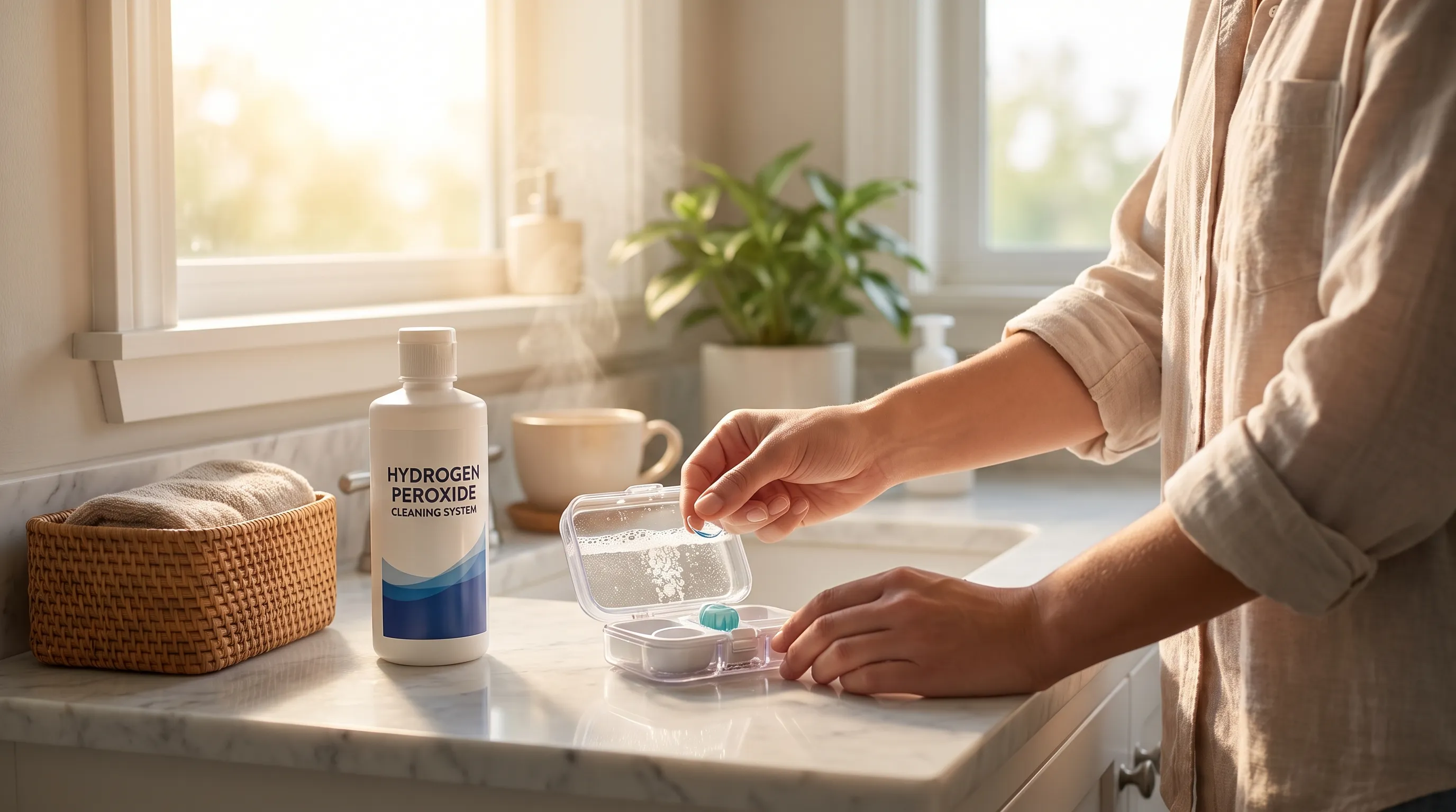
For individuals prone to GPC, those who have previously experienced severe inflammation, or wearers of silicone hydrogel lenses (which are highly susceptible to lipid and protein deposition), transitioning to a hydrogen peroxide-based disinfection system (e.g., 3% H2O2) is strongly advised by clinical guidelines. These systems utilize a platinum or catalase-coated neutralizing disk to convert 3% hydrogen peroxide into pure saline and oxygen gas over a minimum 6-hour period. This process provides exceptional, broad-spectrum disinfection, eliminates preservatives entirely, and effectively loosens and removes stubborn deposits.
Multiple peer-reviewed studies consistently demonstrate that hydrogen peroxide systems reduce papillary recurrence rates, improve end-of-day comfort, and decrease microbial binding compared to traditional multipurpose solutions. It is absolutely crucial to follow the exact neutralization time; inserting lenses before complete neutralization will cause immediate, severe corneal epithelial burns and intense pain. Modern systems feature clear visual indicators (color-changing catalysts or bubbling cessation) to ensure patient safety, making these systems highly effective when protocols are meticulously followed.
Integrating Lid Hygiene and Systemic Allergy Control
The ocular surface does not exist in isolation. Systemic atopy, uncontrolled seasonal allergies, and blepharitis (chronic eyelid margin inflammation) significantly lower the threshold for developing GPC by increasing the baseline concentration of inflammatory mediators, histamine, and proteolytic enzymes in the tear film. Incorporating daily warm compresses (applied at 40-45°C for 8-10 minutes) helps liquefy meibum, stabilize the tear film lipid layer, and reduce evaporative dryness. Gentle lid margin cleansing using hypochlorous acid sprays or tea tree oil-based wipes (for Demodex-associated blepharitis) reduces bacterial load and staphylococcal exotoxins that exacerbate conjunctival hypersensitivity.
Patients with known environmental allergies should begin prophylactic oral or topical antihistamine therapy before peak allergy seasons. Managing systemic conditions like eczema, asthma, or allergic rhinitis through dermatological and primary care indirectly supports ocular surface stability by lowering systemic IgE levels and cytokine burden. Dietary considerations also play a role; omega-3 fatty acid supplementation (EPA/DHA) has been shown to modulate inflammatory pathways, improve meibomian gland secretion, and reduce ocular surface inflammation. Regular annual comprehensive eye exams, or biannual for lens wearers, allow practitioners to monitor lens fit parameters (base curve, diameter, movement), assess early micro-papillary changes, evaluate tear osmolarity, and adjust replacement schedules or modalities before symptoms become clinically apparent.
Potential Complications and Prognosis
When managed promptly, correctly, and with full patient compliance, the eye condition GPC carries an excellent prognosis and rarely leads to permanent vision impairment. The conjunctiva is highly regenerative, and papillae can fully flatten within weeks of appropriate therapy. However, chronic neglect, self-medication with inappropriate drops, or improper lens management can result in significant, sometimes irreversible, ocular complications.
Corneal Staining, Erosion, and Secondary Keratitis
Persistent mechanical rubbing from enlarged, fibrotic papillae against the superior cornea and limbus can cause superior epithelial arcuate lesions (SEALs) and superficial punctate keratitis (SPK). These are characterized by tiny, linear epithelial defects and micro-abrasions on the superior corneal surface, precisely where the upper eyelid drapes during blinking. These defects compromise the corneal epithelial barrier, the eye's primary defense against pathogens, creating direct entry points for bacteria, fungi, or acanthamoeba. If secondary microbial keratitis develops, patients may experience severe, throbbing pain, marked photophobia, purulent discharge, corneal ulcers, and potential stromal scarring that permanently affects visual acuity and may require penetrating keratoplasty (corneal transplant) in extreme cases.
Additionally, thick mucous discharge can physically trap debris, allergens, and bacteria against the cornea, creating a localized inflammatory environment that delays healing and exacerbates surface irregularity. Prompt discontinuation of lens wear, strict adherence to prescribed anti-inflammatory protocols, and use of preservative-free lubricants effectively mitigate these risks and promote rapid epithelial migration and repair.
Managing Long-Term Lens Wear and Alternative Vision Correction
A subset of patients, particularly those with severe, chronic GPC combined with underlying atopic keratoconjunctivitis or severe dry eye syndrome, may develop permanent contact lens intolerance. In these cases, the conjunctival and limbal tissue remains chronically hypersensitive, mast cell thresholds remain low, and even strict daily disposable protocols trigger recurrent inflammation. For these individuals, transitioning away from traditional contact lenses is often the safest path to ocular health.
Refractive surgery evaluation (LASIK, PRK, SMILE, or ICL) may be highly appropriate for suitable candidates with stable refractions and adequate corneal thickness. Modern wavefront-guided procedures offer excellent visual outcomes without the chronic ocular surface stress associated with decades of lens wear. Alternatively, orthokeratology (overnight RGP wear) can reshape the cornea temporarily while allowing the conjunctiva to remain uncovered during waking hours, though careful monitoring is required. Permanent transition to high-quality eyeglasses with anti-reflective coatings, specialized filters, and optimized prescriptions remains a highly effective, complication-free alternative. Open, proactive communication with an eye care professional ensures that patients find a sustainable, comfortable vision correction method tailored to their unique ocular health profile, lifestyle, and long-term safety.

Frequently Asked Questions
Can I ever wear contact lenses again after being diagnosed with GPC?
Yes, the vast majority of patients can safely return to contact lens wear after complete symptom resolution and formal clinician clearance. This typically requires a strict minimum two-to-four-week lens holiday, successful treatment with appropriate anti-inflammatory drops, and a confirmed resolution of papillary inflammation upon slit-lamp examination. Resuming wear should follow a graduated, phased approach, starting with just a few hours daily and slowly increasing. Most patients successfully transition to daily disposable silicone hydrogel lenses or switch to rigid gas permeable (RGP) lenses, both of which significantly reduce recurrence risk. Returning too early, ignoring mild symptoms, or reverting to the exact same lens type and care system that triggered the condition will almost certainly lead to rapid relapse and potentially more severe inflammation.
Is giant papillary conjunctivitis contagious or caused by an active infection?
No, GPC is fundamentally a non-infectious, sterile inflammatory and hypersensitivity condition. It arises from a combination of mechanical friction, compromised tear film dynamics, and an immune-mediated reaction (Types I and IV) to accumulated denatured tear proteins, lipid deposits, contact lens materials, or solution preservatives. Because it is not caused by pathogenic bacteria, viruses, or fungi, it cannot be transmitted to other individuals through shared towels, contact, airborne droplets, or environmental surfaces. However, the compromised ocular surface during active GPC is more vulnerable to secondary infections, so strict hygiene remains paramount.
How long does it take for GPC symptoms to completely resolve?
Mild cases (Grade 1-2) typically resolve within two to four weeks once lens wear is completely discontinued and appropriate anti-inflammatory therapy begins. Moderate cases (Grade 3) with noticeable papillary formation and mucous production often require four to six weeks of consistent pharmacological management, including potential steroid tapering. Severe cases (Grade 4) with significant cobblestone papillae, fibroblast activation, and corneal involvement may require six to twelve weeks of comprehensive treatment. Complete tissue normalization, where the conjunctiva returns entirely to its smooth, baseline state without residual hyperemia or subtle fibrosis, depends heavily on strict medication adherence, environmental control, and absolute prevention of re-exposure to the original triggering factors.
Are daily disposable lenses safer than monthly lenses for preventing this condition?
Absolutely. Clinical evidence and consensus guidelines from major ophthalmological organizations strongly support daily disposable lenses as the safest and most effective option for GPC-prone individuals, first-time wearers, and those with allergy histories. Because these lenses are worn once and discarded before significant biofilm or protein deposition can occur, they prevent the gradual buildup of tear proteins, lipids, environmental allergens, and bacterial antigens that serve as the primary catalysts for mast cell activation and papillae formation. Studies consistently show GPC incidence is reduced by over 60% in daily disposable wearers compared to reusable soft lens modalities.
What should I do if my eyelids stick together in the morning due to thick mucus?
This symptom indicates moderate to advanced conjunctival inflammation and goblet cell hyperactivity. Apply a clean, sterile, warm compress to the closed eyelids for 5-10 minutes to soften and liquefy the mucus. Gently wipe the lash margins outward using a sterile, preservative-free eyelid wipe or clean gauze; never use harsh soaps or rough towels. Instill preservative-free artificial tears to flush the ocular surface, improve tear film osmolarity, and gently separate the adhered lids without mechanical trauma. Immediately discontinue all contact lens wear and schedule a prompt appointment with an optometrist or ophthalmologist. Strictly avoid rubbing your eyes, as mechanical friction immediately triggers mast cell degranulation, releases histamine, and worsens conjunctival microtrauma and vascular leakage.
Can makeup worsen GPC or trigger a flare-up?
Yes, certain cosmetics can significantly exacerbate GPC. Eyeliner applied to the inner lid margin (waterline), mascara flakes, and powder eyeshadows can directly deposit particles onto the conjunctiva, act as mechanical irritants, and harbor bacteria. Waterproof formulas often require oil-based removers that can disrupt the meibomian lipid layer, worsening dry eye and lowering the threshold for inflammation. Patients recovering from or prone to GPC should avoid inner-rim eyeliner, replace mascara every three months, use preservative-free or ophthalmologist-tested products, and thoroughly but gently remove all makeup with hypoallergenic micellar water or dedicated lid wipes before sleep.
Conclusion
Navigating the eye condition GPC requires patience, diligence, and a steadfast commitment to evidence-based ocular hygiene and lens management protocols. While the inflammatory response can be highly uncomfortable, visually disruptive, and temporarily debilitating, it remains highly reversible when addressed promptly with complete lens cessation, targeted pharmacological therapy, and strategic lens modification. By understanding the complex immunological and mechanical pathways behind papillary formation, patients can fundamentally transform their approach to contact lens care from reactive symptom management to proactive ocular surface preservation.
Embracing daily disposable lens modalities, utilizing advanced preservative-free or hydrogen peroxide disinfection systems, integrating daily lid hygiene, and managing systemic allergies will drastically reduce recurrence rates and extend comfortable lens tolerance for years. Regular, comprehensive consultations with qualified eye care professionals ensure that subtle conjunctival changes are detected and addressed early, preserving long-term ocular comfort, visual clarity, and corneal integrity. With the right management strategies, individualized fitting approaches, and informed patient participation, individuals can safely enjoy the convenience and lifestyle benefits of modern contact lenses while actively protecting their delicate conjunctival tissue from chronic inflammation.
For further reading on clinical guidelines, pathophysiology, and patient management strategies, consult authoritative, peer-reviewed resources such as the American Academy of Ophthalmology (EyeWiki), the TFOS Contact Lens and Ocular Surface reports, and the comprehensive clinical overview provided by NCBI Bookshelf. Patient-focused guidance and symptom tracking tools are also available through the Cleveland Clinic and Healthline.
To visualize the clinical presentation, grading scales, and advanced management techniques discussed, we recommend exploring expert-led educational content from accredited optometric and ophthalmological institutions. The following curated clinical lectures provide in-depth visual explanations of ocular surface pathology, lens complication management, and modern therapeutic protocols:
Disclaimer: This article is intended strictly for educational and informational purposes only and does not constitute medical advice, diagnosis, or treatment. Always consult a licensed, board-certified optometrist or ophthalmologist for comprehensive eye examinations, accurate diagnosis, individualized treatment planning, and ongoing management of ocular surface conditions and contact lens wear.
How we work
Researched and drafted with AI assistance, then reviewed by a human editor against the cited sources.
This article is general health information, not medical advice. Consult a qualified healthcare professional about your own situation.