Calf Pain with Back Pain: Causes, Diagnosis, and Evidence-Based Relief Strategies
Experiencing discomfort that travels from your spine down to your lower leg can be both alarming and profoundly disruptive to your daily routine. When nerve pathways become irritated or structurally compressed, the resulting symptoms often create a distinct clinical picture that requires careful evaluation and targeted intervention. Understanding the underlying mechanisms behind calf pain with back pain is essential for developing an effective, evidence-based recovery plan. While many individuals initially assume they are dealing with a simple muscle strain, the interconnected nature of spinal anatomy and peripheral nerve distribution means that what feels like localized leg discomfort is frequently rooted in lumbar spine pathology. This comprehensive guide explores the anatomical foundations, diagnostic pathways, and clinically proven management strategies to help you navigate your symptoms safely and restore functional mobility. By integrating current medical research with practical lifestyle adjustments, patients and caregivers can make informed decisions that prioritize long-term spinal health and neurological preservation.
Understanding the Neurological Connection Between Spinal Discomfort and Leg Pain
The human body operates as an integrated kinetic chain, where structural changes in one region frequently manifest as symptoms in seemingly unrelated areas. The lumbosacral spine serves as the primary weight-bearing foundation of the torso while simultaneously housing delicate neural pathways that extend throughout the lower extremities. When these pathways become compromised, patients frequently report experiencing calf pain with back pain as a coordinated symptom cluster rather than two isolated issues. Recognizing this neurological linkage is the first step toward accurate diagnosis and appropriate therapeutic intervention.
Free calculatorBMI CalculatorCalculate Body Mass IndexOpen the calculator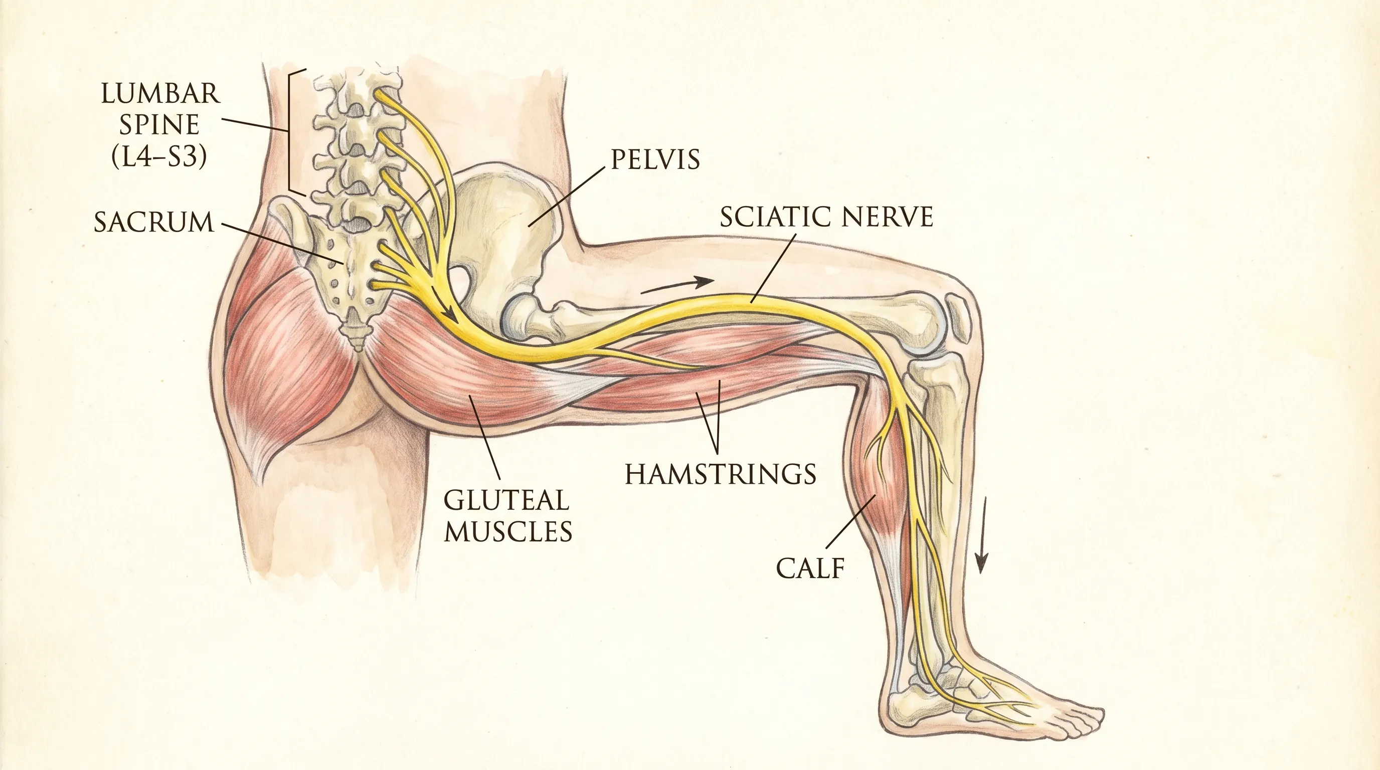
The Anatomy of the Sciatic Nerve
The sciatic nerve is the longest and thickest peripheral nerve in the human body, measuring up to two centimeters in diameter. It originates from the lumbosacral plexus, specifically drawing nerve roots from spinal levels L4 through S3. These nerve fibers converge to form a thick neural bundle that exits the pelvis through the greater sciatic foramen, courses beneath the piriformis muscle, travels posteriorly through the thigh, and bifurcates into the tibial and common fibular nerves just above the knee. The tibial branch continues down into the posterior calf, ankle, and foot, providing both motor innervation to the posterior leg muscles and sensory feedback from the skin and deep tissues. According to peer-reviewed clinical references like StatPearls from the National Institutes of Health, irritation at any point along this extensive pathway can generate radiating symptoms that patients perceive as distinct from the original source of compression. This anatomical reality explains why lumbar pathology frequently presents as distal leg discomfort rather than localized spinal pain.
How Nerve Compression Creates Radiating Pain
Nerve tissue is highly sensitive to mechanical deformation and inflammatory mediators. When a spinal structure exerts sustained pressure on a nerve root, it disrupts normal axonal transport, compromises blood flow to the neural tissue, and triggers a localized inflammatory cascade. This process releases cytokines and prostaglandins that sensitize nociceptors, resulting in pain signals that travel along the entire length of the affected nerve. Patients often describe this phenomenon as electric shocks, burning sensations, or deep aching that follows a predictable dermatomal pattern. The presence of calf pain with back pain strongly suggests that the irritation is occurring at the lumbar level before the nerve exits the spinal column. Mayo Clinic clinical guidelines emphasize that true radicular pain typically radiates below the knee, distinguishing it from localized musculoskeletal strain that generally remains confined to the lumbar region.
Primary Causes of Calf Pain with Back Pain
Identifying the precise etiology of spinal-related leg discomfort requires a systematic evaluation of structural, mechanical, and inflammatory factors. Medical literature consistently demonstrates that the majority of cases originate from degenerative changes, traumatic injuries, or postural imbalances that gradually compromise the lumbar intervertebral spaces. Each underlying condition follows a distinct pathophysiological trajectory, yet they ultimately converge on a shared clinical endpoint: mechanical impingement or chemical irritation of the exiting nerve roots.
Lumbar Disc Herniation and Degeneration
Herniated intervertebral discs represent the most prevalent cause of radicular symptoms affecting the lower extremities. Each spinal disc consists of a gelatinous nucleus pulposus encased within a tough, fibrous annulus fibrosus. Repetitive microtrauma, age-related dehydration, or acute heavy lifting can create fissures in the annulus, allowing nuclear material to extrude and press directly against adjacent nerve roots. When a herniation occurs at the L4-L5 or L5-S1 levels, the resulting neural compression frequently produces calf pain with back pain, often accompanied by sensory alterations in the foot or ankle. As outlined by the Cleveland Clinic, degenerative disc disease accelerates this process by reducing disc height, altering spinal biomechanics, and decreasing the natural shock-absorbing capacity of the vertebral column. Clinical imaging studies consistently correlate disc herniation size and location with symptom severity and distribution patterns.
Spinal Stenosis and Structural Narrowing
Lumbar spinal stenosis involves the progressive narrowing of the central spinal canal or lateral recesses where nerve roots travel to exit the vertebral column. This condition typically develops secondary to facet joint hypertrophy, ligamentum flavum thickening, and osteophyte formation. Unlike acute disc herniations, stenosis symptoms often manifest gradually and are heavily influenced by positional changes. Patients frequently report symptom exacerbation during prolonged standing or walking downhill, with notable relief when sitting or leaning forward, a posture that temporarily increases canal diameter and reduces neural compression. Research from the National Institute of Neurological Disorders and Stroke (NIH) notes that when stenosis affects the L4 or L5 nerve roots, the clinical presentation closely mirrors the classic pattern of calf pain with back pain, though it may also involve cramping, heaviness, or neurogenic claudication during ambulation.
Secondary Mechanical and Muscular Triggers
Beyond direct spinal compression, several auxiliary mechanisms can reproduce similar symptom clusters. Spondylolisthesis occurs when a vertebral body slips anteriorly relative to the adjacent level, reducing neural foramen space and potentially entrapping exiting nerve roots. Piriformis syndrome, though less common, involves hypertrophy or spasm of the deep gluteal muscle, which can compress the sciatic nerve as it passes beneath or through the muscular fibers. Additionally, sacroiliac joint dysfunction and myofascial trigger points in the posterior chain can refer pain distally, creating diagnostic overlap that requires careful clinical differentiation. Healthcare providers must systematically evaluate these mechanical variables to ensure treatment targets the actual pain generator rather than superficially addressing secondary muscular tension.
The Critical Vascular Differential: Deep Vein Thrombosis (DVT)
While neurological compression accounts for the vast majority of cases presenting with spinal and lower leg discomfort, clinical best practices mandate ruling out vascular pathology before initiating conservative spinal management. Deep vein thrombosis represents a potentially life-threatening condition that requires immediate recognition and distinct therapeutic protocols. Misdiagnosis or delayed intervention can result in pulmonary embolism, making accurate differential assessment a critical component of patient safety.
Differentiating Nerve Pain from Vascular Clots
DVT occurs when a blood clot forms within the deep venous system, most commonly in the proximal calf veins or popliteal region. The clinical presentation differs significantly from radicular pain in several key parameters. CDC clinical resources indicate that vascular clots typically produce localized swelling, a sensation of deep cramping or soreness, visible skin erythema, and noticeable warmth compared to the contralateral limb. Unlike sciatica, DVT pain generally does not follow a dermatomal pathway and rarely includes tingling, numbness, or shooting electrical sensations. Furthermore, vascular symptoms typically worsen with dependency and improve with leg elevation, whereas nerve pain often fluctuates with spinal positioning, coughing, or sneezing. Recognizing these distinctions is vital when evaluating patients experiencing calf pain with back pain, as vascular emergencies require anticoagulation therapy rather than spinal decompression strategies.
Risk Factors and Vascular Warning Signs
Several physiological and lifestyle variables significantly increase the likelihood of thrombotic events. Advanced age, prolonged immobilization, recent surgical procedures, pregnancy, malignancy, obesity, and inherited hypercoagulable states all elevate baseline risk profiles. Tobacco use and certain hormonal medications further compromise vascular endothelial integrity and promote stasis. Patients presenting with unilateral leg edema, unexplained skin discoloration, or palpable cord-like veins alongside lower extremity discomfort should undergo prompt duplex ultrasonography to confirm or exclude thrombus formation. Clinicians emphasize that while DVT rarely coexists with true lumbar radiculopathy, the combination of symptoms warrants comprehensive vascular screening to prevent catastrophic embolic complications.
Clinical Diagnosis and Professional Evaluation
Accurate diagnosis of spinal-related lower extremity symptoms relies on a structured, multi-modal assessment approach that integrates patient history, targeted physical examination, and selective diagnostic imaging. Relying solely on symptom description often leads to misclassification, which delays appropriate treatment and potentially worsens underlying pathology. Healthcare providers utilize standardized protocols to isolate pain generators, assess neurological integrity, and determine the most effective intervention pathway.
Physical Examination Protocols
The initial clinical evaluation focuses on functional mobility, muscle strength, reflex integrity, and provocative maneuvers designed to reproduce or alleviate symptoms. The straight leg raise test remains the most widely validated screening tool for lumbar radiculopathy. With the patient supine, the examiner passively elevates the affected limb between thirty and seventy degrees. Reproduction of shooting pain below the knee suggests nerve root tension, typically at the L5 or S1 level. Additional assessments include heel and toe walking to evaluate peroneal and posterior tibial nerve function, deep tendon reflex testing at the patellar and Achilles tendons, and sensory mapping to identify specific dermatomal deficits. Providers also assess postural alignment, palpate paraspinal musculature for hypertonicity, and observe gait mechanics for antalgic patterns that indicate compensatory movement strategies.
Advanced Imaging and Neurological Testing
Imaging studies are generally reserved for cases presenting with severe motor weakness, progressive neurological decline, or symptoms persisting beyond six weeks despite conservative management. Plain radiographs effectively identify bony abnormalities such as osteophytes, vertebral misalignment, and significant disc space narrowing. Magnetic resonance imaging (MRI) provides superior soft tissue resolution, clearly visualizing disc herniations, ligament thickening, nerve root compression, and spinal canal dimensions. Computed tomography (CT) myelography may be utilized when MRI is contraindicated or when detailed bony anatomy is required for surgical planning. Electromyography (EMG) and nerve conduction studies offer objective data regarding the degree of axonal injury, differentiate peripheral neuropathies from radiculopathies, and help localize compression sites when imaging findings remain inconclusive. These diagnostic modalities work synergistically to construct a comprehensive clinical picture that guides targeted therapeutic decision-making.
Evidence-Based Treatment Pathways
Managing spinal-related leg discomfort requires a phased approach that progresses from conservative self-care to targeted medical interventions based on symptom severity, functional limitation, and underlying etiology. Current clinical guidelines strongly emphasize active recovery over passive rest, highlighting the importance of movement modification, pain modulation, and structured rehabilitation. Treatment plans should be individualized, incorporating patient preferences, comorbid conditions, and realistic functional goals.
Initial Self-Care and Conservative Management
During the acute inflammatory phase, short-term symptom control focuses on reducing neural irritation and preventing further mechanical stress. Contrary to historical practices, prolonged bed rest has been shown to delay recovery, promote muscle atrophy, and increase the risk of chronic pain development. Instead, Mayo Clinic recommendations encourage patients to maintain gentle daily movement, incorporating short walking intervals and avoiding positions that exacerbate symptoms. Cold therapy applied for fifteen to twenty minutes during the first forty-eight hours effectively reduces localized inflammation and provides temporary analgesia. Transitioning to moist heat after the acute phase promotes tissue relaxation and improves regional circulation. Over-the-counter nonsteroidal anti-inflammatory drugs (NSAIDs) may offer symptomatic relief, though clinical evidence suggests their efficacy for radicular pain remains moderate. Paracetamol is generally considered insufficient for nerve-related discomfort. Gentle, static stretching held for thirty seconds without bouncing helps maintain tissue elasticity and reduces muscular guarding that often accompanies protective spinal postures.
Medical Interventions and Procedural Options
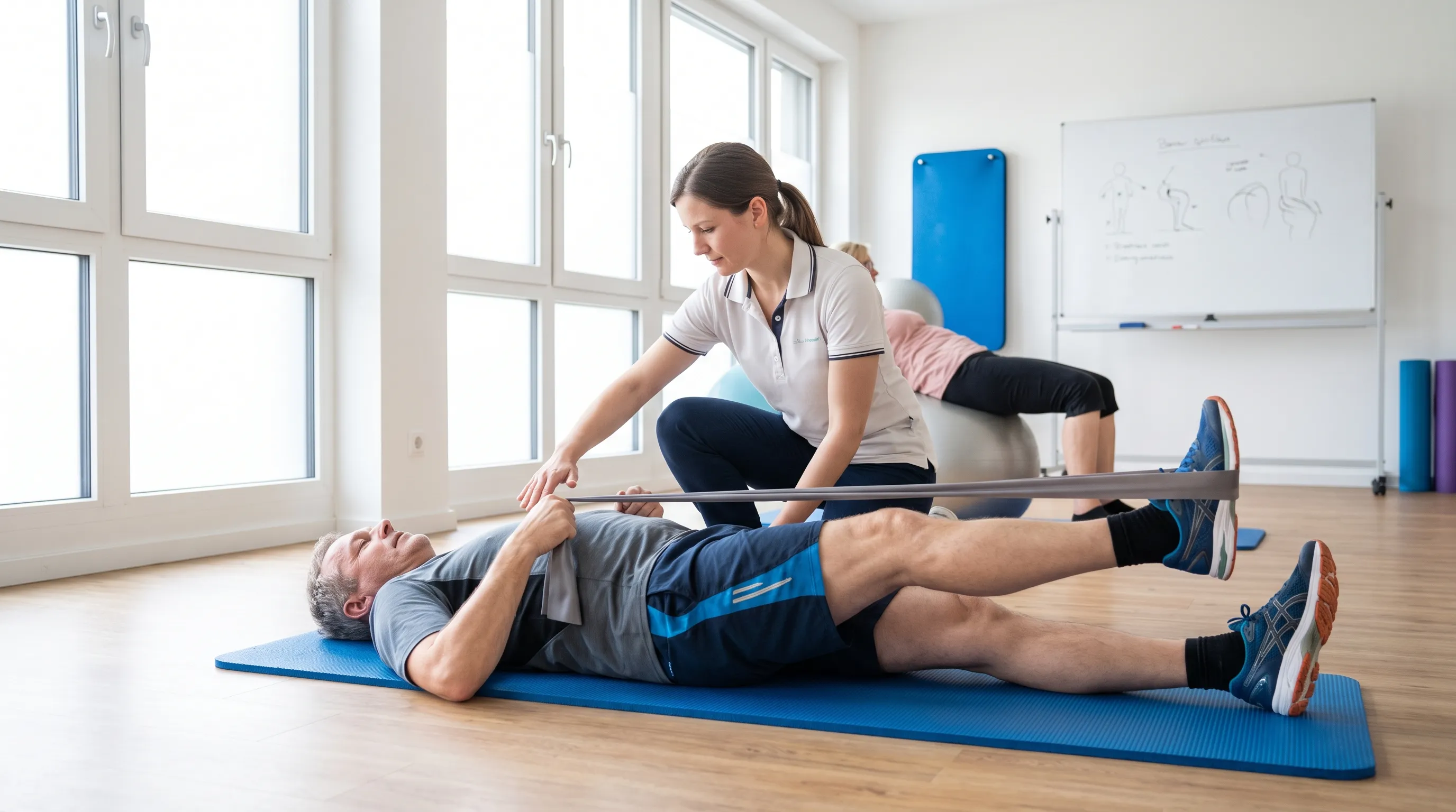
When conservative measures fail to produce meaningful improvement, structured physical therapy becomes the cornerstone of functional restoration. Licensed therapists design progressive exercise programs targeting core stabilization, hip mobility, and neural gliding techniques that improve axonal transport and reduce mechanical sensitivity. Cleveland Clinic specialists note that corticosteroid epidural injections deliver potent anti-inflammatory medication directly to the affected nerve root, typically providing temporary relief that enables patients to participate more effectively in rehabilitation. Prescription medications, including neuropathic pain modulators, muscle relaxants, and in select short-term cases, opioid analgesics, may be utilized under strict medical supervision to manage severe symptom flares. Surgical decompression or microdiscectomy is considered a definitive option when patients experience progressive neurological deficits, bowel or bladder dysfunction, or debilitating pain that severely compromises quality of life after exhausting non-operative pathways.
| Treatment Modality | Primary Mechanism of Action | Typical Duration | Best Suited For |
|---|---|---|---|
| Activity Modification & Ice/Heat Therapy | Reduces inflammation, prevents mechanical aggravation | Days to weeks | Acute symptom flare-ups |
| Structured Physical Therapy | Restores mobility, strengthens core, improves neural glide | 6–12 weeks | Subacute recovery & prevention |
| Epidural Corticosteroid Injection | Localized anti-inflammatory effect on nerve roots | Relief for weeks to months | Moderate to severe radicular pain |
| Neuropathic Pain Medications | Modulates nerve signal transmission, reduces hypersensitivity | Ongoing, as prescribed | Chronic nerve irritation & burning pain |
| Surgical Decompression | Removes structural compression on affected neural tissue | Permanent structural relief | Progressive weakness or cauda equina syndrome |
Complementary and Alternative Therapies
Integrative approaches can serve as valuable adjuncts to conventional medical management when implemented safely and under professional guidance. Acupuncture has demonstrated moderate efficacy in reducing chronic musculoskeletal and neuropathic discomfort by stimulating endogenous opioid release and modulating pain pathway signaling. Clinical trials indicate that chiropractic spinal manipulation, when performed by licensed practitioners on appropriately screened patients, offers comparable outcomes to standard medical care for mechanical back pain without significant adverse event rates. Mind-body techniques such as progressive muscle relaxation, mindfulness meditation, and paced breathing exercises effectively reduce central pain sensitization and improve coping mechanisms during prolonged recovery periods. Patients considering complementary modalities should verify practitioner credentials, maintain open communication with their primary care providers, and avoid aggressive manual techniques during acute inflammatory phases.
Prevention, Rehabilitation, and Long-Term Spine Care
Sustained spinal health relies on proactive lifestyle habits, consistent physical conditioning, and ergonomic optimization that collectively reduce cumulative mechanical stress on the lumbar region. Preventative strategies extend beyond symptom management, focusing on structural resilience and neuromuscular efficiency that minimize recurrence risk. Implementing evidence-based practices early in the recovery phase significantly improves long-term functional outcomes and reduces dependency on pharmacological interventions.
Ergonomic and Postural Optimization
Proper biomechanics during daily activities play a decisive role in maintaining intervertebral disc integrity and preventing progressive degeneration. When lifting objects, individuals should hinge at the hips and knees, maintain a neutral spinal alignment, and avoid rotational movements while bearing weight. Prolonged sitting increases intradiscal pressure and promotes anterior pelvic tilt, which strains posterior spinal structures. Implementing CDC-recommended ergonomic adjustments effectively counteract sedentary-related spinal loading. Standing desks, ergonomic keyboard positioning, and supportive footwear further contribute to optimal kinetic chain alignment throughout the workday.
Lifestyle Modifications and Activity Guidelines
Regular aerobic exercise, particularly low-impact modalities such as walking, swimming, or stationary cycling, promotes systemic circulation, facilitates nutrient delivery to avascular disc tissues, and supports healthy metabolic function. NIH-supported research strongly advocates for targeted core strengthening programs that activate the transversus abdominis, multifidus, and pelvic floor musculature to create a natural corset effect that stabilizes spinal segments. Weight management remains a critical factor, as excess adipose tissue significantly increases compressive forces on the lumbar spine and accelerates degenerative processes. Smoking cessation is strongly recommended, as nicotine impairs disc vascularization, reduces oxygen delivery, and delays tissue healing. Patients experiencing recurrent episodes of calf pain with back pain should prioritize consistency over intensity, gradually progressing activity levels under professional supervision while monitoring for early warning signs of symptom exacerbation.
Red Flags: When Symptoms Require Emergency Intervention
While most cases of radiating lower extremity discomfort respond favorably to conservative management, certain clinical presentations indicate severe neurological compromise that demands immediate medical evaluation. Recognizing emergency warning signs prevents permanent nerve damage and facilitates timely surgical intervention when anatomical decompression becomes medically necessary. Delaying care during these acute neurological events can result in irreversible functional deficits.
Recognizing Cauda Equina Syndrome
Cauda equina syndrome occurs when massive compression affects the bundle of lumbar and sacral nerve roots residing below the conus medullaris. Mayo Clinic emergency protocols warn that this medical emergency typically presents with bilateral lower extremity weakness, profound saddle anesthesia affecting the perineal region, and acute dysfunction of bowel or bladder control, including urinary retention or incontinence. Patients may also experience sudden, severe numbness in both legs, loss of deep tendon reflexes, or symptoms following high-velocity trauma such as motor vehicle collisions or falls from height. Immediate emergency department evaluation, urgent MRI confirmation, and surgical decompression within twenty-four to forty-eight hours are critical to preserving neurological function and preventing permanent disability. Individuals experiencing any of these warning signs alongside persistent calf pain with back pain must bypass routine outpatient evaluation and seek emergency medical services without delay.
Frequently Asked Questions
Why does the pain travel down to the calf instead of staying in the lower back?
The lumbar nerve roots that form the sciatic nerve serve dual functions, transmitting sensory information and motor signals to specific regions of the lower extremities. When compression or inflammation occurs at the spinal origin, the nervous system interprets the signal along the entire pathway, a phenomenon known as referred pain. This explains why structural issues in the spine frequently manifest as distal leg discomfort rather than localized spinal pain.
Can calf pain with back pain occur on both sides simultaneously?
Yes, bilateral symptoms typically indicate central canal pathology affecting multiple nerve roots or bilateral foraminal narrowing. While unilateral presentation remains most common, symmetrical discomfort often correlates with advanced spinal stenosis, large central disc herniations, or inflammatory spondyloarthropathies. Bilateral involvement generally warrants more aggressive diagnostic imaging and closer neurological monitoring.
Is it safe to continue exercising when experiencing radiating leg discomfort?
Low-impact, controlled movement is generally encouraged as it promotes circulation, reduces inflammatory mediators, and prevents deconditioning. Patients should avoid exercises involving heavy axial loading, extreme spinal flexion, or rapid twisting motions that increase intradiscal pressure. Modifying intensity, prioritizing neutral spine positioning, and stopping immediately if symptoms worsen below the knee ensures safe progression during recovery.
How long should conservative self-care continue before seeking professional help?
Clinical guidelines recommend a four-to-six-week trial of conservative management for uncomplicated cases. If symptoms progressively worsen, significantly impair mobility, fail to respond to activity modification, or are accompanied by sensory loss or muscle weakness, early referral to a spine specialist or physical therapist is warranted. Persistent discomfort beyond this window typically indicates the need for advanced imaging and targeted interventions.
What is the most effective daily habit for preventing recurrence after recovery?
Consistent engagement in structured core stabilization exercises, combined with mindful posture maintenance during daily activities, demonstrates the strongest evidence for long-term prevention. Additionally, maintaining a healthy body weight, staying aerobically active, and avoiding prolonged static postures significantly reduce cumulative spinal stress. Integrating neural gliding techniques and hip mobility drills into a weekly routine further preserves nerve mobility and prevents future impingement episodes.
Conclusion
Navigating the complexities of calf pain with back pain requires a balanced approach that integrates accurate anatomical understanding, evidence-based symptom management, and proactive lifestyle modifications. By recognizing the neurological pathways that connect spinal health to lower extremity function, individuals can make informed decisions that prioritize safe recovery and prevent unnecessary complications. Distinguishing between common radicular patterns and serious vascular or neurological emergencies remains essential for appropriate care triage. Consistent adherence to professional guidance, combined with sustainable ergonomic practices and targeted physical conditioning, provides the strongest foundation for lasting mobility and pain reduction. Patients should view symptom management not as a temporary fix, but as an ongoing commitment to structural resilience and functional independence. With proper education and strategic intervention, most individuals successfully return to their daily activities while maintaining long-term spinal wellness and optimal quality of life.
How we work
Researched and drafted with AI assistance, then reviewed by a human editor against the cited sources.
This article is general health information, not medical advice. Consult a qualified healthcare professional about your own situation.