Abdominal Muscle Strain: Causes, Recovery Protocols, and Prevention Strategies

Key points
- NIH StatPearls: Muscle Strains
- Cleveland Clinic: Abdominal Strain Overview
- Mayo Clinic: Muscle Strain Symptoms & Causes
- CDC: Physical Activity & Injury Prevention Guidelines
An abdominal muscle strain is one of the most common yet frequently misunderstood injuries in both athletic populations and everyday life. Whether you felt a sudden, sharp tear while lifting heavy furniture, experienced a dull ache after an intense core workout, or noticed lingering discomfort following a violent coughing spell, you know exactly how debilitating this condition can become. The core musculature serves as the central pillar of human movement, responsible for everything from spinal stabilization and respiratory function to posture control and force transfer during athletic performance. When these vital fibers sustain structural damage, the ripple effects extend far beyond localized discomfort, often limiting daily mobility, disrupting sleep cycles, and halting fitness progress entirely. Understanding the underlying biomechanics, recognizing early clinical warning signs, and implementing evidence-based recovery protocols are absolutely essential for restoring full function and preventing chronic complications, as outlined in Mayo Clinic's comprehensive muscle strain overview. In this comprehensive guide, we will explore the detailed anatomy, primary causes, diagnostic pathways, and scientifically backed rehabilitation strategies needed to safely heal an abdominal muscle strain. You will discover actionable advice, clinical insights, progressive loading frameworks, and nutritional support guidelines designed to guide you through every critical stage of tissue repair.
What Is an Abdominal Muscle Strain?
An abdominal muscle strain occurs when the muscle fibers or their connecting tendons in the midsection are overstretched or partially torn. This injury disrupts the microscopic structure of the sarcomeres, triggering an immediate inflammatory response that serves as the body's initial healing signal. While colloquially referred to as a pulled stomach muscle, the clinical reality involves complex interactions between connective tissue, neuromuscular signaling, and localized vascular changes. The severity of the injury depends entirely on the magnitude of force applied, the velocity of the movement, and the preexisting condition of the muscular architecture, consistent with Cleveland Clinic's clinical guidance on abdominal injuries.
Free calculatorLab Values Reference ChartComplete reference chart for medical lab valuesOpen the calculatorUnderstanding Abdominal Anatomy
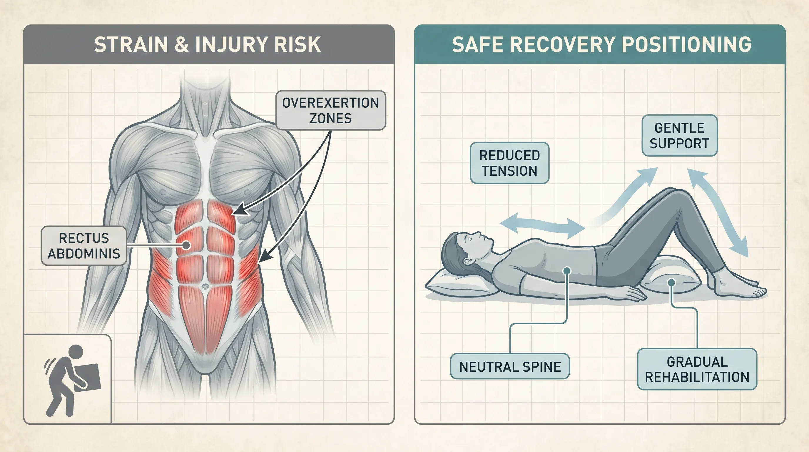
The abdominal wall consists of multiple overlapping muscle groups, each serving distinct functional roles. The rectus abdominis runs vertically along the front of the abdomen, separated into segments by the linea alba, and is primarily responsible for spinal flexion and intra-abdominal pressure regulation. Flanking this central band are the external and internal obliques, which work in tandem to produce trunk rotation and lateral flexion. Deeper still lies the transversus abdominis, the most crucial stabilizer in the entire core system, functioning like a natural corset by compressing the viscera and maintaining lumbar spine alignment. Beneath these muscular layers rests the abdominal fascia, a dense sheet of collagenous tissue that distributes mechanical tension across the entire midsection. Strains most frequently occur at the musculotendinous junction, where muscle fibers transition into tendon, due to the sudden disparity in elasticity between these two tissue types. Understanding this layered architecture explains why certain movements trigger specific pain patterns and why rehabilitation must address the entire kinetic chain rather than isolated fiber clusters.
Common Causes and Risk Factors
The mechanism of injury typically follows one of three pathways: sudden eccentric loading, explosive concentric contraction, or repetitive microtrauma. In athletic settings, sports requiring rapid rotational forces like golf, tennis, baseball pitching, and hockey are notorious for precipitating abdominal tears. Weightlifters often sustain damage during deadlifts, cleans, or overhead presses when core bracing breaks down under maximal load. Beyond athletics, everyday activities pose significant risks. Improper lifting techniques, chronic postural deviations, prolonged sitting followed by sudden exertion, and persistent violent coughing from respiratory infections all strain the abdominal fibers. Certain populations face elevated vulnerability due to physiological or lifestyle factors. Individuals with weak core stabilization, inadequate warm-up routines, poor flexibility, previous abdominal injuries, or nutritional deficiencies exhibit higher susceptibility. Hormonal fluctuations, particularly during pregnancy when the linea alba naturally stretches and the abdominal wall expands, also predispose individuals to strain. Chronic dehydration reduces fascial pliability, while insufficient sleep impairs cellular repair mechanisms, compounding the risk profile.
Recognizing the Symptoms and Severity Grading
Clinical presentation varies significantly based on tissue damage extent, making accurate self-assessment crucial for appropriate intervention. Pain is the universal hallmark, typically described as sharp, pulling, or burning during movement, with noticeable tenderness upon direct palpation. Swelling, localized warmth, and muscle spasms frequently accompany the initial injury phase. Functional limitations emerge as protective guarding mechanisms activate, causing reflexive hesitation during bending, twisting, coughing, or even deep breathing. In severe cases, visible bruising may appear along the lower rib margin or pelvic girdle as microvascular rupture occurs. Recognizing these patterns helps distinguish isolated muscular trauma from more complex visceral or structural conditions.
Grading Severity: Mild to Severe Tears
Medical professionals classify abdominal injuries into three distinct grades to guide treatment intensity and recovery projections, following National Institutes of Health (NIH) clinical grading standards. Grade I strains represent mild microtearing with intact muscle architecture. Patients experience minimal pain, negligible strength loss, and can typically continue modified daily activities without major disruption. Inflammation remains localized, and recovery usually spans fourteen to twenty-one days with basic conservative care. Grade II injuries involve partial fiber disruption. Pain intensifies significantly during contraction or stretching, swelling becomes more pronounced, and functional capacity drops noticeably. Patients often adopt a guarded posture to minimize discomfort, and bruising may emerge within forty-eight hours. Rehabilitation requires structured intervention, typically lasting four to six weeks, with progressive loading carefully monitored to avoid secondary damage. Grade III tears constitute complete muscular or tendinous rupture. These present with immediate, severe pain, profound weakness, palpable gaps in the muscle belly, and extensive swelling. Functional impairment is substantial, often requiring extended immobilization, advanced physical therapy, and occasionally surgical repair. Recovery extends beyond three months, demanding meticulous tissue remodeling protocols.
When to Seek Immediate Medical Attention
While most abdominal strains resolve with appropriate self-care, specific red flags demand prompt clinical evaluation. Unrelenting pain that does not improve with rest, difficulty breathing, or chest discomfort may indicate cardiac or pulmonary involvement rather than isolated musculoskeletal trauma. Nausea, vomiting, fever, or changes in bowel function suggest visceral pathology like appendicitis, cholecystitis, or bowel obstruction. Visible bulging that enlarges with coughing or straining strongly indicates a hernia requiring surgical assessment. Sudden, extensive bruising without direct trauma could signal a rectus sheath hematoma, particularly in individuals taking anticoagulant medications. Neurological symptoms such as numbness, tingling, or radiating pain down the legs warrant immediate imaging to rule out spinal nerve compression. Persistent symptoms beyond ten days despite conservative management also justify professional evaluation to rule out underlying tissue degeneration or improper healing trajectories.
Diagnosis and Medical Evaluation
Accurate diagnosis begins with a thorough clinical history followed by systematic physical assessment. Healthcare providers document injury mechanism, symptom onset, aggravating factors, and prior medical history to establish a comprehensive baseline. Physical examination focuses on palpation mapping, resisted muscle contraction testing, and dynamic movement observation to isolate the precise tissue involved and quantify functional impairment.
Physical Examination Techniques
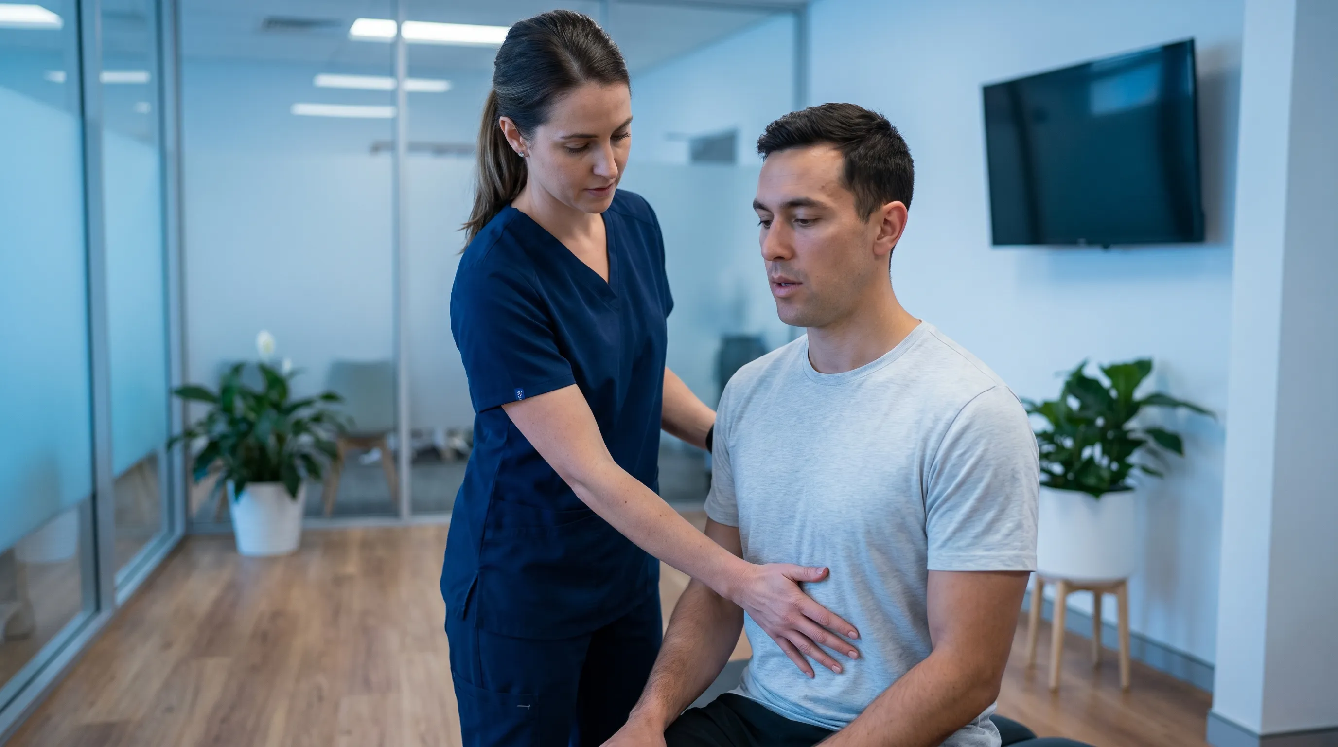
The clinician begins with visual inspection, noting asymmetry, swelling, or skin discoloration. Palpation follows, systematically mapping tender zones while assessing muscle tone and tissue density. Patients are asked to perform active trunk flexion against manual resistance, revealing pain localization and strength deficits. The cough test serves as a functional stressor, reproducing symptoms through sudden intra-abdominal pressure spikes. Range of motion assessments measure flexion, extension, rotation, and lateral bending capabilities, documenting limitations that guide rehabilitation planning. Special orthopedic tests like the Carnett sign help differentiate muscular pain from intra-abdominal organ pathology by asking patients to tense their abdominal muscles during palpation; worsening pain with muscle contraction indicates musculoskeletal origin, while improvement suggests visceral referral. Neurological screening ensures nerve pathways remain intact, ruling out thoracic radiculopathy or intercostal neuralgia that can mimic abdominal strain presentations.
Imaging and Diagnostic Tests
Most uncomplicated cases require no advanced imaging, as clinical evaluation provides sufficient diagnostic clarity. However, persistent or ambiguous symptoms justify further investigation. Ultrasound emerges as the primary imaging modality, offering real-time visualization of muscle fiber continuity, fluid accumulation, and dynamic contraction patterns. It effectively identifies partial tears, hematoma formation, and fascial disruption while remaining non-invasive and radiation-free. Magnetic resonance imaging provides exceptional soft tissue contrast, particularly valuable for grading severe injuries, detecting occult tears, or differentiating between muscular and tendon pathology. X-rays occasionally accompany initial assessments to exclude costochondral fractures, spinal abnormalities, or calcifications, though they do not visualize muscle tissue directly. Blood work remains rarely necessary unless systemic infection, inflammatory myopathy, or coagulation disorders enter the differential diagnosis. Combining clinical examination with targeted imaging ensures accurate classification, preventing mismanagement of overlapping conditions like sports hernias or pelvic floor dysfunction.
Evidence-Based Treatment and Recovery
Effective rehabilitation follows a phased approach aligned with the biological timeline of soft tissue healing. The initial phase prioritizes inflammation management and pain control, the intermediate phase focuses on mobility restoration and controlled loading, and the final phase emphasizes strength rebuilding and functional reintegration. Rushing any stage compromises collagen alignment and increases recurrence risk, making patience and consistency the cornerstones of successful recovery.
The Acute Management Protocol
During the first seventy-two hours, implementing the POLICE principle (Protection, Optimal Loading, Ice, Compression, Elevation) establishes a favorable healing environment, aligning with CDC physical activity and injury prevention guidelines. Protection involves avoiding movements that trigger sharp pain, utilizing supportive bracing or taping techniques when necessary, and modifying daily activities to reduce strain on the anterior core. Optimal loading replaces strict rest with gentle, pain-free movements that maintain tissue pliability and prevent excessive stiffness. Gentle walking, diaphragmatic breathing, and pelvic tilts stimulate circulation without stressing damaged fibers. Ice application, delivered in fifteen to twenty minute intervals every two to three hours, effectively reduces metabolic demand and limits secondary tissue damage. Compression garments or elastic wraps provide external support, minimize edema formation, and enhance proprioceptive feedback during early movement. Proper hydration and sleep optimization during this critical window significantly accelerate cellular repair processes. Nutritional support emphasizes high-quality protein intake, vitamin C, zinc, and omega-3 fatty acids to fuel fibroblast activity and collagen synthesis. Avoiding excessive NSAID use beyond three days remains clinically recommended, as prolonged anti-inflammatory medication can inadvertently disrupt the necessary inflammatory cascade that initiates tissue regeneration.
Medications and Professional Therapies
Pharmacological intervention complements conservative care during peak symptom intensity. Acetaminophen serves as a first-line analgesic for mild to moderate pain without interfering with natural healing pathways. Topical NSAIDs like diclofenac gel provide localized relief while minimizing systemic absorption and gastrointestinal side effects. Muscle relaxants are occasionally prescribed short-term when severe guarding or spasm prevents basic mobility. Beyond medication, professional physical therapy accelerates recovery through targeted interventions. Manual therapy techniques, including myofascial release and gentle tissue mobilization, restore fascial glide and reduce adhesions. Therapeutic ultrasound or low-level laser therapy may be applied to enhance local circulation and stimulate mitochondrial activity. Electrical muscle stimulation maintains neuromuscular activation without active contraction, preventing atrophy during the immobilization phase. Scar tissue mobilization techniques guide collagen fibers to align along functional stress lines, preventing rigid, restrictive healing that limits future range of motion. Practitioners tailor each modality to individual response patterns, adjusting intensity and frequency as tissue tolerance improves.
Advanced Healing Support and Lifestyle Modifications
Sustained recovery extends beyond clinical interventions, requiring comprehensive lifestyle optimization. Sleep architecture directly influences growth hormone release and cytokine regulation, making seven to nine hours of quality rest non-negotiable for optimal tissue regeneration. Stress management techniques like guided breathing, meditation, and progressive muscle relaxation reduce cortisol levels, which otherwise degrade collagen integrity and prolong inflammation. Hydration maintains extracellular matrix viscosity, ensuring nutrients reach damaged zones and metabolic waste clears efficiently. Alcohol and tobacco use must be minimized, as both substances impair angiogenesis and significantly delay muscle fiber repair. Ergonomic adjustments at work and home prevent compensatory movement patterns that overload healing tissue. Sitting posture correction, standing desk utilization, and proper lifting mechanics form foundational habits that protect the recovering core during the vulnerable remodeling phase.
Rehabilitation Exercises and Safe Return to Activity
Progressive loading forms the backbone of functional restoration, transitioning the tissue from fragile repair to resilient strength. Rehabilitation follows a strict hierarchy: establishing neuromuscular control, restoring pain-free mobility, building foundational endurance, developing dynamic strength, and finally reintroducing sport-specific demands. Each phase requires objective benchmarks before advancing, ensuring tissue capacity matches mechanical demands.
Progressive Loading for Tissue Healing
Phase one begins with pain-free activation drills focused on restoring proper breathing mechanics and core stabilization. Diaphragmatic breathing exercises teach controlled intra-abdominal pressure regulation without recruiting compensatory muscles. Pelvic tilts and gentle abdominal bracing reestablish mind-muscle connection while maintaining joint neutrality. Once acute inflammation subsides and range of motion normalizes, phase two introduces isometric contractions. Dead bug variations, modified planks, and supine marching engage stabilizing fibers while minimizing shear forces across healing tissue. Progression depends entirely on symptom response; pain exceeding a three out of ten scale during or after exercise indicates premature advancement. Phase three incorporates dynamic movement patterns emphasizing controlled eccentric loading. Bird dogs, side planks with hip dips, and stability ball marches challenge coordination while reinforcing fascial continuity. Phase four integrates resistance training and rotational movements, utilizing cable machines, medicine balls, and controlled twisting drills. Load increases gradually by five to ten percent weekly, allowing collagen cross-linking to mature under manageable stress. Throughout all phases, movement quality supersedes intensity, prioritizing proper alignment and breath synchronization over volume or speed.
Preventing Re-injury Long-Term
Sustained injury prevention requires fundamental movement reprogramming and consistent maintenance strategies. Core training must evolve beyond traditional crunches, emphasizing integrated stabilization, anti-extension, anti-rotation, and controlled flexion patterns. Incorporating multi-planar movements ensures obliques and transversus abdominis receive adequate stimulus for daily function. Warm-up routines should always precede exertion, featuring dynamic mobility drills, activation sequences, and gradual intensity ramping to prepare fascial networks for load demands. Cool-down protocols incorporating static stretching and breathing restoration accelerate recovery between sessions. Periodization remains critical; alternating high-intensity weeks with deliberate recovery phases prevents cumulative fatigue and tissue breakdown. Addressing biomechanical imbalances like hip tightness, thoracic mobility restrictions, or pelvic floor dysfunction reduces compensatory abdominal overload. Consistent movement practices like yoga, Pilates, or controlled calisthenics build resilient musculature while enhancing proprioception. Regular self-assessment through pain monitoring, mobility testing, and strength tracking identifies early warning signs before they progress into full tears. Long-term success depends on viewing core training as an ongoing maintenance practice rather than a temporary intervention, embedding protective habits into daily routines permanently.
Abdominal Muscle Strain vs Similar Conditions: A Comparison Guide
Accurate differentiation between overlapping clinical presentations prevents inappropriate treatment selection and ensures optimal recovery pathways. The following comparison highlights key distinguishing features between common conditions mimicking abdominal trauma.
| Condition | Primary Symptoms | Key Differentiator | Typical Recovery | Diagnostic Method |
|---|---|---|---|---|
| Abdominal Muscle Strain | Localized pain, tenderness, swelling, pain worsens with contraction or coughing | Pain intensifies with muscle tensing (positive Carnett sign) | 2-8 weeks with conservative care | Clinical exam, ultrasound |
| Inguinal Hernia | Groin/abdominal bulge, heaviness, pain during straining | Visible or palpable protrusion that reduces when lying flat | Surgical repair required for most cases | Physical exam, ultrasound, CT scan |
| Rectus Sheath Hematoma | Sudden severe pain, palpable mass, extensive bruising | History of coughing or anticoagulant use, fixed mass that does not reduce | 4-12 weeks, sometimes drainage | Ultrasound, CT imaging |
| Osteitis Pubis | Deep pelvic pain, tenderness at pubic symphysis, pain with running or kicking | Inflammation of joint rather than muscle, chronic onset | Months to year with graded loading | MRI, clinical provocation tests |
| Gastrointestinal Reflux | Burning upper abdominal pain, acid regurgitation, nausea | Correlates with meals and lying down, no pain with core contraction | Days to weeks with dietary modification | Endoscopy, pH monitoring |
Understanding these distinctions ensures appropriate medical routing and prevents dangerous self-diagnosis. When symptoms deviate from classic muscular patterns or fail to respond to conservative protocols, immediate professional evaluation safeguards against missed visceral pathology or structural compromise.
Frequently Asked Questions
How long does it take for an abdominal muscle strain to heal?
Recovery timelines depend heavily on severity. Grade I strains typically resolve within two to three weeks with proper rest and gentle movement. Grade II injuries often require four to six weeks of structured rehabilitation. Complete tears or Grade III injuries may take three months or longer, sometimes requiring surgical consultation and extended physical therapy. Healing speed varies based on age, nutrition, sleep quality, and adherence to phased rehabilitation protocols.
Can I continue working out with a pulled stomach muscle?
You should immediately stop activities that trigger sharp pain or involve heavy trunk flexion and rotation. Light, pain-free movements like walking or gentle mobility drills are generally acceptable, but pushing through pain can worsen microtears and delay tissue remodeling. Always follow a phased return-to-training protocol under professional guidance, prioritizing movement quality over intensity until full functional capacity returns.
Is heat or ice better for treating an abdominal strain?
During the first forty-eight to seventy-two hours, ice application helps reduce acute inflammation, swelling, and pain. After the acute phase subsides, switching to heat therapy promotes blood flow, relaxes tightened fascia, and prepares the tissue for stretching. Contrast therapy or alternating modalities can also support the transition between healing phases. Always use protective barriers to prevent skin damage during application.
What is the difference between an abdominal strain and a hernia?
An abdominal muscle strain involves damage to the muscle fibers or tendons themselves, causing localized pain, swelling, and tenderness without visible protrusion. A hernia occurs when an internal organ or fatty tissue pushes through a weakened spot in the abdominal wall, often presenting as a visible bulge that worsens with straining or coughing. Clinical examination and imaging are necessary to differentiate the two and guide appropriate treatment pathways.
When should I see a doctor for abdominal pain after exercise?
Seek immediate medical evaluation if you experience severe, unrelenting pain, visible deformity, difficulty breathing, fever, nausea, vomiting, or pain that radiates to your chest or groin. Sudden bruising without trauma, inability to contract the core, or symptoms persisting beyond a week despite conservative care also warrant professional assessment to rule out serious underlying conditions.
Conclusion
Navigating the recovery journey from an abdominal muscle strain requires a blend of clinical knowledge, disciplined rehabilitation, and consistent self-care. By understanding the anatomical complexity of the core system, recognizing early warning signs, and adhering to evidence-based treatment protocols, you establish the strongest possible foundation for complete tissue restoration. Progressive loading, nutritional optimization, sleep prioritization, and biomechanical correction work synergistically to rebuild resilient musculature that withstands future demands. Avoiding premature return to high-intensity activities prevents secondary damage and ensures long-term functional capacity. Whether you are an elite athlete, a weekend enthusiast, or someone navigating daily physical demands, treating your core with respect and attention yields compounding returns in mobility, strength, and injury resilience. Implement the strategies outlined in this guide, monitor your response carefully, and consult healthcare professionals when symptoms deviate from expected healing trajectories. Your abdominal wall supports virtually every human movement, making its preservation a foundational priority for lifelong wellness and sustained physical performance.
Additional Clinical Resources
- NIH StatPearls: Muscle Strains
- Cleveland Clinic: Abdominal Strain Overview
- Mayo Clinic: Muscle Strain Symptoms & Causes
- CDC: Physical Activity & Injury Prevention Guidelines
How we work
Researched and drafted with AI assistance, then reviewed by a human editor against the cited sources.
This article is general health information, not medical advice. Consult a qualified healthcare professional about your own situation.