Why Is My Knee Popping With Every Step: Causes, Solutions & Complete Guide
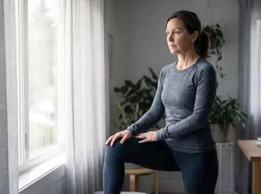
If you have been wondering why is my knee popping with every step, you are not alone. Millions of people experience audible joint sounds during routine movements like walking, climbing stairs, or rising from a seated position (CDC). The human knee is an intricate biomechanical hinge that bears substantial weight while allowing dynamic multi-planar motion. When the delicate balance of cartilage, ligaments, tendons, muscles, and joint fluid is disrupted, the result is often a noticeable click, crack, or pop that echoes with each stride. While many assume joint noises automatically signal damage or inevitable arthritis, the reality is far more nuanced. Some popping represents entirely benign physiological processes, while other instances serve as early warning flags for structural imbalances or progressive wear. Understanding the difference requires a closer look at anatomy, biomechanics, and lifestyle factors that influence joint health over time (Mayo Clinic). This comprehensive guide explores the science behind knee sounds, distinguishes harmless crepitus from clinically significant symptoms, and outlines evidence-based strategies to restore smooth, quiet movement without relying on guesswork or outdated remedies.
Understanding the Mechanics Behind Knee Sounds
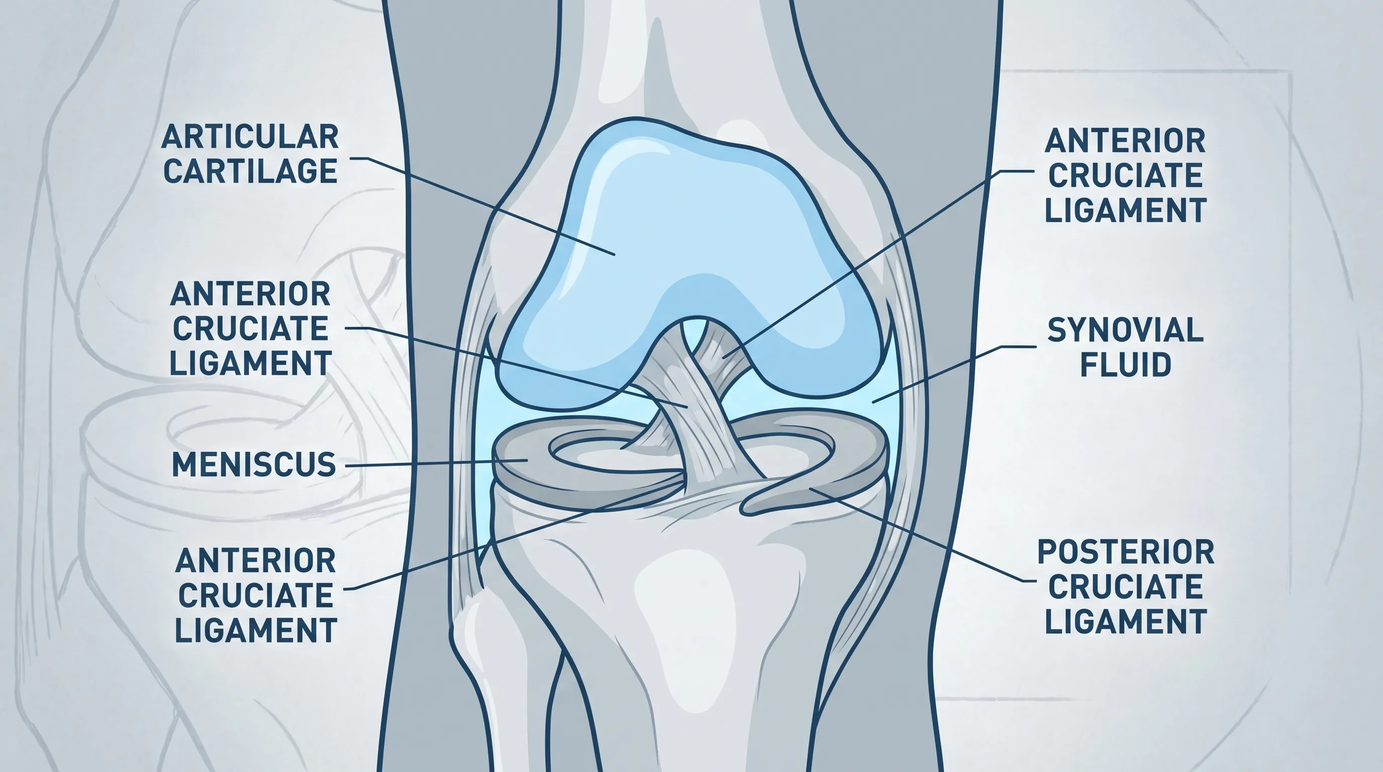
The knee joint operates as a modified synovial hinge, combining elements of both rolling and sliding motion to accommodate complex functional demands. Beneath the surface, three primary bones interact: the femur above, the tibia below, and the patella (kneecap) sitting anteriorly. These bones do not touch directly. Instead, they are separated by articular cartilage, a smooth, rubbery connective tissue that reduces friction and absorbs shock. The joint capsule encloses everything and secretes synovial fluid, which acts as both a lubricant and a nutrient delivery system for avascular cartilage. When any of these components experience altered loading, inflammation, or structural changes, the acoustic environment of the joint shifts dramatically.
Free calculatorBMI CalculatorCalculate Body Mass IndexOpen the calculatorWhat Is Crepitus and How Does It Develop?
Crepitus is the clinical term for the grinding, crunching, or popping sounds and sensations that occur within joints. It is not a disease itself but rather a symptom of underlying mechanical changes (Mayo Clinic). In the knee, crepitus typically emerges from one of three mechanisms. First, nitrogen bubbles within synovial fluid can rapidly form and collapse during joint movement, producing a harmless pop similar to knuckle cracking. Second, tight or inflamed tendons and ligaments may momentarily catch and then snap over bony landmarks as the knee flexes and extends. Third, and most relevant to degenerative concerns, articular cartilage can soften, thin, or develop surface irregularities over time. When the patella glides across the femoral trochlea or when the tibial plateau bears weight on compromised surfaces, these roughened areas create friction that translates into audible grinding or popping. Recognizing which mechanism applies to your specific situation requires careful symptom tracking and, when necessary, professional evaluation.
Normal Joint Sounds Versus Pathological Indicators
Not all knee noises warrant clinical intervention. Many healthy individuals experience occasional pops during deep squats, prolonged sitting followed by standing, or changes in exercise intensity. These transient sounds typically lack pain, swelling, warmth, or functional limitation. Pathological popping, by contrast, tends to be consistent, reproducible, and tied to specific movement patterns that previously felt comfortable. If the sound correlates with mechanical catching, sudden giving way, or progressive stiffness that worsens throughout the day, it suggests an internal derangement or inflammatory process rather than benign gas release. Listening to your body requires balancing awareness without developing kinesiophobia, the fear of movement that often exacerbates joint stiffness through disuse and muscle guarding.
Why Is My Knee Popping With Every Step? Common Causes
When the question why is my knee popping with every step transitions from occasional curiosity to daily reality, several physiological and structural culprits emerge. Each cause carries distinct characteristics, progression patterns, and treatment responses. Identifying the root driver is essential for selecting interventions that address mechanics rather than merely masking noise.
Cartilage Breakdown and Early Osteoarthritis
Articular cartilage relies on consistent compressive loading and synovial fluid diffusion to maintain cellular health. Repetitive microtrauma, previous ligament injuries, congenital alignment variations like genu valgum or varum, and natural aging all contribute to gradual cartilage thinning. As the surface loses its glassy smoothness, it develops fibrillation, pits, and exposed subchondral bone. Weight-bearing then produces audible friction with each step. Early osteoarthritis often presents with morning stiffness lasting under thirty minutes, mild swelling after prolonged activity, and a dull ache that improves with gentle movement but worsens after excessive loading. Research consistently demonstrates that early-stage cartilage changes respond well to load management, targeted strengthening, and anti-inflammatory lifestyle modifications before irreversible structural degradation occurs (NIAMS/NIH).
Patellar Tracking Dysfunction and Biomechanical Imbalances
The patella functions as a floating sesamoid bone embedded within the quadriceps tendon, designed to improve the mechanical advantage of knee extension. For optimal tracking, the kneecap must glide smoothly within the femoral trochlear groove throughout flexion and extension cycles. Imbalances in vastus medialis obliquus strength, tightness in the lateral retinaculum, iliotibial band friction, or hip external rotator weakness frequently pull the patella laterally, creating increased pressure and audible grinding during weight acceptance phases of gait. This maltracking condition, sometimes termed chondromalacia patellae or runner’s knee, often develops in active individuals who experience sudden training spikes, inadequate warm-ups, or prolonged periods of seated desk work that shortens anterior thigh musculature while deactivating posterior chain stabilizers (Cleveland Clinic).
Meniscal Tears and Internal Derangements
The medial and lateral menisci are C-shaped fibrocartilaginous wedges that distribute load, absorb shock, and enhance joint congruity. Acute twisting motions under weight-bearing can produce radial, flap, or bucket-handle tears, while degenerative meniscus changes often appear as horizontal cleavages in older adults. A torn meniscus fragment can shift during knee motion, catching between articular surfaces and producing sharp pops, clicks, or transient locking sensations. Unlike crepitus that sounds uniformly across all ranges of motion, meniscal-related popping typically localizes to a specific joint line angle and often coincides with deep knee flexion or rotational stress. While not all meniscal tears require surgery, those causing mechanical symptoms or persistent instability frequently benefit from orthopedic assessment and tailored rehabilitation protocols (WHO).
Synovial Fluid Alterations and Hyaluronic Acid Changes
Synovial fluid viscosity depends largely on hyaluronic acid concentration and molecular weight. Inflammation, dehydration, metabolic conditions like diabetes, or chronic joint overuse can thin this lubricant, reducing its shock-absorbing capacity. Thinner fluid allows more direct tissue contact during movement, amplifying friction sounds. Restoring optimal joint hydration through consistent low-impact motion, proper systemic hydration, and potentially viscosupplementation injections under medical guidance helps reestablish smoother articulation. The body also naturally cycles synovial fluid during dynamic activity, meaning prolonged immobility paradoxically reduces joint lubrication quality while consistent gentle movement promotes nutrient exchange and waste clearance from the articular environment.
Distinguishing Harmless Noises From Warning Signs
Determining whether popping requires intervention hinges on associated clinical features rather than sound intensity alone. Medical professionals evaluate pain patterns, swelling characteristics, mechanical behavior, and functional limitations to separate physiological noise from structural compromise. Understanding these distinctions empowers individuals to make informed decisions about activity modification and when to seek professional guidance.
The Presence of Pain and Discomfort Patterns
Pain remains the primary differentiator. Benign crepitus occurs without discomfort or post-activity soreness. When popping coincides with sharp, burning, or aching sensations localized around the patella, joint lines, or posterior knee, it suggests tissue stress or inflammation. Activity-related pain that improves after five to ten minutes of movement often indicates tendinopathy or mild synovitis, whereas pain that worsens progressively with continued weight-bearing points toward cartilage stress or meniscal involvement. Nighttime discomfort or rest pain warrants prompt evaluation, as it may reflect advanced joint changes or inflammatory arthropathies.
Swelling, Warmth, and Morning Stiffness
Effusion, commonly known as water on the knee, develops when the synovial membrane reacts to irritation by producing excess fluid. Acute swelling appearing within hours of injury suggests ligament rupture, fracture, or acute meniscal tear. Chronic, low-grade swelling developing over days typically reflects osteoarthritis flare-ups, bursitis, or inflammatory synovitis. Palpating for warmth helps differentiate infectious or inflammatory processes from mechanical issues. Morning stiffness under thirty minutes aligns with degenerative changes, while stiffness persisting beyond sixty minutes often signals systemic inflammatory arthritis requiring specialized rheumatologic management.
Mechanical Catching, Locking, and Instability
A truly alarming sign occurs when the knee physically catches, locks, or gives way during weight-bearing. Locking indicates a displaced tissue fragment blocking full extension, commonly from a meniscal flap or loose osteochondral body. Instability or buckling suggests ligamentous insufficiency, quadriceps inhibition, or severe proprioceptive deficits. These mechanical symptoms rarely resolve spontaneously and typically require diagnostic imaging and structured rehabilitation to restore joint stability and prevent secondary injuries to compensatory joints.
The Clinical Diagnostic Pathway
When home monitoring fails to clarify symptoms or when red flags emerge, professional evaluation becomes essential. Orthopedic and physical therapy specialists utilize systematic assessment frameworks to isolate pain generators, reproduce mechanical symptoms safely, and establish baseline function before initiating treatment.
Orthopedic Assessment and Special Movement Tests
A comprehensive knee examination begins with observation, palpation, and range-of-motion measurement. Clinicians assess gait symmetry, quadriceps atrophy, joint effusion presence, and patellar mobility. Special tests like the McMurray maneuver, Thessaly test, patellar grind test, and Lachman exam help isolate meniscal, patellofemoral, and ligamentous structures. Functional assessments including single-leg squats, step-down tests, and hop mechanics reveal biomechanical compensations that contribute to repetitive joint stress. Movement quality analysis identifies faulty loading patterns that sustain popping and discomfort during routine activities.
Imaging Studies and Advanced Diagnostic Modalities
While physical examination remains foundational, imaging clarifies structural realities when symptoms persist or worsen. Plain radiographs evaluate bone alignment, joint space narrowing, osteophyte formation, and calcific deposits. Magnetic resonance imaging provides detailed visualization of soft tissues, revealing meniscal tear patterns, cartilage thickness variations, ligament integrity, and synovial inflammation. Ultrasound dynamically assesses tendon movement, fluid collections, and real-time tracking abnormalities during flexion. Selecting the appropriate modality depends on symptom duration, trauma history, age factors, and response to initial conservative care. Imaging alone never dictates treatment; clinical correlation remains paramount to avoid overdiagnosing incidental findings common in asymptomatic populations.
Evidence-Based Treatment and Management Approaches
Effective management of knee popping requires addressing underlying causes rather than suppressing symptoms. Modern orthopedic rehabilitation emphasizes active recovery, neuromuscular retraining, and lifestyle optimization over passive interventions that offer temporary relief without structural improvement.
Conservative Care and Activity Modification
Initial management centers on load management and inflammation control. The traditional RICE protocol has evolved into PEACE and LOVE frameworks, emphasizing protection, elevation, avoidance of anti-inflammatories in acute phases, compression, education, followed by optimal loading, vascularization, and exercise (Mayo Clinic). Activity modification involves temporarily reducing high-impact stressors, substituting painful movements with biomechanically sound alternatives, and pacing daily tasks to prevent cumulative joint overload. Cross-training with swimming, cycling, or elliptical machines maintains cardiovascular fitness while allowing articular tissues to recover. Gradual exposure to previously aggravating activities rebuilds tolerance without triggering symptom recurrence.
Targeted Physical Therapy Protocols
Physical therapy remains the cornerstone of functional knee restoration. Programs prioritize quadriceps and gluteal strengthening to normalize patellar tracking and reduce tibiofemoral shear forces. Eccentric loading protocols improve tendon resilience, while proprioceptive retraining restores neuromuscular control essential for dynamic stability. Manual therapy techniques address joint capsule restrictions, soft tissue adhesions, and mobility deficits limiting smooth articulation. Therapists utilize cueing strategies to modify squat depth, foot progression angle, and trunk lean, directly influencing joint reaction forces. Consistency with home exercise programs determines long-term outcomes, making adherence strategies and progressive overload principles critical components of successful rehabilitation.
Pharmacological and Procedural Interventions
When conservative measures plateau, adjunctive therapies may provide additional relief. Topical NSAIDs reduce localized inflammation with minimal systemic absorption. Oral anti-inflammatories and acetaminophen manage moderate discomfort during rehabilitation phases. Intra-articular corticosteroid injections temporarily suppress acute synovitis, facilitating participation in physical therapy. Hyaluronic acid injections aim to restore synovial viscosity, though evidence varies regarding long-term efficacy. Platelet-rich plasma and stem cell therapies remain under active investigation, showing promise for early cartilage preservation but lacking standardized protocols and insurance coverage. Surgical intervention, including arthroscopic debridement, meniscal repair, or joint replacement, reserves for severe mechanical blockages, refractory symptoms, or advanced degenerative changes unresponsive to exhaustive non-operative care (Mayo Clinic).
Daily Lifestyle Modifications for Long-Term Joint Health
Joint longevity depends heavily on daily habits that either protect or accelerate articular wear. Sustainable knee health requires attention to movement mechanics, footwear selection, body composition, nutritional support, and recovery practices that collectively influence tissue resilience and inflammatory status.
Biomechanics, Footwear, and Gait Retraining
Footwear directly influences ground reaction forces transmitted upward through the kinetic chain. Shoes with adequate arch support, heel cushioning, and appropriate toe-splay capacity maintain natural foot mechanics, reducing excessive internal tibial rotation that strains the knee joint. Worn-out midsoles lose shock absorption within three to six months of regular use, silently increasing joint loading. Gait analysis identifies overstriding, heel-striking dominance, or asymmetrical weight distribution that amplifies patellofemoral stress. Retraining toward midfoot strike patterns, shorter stride lengths, and higher cadence reduces peak knee flexion moments, directly addressing why is my knee popping with every step during walking routines.
Weight Management and Anti-Inflammatory Nutrition
Every additional pound of body weight translates to approximately three to four extra pounds of compressive force across the knee during stance phase (Cleveland Clinic). Gradual, sustainable weight reduction significantly decreases joint stress while improving metabolic markers linked to systemic inflammation. Dietary patterns emphasizing omega-3 fatty acids, leafy greens, berries, turmeric, and high-quality protein support collagen synthesis and modulate inflammatory pathways. Hydration maintains synovial fluid volume and viscosity. Avoiding excessive refined sugars, processed oils, and chronic alcohol consumption prevents glycation end-product accumulation that stiffens connective tissues and accelerates degenerative changes. Nutritional timing combined with movement creates a synergistic environment for joint preservation (CDC).
Comparison of Activity Impact Levels on Knee Joint Loading
| Activity Type | Joint Stress Level | Recommended Frequency | Best Use Cases |
|---|---|---|---|
| Walking on soft terrain | Low to Moderate | 5-7 days weekly | Baseline mobility, recovery days |
| Cycling with proper fit | Low to Moderate | 3-5 days weekly | Cardio conditioning without impact |
| Swimming / Aquatic exercise | Very Low | 2-4 days weekly | Rehabilitation, high body mass individuals |
| Jogging on pavement | High | 2-3 days weekly | Conditioned athletes, gradual progression |
| Deep squats with heavy load | Very High | 1-2 days weekly | Strength training, advanced mobility |
Safe At-Home Rehabilitation and Mobility Drills
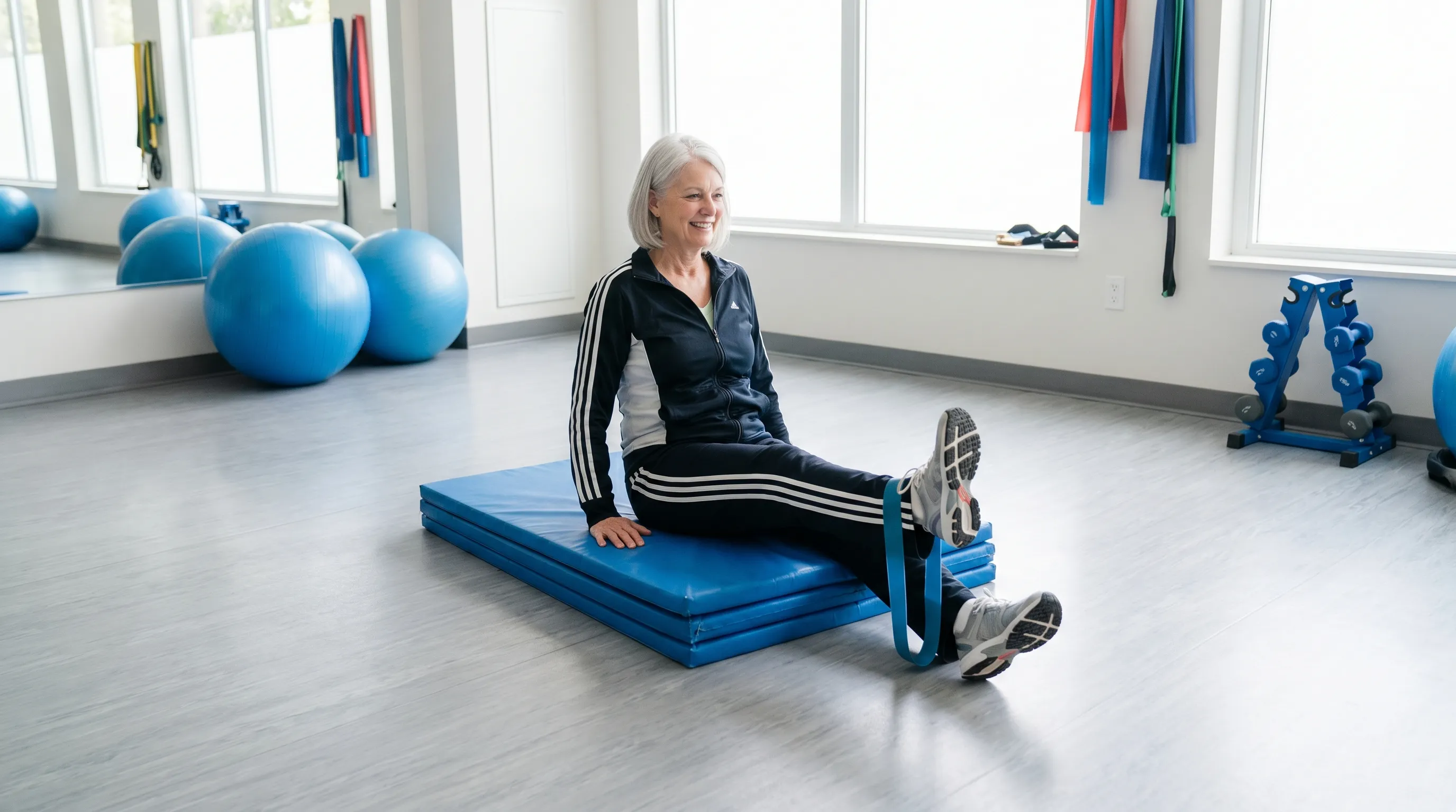
Implementing consistent, progressive exercises builds tissue tolerance while restoring optimal movement patterns. Home rehabilitation bridges clinical sessions and daily function, requiring proper technique, gradual intensity progression, and attentive monitoring of symptom responses.
Foundational Strengthening Movements
Quadriceps setting involves tightening the front thigh while keeping the knee straight, holding for five seconds before relaxing. This movement reactivates neuromuscular pathways without joint loading. Straight leg raises progress this activation by lifting the heel four inches off the floor while maintaining full extension. Clamshells and side-lying leg lifts target gluteus medius, stabilizing the pelvis and preventing dynamic knee valgus during gait. Mini-squats limited to thirty degrees of flexion build tolerance before progressing to full range as symptoms permit. Consistency with three to four sets of ten to fifteen repetitions daily establishes a foundation for joint protection and functional strength.
Flexibility and Neural Mobilization
Tight hip flexors, hamstrings, and gastrocnemius muscles alter lower extremity alignment, increasing compressive forces on the patellofemoral compartment. Gentle static stretching held for thirty seconds after warm-up improves tissue extensibility without triggering protective muscle guarding. Foam rolling addresses myofascial restrictions in the iliotibial band, quadriceps, and calves, temporarily reducing perceived stiffness and preparing tissues for strengthening. Ankle mobility drills including knee-to-wall lunges and dorsiflexion stretches ensure proper tibia tracking, preventing compensatory knee valgus that frequently contributes to tracking dysfunction and audible friction during stance phase.
Integrating these drills requires patience and progressive overload principles. Tissue adaptation occurs over weeks, not days. Initial mild muscle fatigue indicates appropriate stimulus, whereas sharp joint pain, increased swelling, or mechanical symptoms signal the need to reduce volume, modify technique, or consult a healthcare provider. Tracking repetitions, ranges of motion, and symptom responses in a simple journal establishes objective progress markers, replacing subjective anxiety with measurable improvement. Remember that addressing why is my knee popping with every step requires viewing rehabilitation as a long-term investment rather than a quick fix. Sustainable movement habits built through consistency yield greater joint longevity than intermittent intensive sessions that provoke flare-ups.
Frequently Asked Questions
Is it normal for my knees to pop when I walk?
Occasional popping without accompanying pain, swelling, or instability typically represents benign crepitus from gas bubble dynamics or tendon gliding over bone. However, consistent popping synchronized with every step warrants attention, especially if you notice progressive stiffness, altered gait patterns, or reduced endurance during previously manageable activities. Monitoring symptom patterns over two to three weeks helps distinguish normal joint acoustics from early structural changes requiring intervention.
Can weak muscles cause knee popping?
Absolutely. The quadriceps, gluteals, and core muscles function as dynamic stabilizers that control joint kinematics and distribute compressive loads evenly. When these muscles weaken or activate inefficiently, the patella tracks abnormally, and tibiofemoral alignment shifts under weight-bearing. This mechanical misalignment increases friction between articular surfaces, producing audible grinding or clicking sounds. Targeted strengthening restores proper tracking mechanics, often eliminating popping without surgical or pharmacological intervention.
How do I stop my knee from popping with every step?
Begin with conservative strategies including supportive footwear, activity pacing, and consistent low-impact mobility work. Implement a structured rehabilitation program emphasizing quadriceps strengthening, hip stabilizer activation, and soft tissue flexibility. Address modifiable factors like excess body weight, prolonged sitting posture, and repetitive high-impact loading. If symptoms persist beyond four to six weeks of consistent self-care, seek evaluation from a physical therapist or orthopedic specialist to identify biomechanical deficits and receive individualized treatment guidance (Mayo Clinic).
Should I be worried if my knee cracks when I squat?
Cracking during deep knee flexion can be normal if completely pain-free and mechanically smooth. Many individuals experience harmless crepitus at end-range positions where joint capsules and tendons experience maximum tension. Concern increases when cracking occurs at mid-range flexion, coincides with sharp pain, causes the knee to catch or lock, or follows a specific injury event. In these scenarios, professional assessment differentiates benign physiological sounds from meniscal involvement, cartilage softening, or ligament strain requiring targeted management.
Will knee popping go away on its own?
Gas-related crepitus fluctuates naturally and often resolves spontaneously with hydration and gentle movement. Popping stemming from muscular imbalances, tracking dysfunction, or early cartilage changes rarely disappears without addressing underlying causes. Ignoring symptomatic noise allows compensatory patterns to solidify, accelerating joint wear and reducing functional capacity. Proactive management through consistent exercise, biomechanical optimization, and lifestyle adjustments significantly improves outcomes, often eliminating popping entirely or reducing it to occasional, asymptomatic occurrences (NIAMS/NIH).
Conclusion
Navigating the question of why is my knee popping with every step requires moving beyond fear-based assumptions and embracing evidence-based understanding. Joint sounds exist on a continuum ranging from harmless physiological acoustics to early indicators of structural imbalance or tissue stress. By recognizing associated symptoms, tracking functional patterns, and implementing progressive rehabilitation strategies, most individuals restore smooth, quiet movement while protecting long-term joint integrity. The knee responds remarkably well to consistent loading management, targeted strengthening, and lifestyle optimization when addressed proactively rather than reactively. Avoid prolonged immobilization that stiffens connective tissues, reject passive treatments that ignore underlying mechanics, and prioritize sustainable movement habits that build resilience over time. Professional guidance accelerates recovery when symptoms persist, mechanical limitations emerge, or self-management plateaus. Ultimately, joint health thrives on informed action, consistent effort, and the understanding that audible movement alone does not equal damage. Addressing biomechanical inefficiencies, maintaining healthy body composition, and committing to regular mobility work ensures your knees continue supporting an active, fulfilling lifestyle for years ahead (WHO).
How we work
Researched and drafted with AI assistance, then reviewed by a human editor against the cited sources.
This article is general health information, not medical advice. Consult a qualified healthcare professional about your own situation.